. 2022 Oct 31;10(1):157.
doi: 10.1186/s40478-022-01457-x.

Artificial intelligence-derived neurofibrillary tangle burden is associated with antemortem cognitive impairment

Affiliations

Artificial intelligence-derived neurofibrillary tangle burden is associated with antemortem cognitive impairment

Gabriel A Marx et al. Acta Neuropathol Commun. .

Abstract

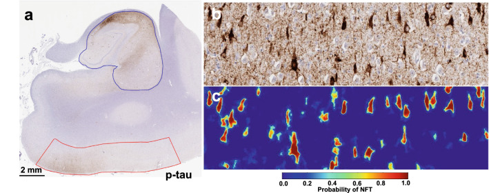
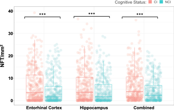
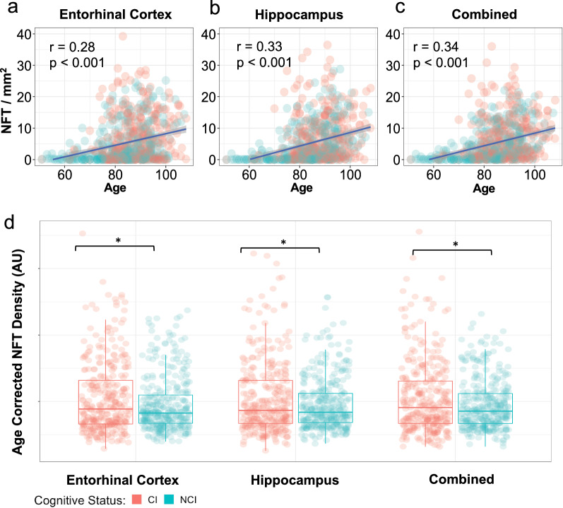
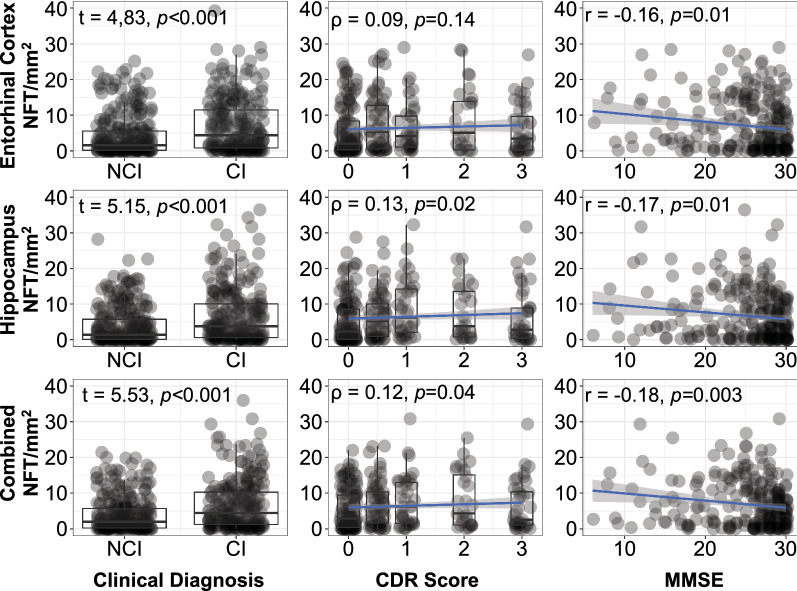
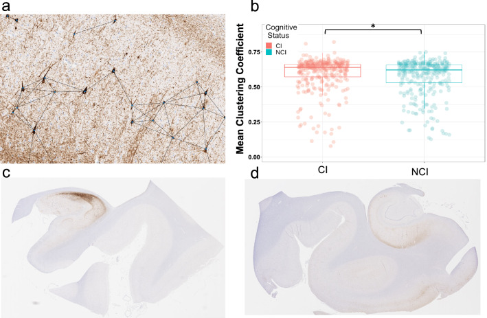
Tauopathies are a category of neurodegenerative diseases characterized by the presence of abnormal tau protein-containing neurofibrillary tangles (NFTs). NFTs are universally observed in aging, occurring with or without the concomitant accumulation of amyloid-beta peptide (Aβ) in plaques that typifies Alzheimer disease (AD), the most common tauopathy. Primary age-related tauopathy (PART) is an Aβ-independent process that affects the medial temporal lobe in both cognitively normal and impaired subjects. Determinants of symptomology in subjects with PART are poorly understood and require clinicopathologic correlation; however, classical approaches to staging tau pathology have limited quantitative reproducibility. As such, there is a critical need for unbiased methods to quantitatively analyze tau pathology on the histological level. Artificial intelligence (AI)-based convolutional neural networks (CNNs) generate highly accurate and precise computer vision assessments of digitized pathology slides, yielding novel histology metrics at scale. Here, we performed a retrospective autopsy study of a large cohort (n = 706) of human post-mortem brain tissues from normal and cognitively impaired elderly individuals with mild or no Aβ plaques (average age of death of 83.1 yr, range 55-110). We utilized a CNN trained to segment NFTs on hippocampus sections immunohistochemically stained with antisera recognizing abnormal hyperphosphorylated tau (p-tau), which yielded metrics of regional NFT counts, NFT positive pixel density, as well as a novel graph-theory based metric measuring the spatial distribution of NFTs. We found that several AI-derived NFT metrics significantly predicted the presence of cognitive impairment in both the hippocampus proper and entorhinal cortex (p < 0.0001). When controlling for age, AI-derived NFT counts still significantly predicted the presence of cognitive impairment (p = 0.04 in the entorhinal cortex; p = 0.04 overall). In contrast, Braak stage did not predict cognitive impairment in either age-adjusted or unadjusted models. These findings support the hypothesis that NFT burden correlates with cognitive impairment in PART. Furthermore, our analysis strongly suggests that AI-derived metrics of tau pathology provide a powerful tool that can deepen our understanding of the role of neurofibrillary degeneration in cognitive impairment.

Keywords: Alzheimer’s disease; Computer vision; Convolutional neural network; Deep learning; Digital pathology; Neurofibrillary tangle; Primary age-related tauopathy; Tauopathy.


Conflict of interest statement

G.F., J.Z., and C.C-C., serve as executive leadership for PerciseDx a private company.

Figures

Fig. 1
Fig. 1
Detection of neurofibrillary tangles (NFT) in phospho-tau (AT8) immunohistochemically stained whole slide images (WSI). a Example of a hippocampal WSI immunohistochemically stained for phosphorylated-tau (AT8). The hippocampus proper (blue) and entorhinal region (red) were manually segmented. b High-power (20x) representative image of the hippocampal CA2 subfield showing p-tau positive neurofibrillary tangles. c Corresponding output of above image passed through semantic segmentation model that identifies NFT. Each pixel value corresponds to the probability that it represents an NFT
Fig. 2
Fig. 2
Increased neurofibrillary tangle (NFT) counts in cognitively impaired subjects. NFT densities are shown split by cognitive status, cognitively impaired (CI) and no cognitive impairment (NCI). NFT counts of the entorhinal cortex, hippocampus, and both regions combined are presented. Triple asterisks (***) denote p < 0.0001 based on a two-sample t-test between groups. Two-way analysis of variance yielded a F-statistic of 58.99
Fig. 3
Fig. 3
AI-detected NFT counts by region with respect to age and cognitive status. ac Relationship between NFT counts and age, grouped by cognitive status in Entorhinal Cortex (a), Hippocampus (b), and combined (c). Pearson correlation values between age and region’s NFT density are shown with associated p value. d Age adjusted NFT density group difference by region. Asterisks denote p < 0.05 based on a two-sample t-test between groups. Two-way analysis of variance yielded a F-statistic of 4.23
Fig. 4
Fig. 4
Relationship between NFT counts by region and each individual cognitive variable. In this analysis we used a loose label of cognitive impairment as a composite metric based on MMSE, CDR, or documented clinical history of cognitive impairment. This figure shows the relationships between AI-detected NFT counts by region and each individual clinical variable. (Left column) Two-sample t-tests were performed for documented clinical history of cognitive impairment. (Middle column) Spearman rho correlation was performed between NFT count and CDR score. (Right column) Pearson r correlation was performed between NFT count and MMSE
Fig. 5
Fig. 5
NFT position as a geometric network and subsequent graph metrics. a A representation of NFT position as a geometric network. Each NFT is represented as a node in a unidirectional binary graph, where an edge exists between two nodes if the Euclidean distance between them is less than some value r. In this figure r = 250 μm. b Group comparison of non-cognitively impaired (NCI) vs cognitively impaired (CI) mean clustering coefficient. Asterisk denotes p < 0.05. Two-sample t-test between groups yielded a t-statistic of  − 2.97 and p = 0.0031. c: Example of hippocampal whole slide image with high mean clustering coefficient (0.75). d Example of hippocampal whole slide image with low mean clustering coefficient (0.47)
Fig. 6
Fig. 6
Odds ratio of cognitive impairment on mean NFT clustering coefficient for a range of given distance thresholds. Since the cutoff of r in our mean NFT clustering coefficient metric has no ground truth, we tested it across a large range of values. Red lines bounding shaded areas demark the upper and lower bounds of the 95% confidence interval. Mean NFT clustering coefficient significantly predicts cognitive impairment for distance thresholds between 300 px (151.98 microns) and 1200 px (607.92 microns)

References

    1. Arriagada PV, Growdon JH, Hedley-Whyte ET, Hyman BT. Neurofibrillary tangles but not senile plaques parallel duration and severity of Alzheimer’s disease. Neurology. 1992;42:631–631. doi: 10.1212/WNL.42.3.631. - DOI - PubMed
    1. Hernández F, Avila J. Tauopathies. Cell Mol Life Sci. 2007;64:2219–2233. doi: 10.1007/s00018-007-7220-x. - DOI - PMC - PubMed
    1. Crary JF, Trojanowski JQ, Schneider JA, Abisambra JF, Abner EL, Alafuzoff I, et al. Primary age-related tauopathy (PART): a common pathology associated with human aging. Acta Neuropathol (Berl) 2014;128:755–766. doi: 10.1007/s00401-014-1349-0. - DOI - PMC - PubMed
    1. Rodriguez RD, Grinberg LT. Argyrophilic grain disease: an underestimated tauopathy. Dement Neuropsychol. 2015;9:2–8. doi: 10.1590/S1980-57642015DN91000002. - DOI - PMC - PubMed
    1. Mohandas E, Rajmohan V. Frontotemporal dementia: an updated overview. Indian J Psychiat. 2009;51:S65–S69. - PMC - PubMed

Publication types