Review
. 2022 Dec 1;323(6):C1757-C1776.
doi: 10.1152/ajpcell.00147.2022. Epub 2022 Nov 1.

Neuroendocrine signaling in the skin with a special focus on the epidermal neuropeptides

Affiliations
Review

Neuroendocrine signaling in the skin with a special focus on the epidermal neuropeptides

Andrzej T Slominski et al. Am J Physiol Cell Physiol. .

Abstract

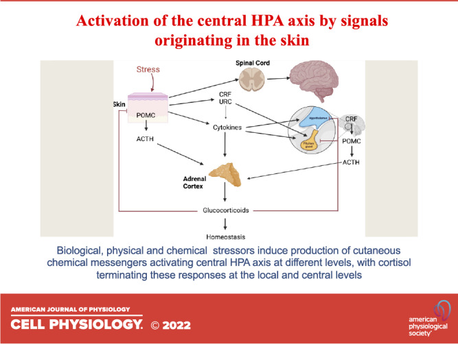
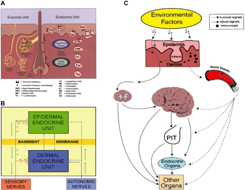
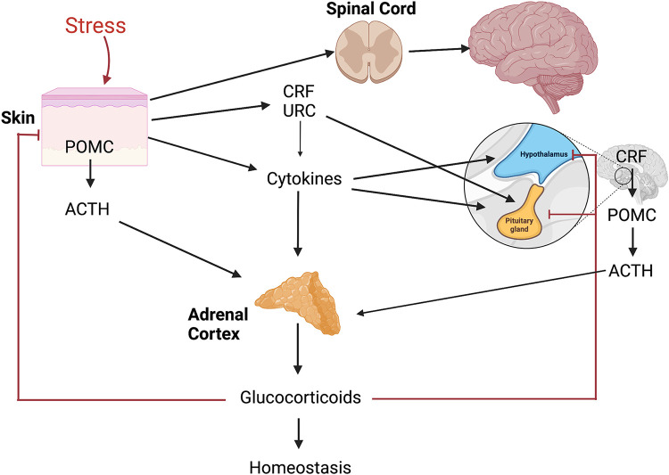
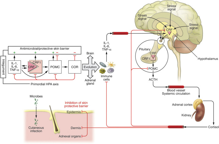
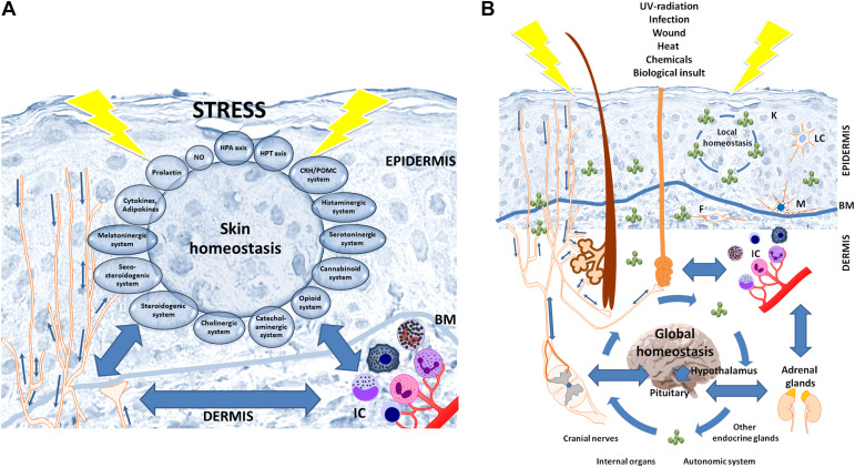
The skin, which is comprised of the epidermis, dermis, and subcutaneous tissue, is the largest organ in the human body and it plays a crucial role in the regulation of the body's homeostasis. These functions are regulated by local neuroendocrine and immune systems with a plethora of signaling molecules produced by resident and immune cells. In addition, neurotransmitters, endocrine factors, neuropeptides, and cytokines released from nerve endings play a central role in the skin's responses to stress. These molecules act on the corresponding receptors in an intra-, juxta-, para-, or autocrine fashion. The epidermis as the outer most component of skin forms a barrier directly protecting against environmental stressors. This protection is assured by an intrinsic keratinocyte differentiation program, pigmentary system, and local nervous, immune, endocrine, and microbiome elements. These constituents communicate cross-functionally among themselves and with corresponding systems in the dermis and hypodermis to secure the basic epidermal functions to maintain local (skin) and global (systemic) homeostasis. The neurohormonal mediators and cytokines used in these communications regulate physiological skin functions separately or in concert. Disturbances in the functions in these systems lead to cutaneous pathology that includes inflammatory (i.e., psoriasis, allergic, or atopic dermatitis, etc.) and keratinocytic hyperproliferative disorders (i.e., seborrheic and solar keratoses), dysfunction of adnexal structure (i.e., hair follicles, eccrine, and sebaceous glands), hypersensitivity reactions, pigmentary disorders (vitiligo, melasma, and hypo- or hyperpigmentary responses), premature aging, and malignancies (melanoma and nonmelanoma skin cancers). These cellular, molecular, and neural components preserve skin integrity and protect against skin pathologies and can act as "messengers of the skin" to the central organs, all to preserve organismal survival.

Keywords: epidermis; keratinocytes; melanocytes; neurohormones; neuropeptides.


Conflict of interest statement

No conflicts of interest, financial or otherwise, are declared by the authors.

Figures

None
Graphical abstract
Figure 1.
Figure 1.
Neuroendocrine organization of the skin. A: components of the exocrine and endocrine units of the skin produce hormones, neural mediators, and cytokines that act upon the corresponding receptors expressed on skin cells. B: the communication between epidermal and dermal neuroendocrine units and the systemic level. C: the cutaneous neuroendocrine system communicates with the central nervous, endocrine, and immune system or other organs through humoral or neural pathways. Reprinted from Ref. with the permission of the publisher.
Figure 2.
Figure 2.
The organization of central hypothalamus-pituitary-adrenal (HPA) axis that can be activated at different levels by stress originating in the skin. CRH, corticotropin releasing hormone; POMC, proopiomelanocortin; URC, urocortin. Figure was created using BioRender.
Figure 3.
Figure 3.
The evolutionary origin of the hypothalamus-pituitary-adrenal (HPA) axis. Right: organization of the central HPA axis; left: primordial integumental HPA axis. CRH, corticotropin releasing hormone; POMC, proopiomelanocortin. Reprinted from Ref. with the permission of the publisher.
Figure 4.
Figure 4.
Skin neuroimmunoendocrine system encompassing the epidermal neuroendocrine system (A), and interactions with the central systems and organs (B) in a coordinated stress response mode. A: epidermal cells are not only are sensitive to neurohormonal regulation but also produce elements of hypothalamus-pituitary-adrenal (HPA) or hypothalamus-pituitary-thyroid (HPT) axes, other neuropeptides, biogenic amines, serotonin, melatonin, nitric oxide, opioids, cannabinoids, catecholamines, acetylcholine, steroids, secosteroids, neurotrophins, and cytokines. B: the skin neuroimmunoendocrine system can activate the central responses with direct homeostatic, metabolic, and phenotypic consequences. The constant exchange of neuroendocrine mediators between skin and other organs is responsible for the maintenance of local and global homeostasis. BM, basement membrane; F, fibrocytes/fibroblasts; IC, immune cells; K, keratinocytes; LC, Langerhans cells; M, melanocytes. Reprinted from Ref. with the permission of the publisher.
Figure 5.
Figure 5.
Time course of the changes of intracellular Ca2+ after corticotropin-releasing hormone (CRH) or urocortin (URC) stimulation. I): time galleries of fluorescence images of the Ca2+ indicators in the HaCaT cells after stimulation with CRF (10−7 M, left) and urocortin (URC) (10−7 M, right). The intensity range (8 bit) is indicated below the image galleries. II), A and B: time course of the relative fluorescence intensities (unbroken line, cytosol; broken line, nucleus) of the Ca2+ indicators in HaCaT cells after stimulation with CRF (A, 10−7 M) and URC (B, 10−7 M). C: dose-dependent frequency of the oscillations in HaCaT-cells after stimulation with URC (means ± SD). Number of cells recorded (n) is shown in the respective columns. D: dose-dependent numbers of HaCaT cells with oscillations after stimulation with URC (percentage of total number of cells recorded). Number of cells recorded with oscillations (n) is shown in the respective columns. Reprinted from Ref. with the permission of the publisher.
Figure 6.
Figure 6.
Differential and overlapping phenotypic effects secondary to activation of corticotropin-releasing hormone receptor 1 (CRHR1) in human keratinocytes and melanocytes. A: activation of CRHR1 in normal epidermal keratinocytes and melanocytes, respectively stimulates and inhibits NF-κB activity. In immortalized HaCaT keratinocytes, CRF both inhibits and stimulates NF-κB activity, depending on the environmental context (171). B: in keratinocytes, activation of CRHR1 directly inhibits proliferation and stimulates differentiation and immune activity via stimulation of NF-κB. This enhances protective epidermal barrier function. In melanocytes, CRHR1 directly and indirectly [through proopiomelanocortin (POMC) peptides] stimulates differentiation and melanin production leading to enhancement of protective barrier function. In contrast to keratinocytes, there is an indirect (through POMC peptides) inhibition of NF-κB with subsequent suppression of immune activity, which can be amplified by production of cortisol by melanocytes. Reprinted from Ref. with the permission of the publisher.

References

    1. Fuchs E. Epithelial skin biology: three decades of developmental biology, a hundred questions answered and a thousand new ones to address. Curr Top Dev Biol 116: 357–374, 2016. doi:10.1016/bs.ctdb.2015.11.033. - DOI - PMC - PubMed
    1. Ramot Y, Bohm M, Paus R. Translational neuroendocrinology of human skin: concepts and perspectives. Trends Mol Med 27: 60–74, 2021. doi:10.1016/j.molmed.2020.09.002. - DOI - PubMed
    1. Elias PM. Structure and function of the stratum corneum extracellular matrix. J Invest Dermatol 132: 2131–2133, 2012. doi:10.1038/jid.2012.246. - DOI - PMC - PubMed
    1. Slominski AT, Zmijewski MA, Skobowiat C, Zbytek B, Slominski RM, Steketee JD. Sensing the environment: regulation of local and global homeostasis by the skin’s neuroendocrine system. Adv Anat Embryol Cell Biol 212: v, vii, 1–115, 2012. doi:10.1007/978-3-642-19683-6_1. - DOI - PMC - PubMed
    1. Slominski A, Wortsman J, Paus R, Elias PM, Tobin DJ, Feingold KR. Skin as an endocrine organ: implications for its function. Drug Discov Today Dis Mech 5: 137–144, 2008. doi:10.1016/j.ddmec.2008.04.004. - DOI - PMC - PubMed

LinkOut - more resources