Review
. 2023 Feb;96(1142):20220164.
doi: 10.1259/bjr.20220164. Epub 2022 Nov 28.

Incidentalomas in the head & neck

Affiliations
Review

Incidentalomas in the head & neck

Andrew McQueen et al. Br J Radiol. 2023 Feb.

Abstract

Incidental findings (IFs) in the head & neck are a frequent challenge to the reporting radiologist. A combination of complex anatomy, widely varied imaging techniques and the high prevalence of benign pathology, makes safe and appropriate management of head & neck IFs problematic. The non-head & neck radiologist is unlikely to have prior personal experience of the relevant specialties or current involvement with the pertinent multidisciplinary teams, creating unfamiliarity with both the clinical aspects of head & neck disease and the value of examination techniques. This triumvirate of complex anatomy, pathology and imaging creates the perfect environment for excessive investigation and overdiagnosis. In this article, the most frequently encountered and clinically relevant head & neck IFs are summarised. To reflect daily clinical practice, we will firstly consider anatomic abnormalities identified on cross-sectional imaging and ultrasound, followed by a review of PET-CT incidental findings.


Figures

Figure 1.
Figure 1.
Showing concerning and suspicious US features. (a) Multiple punctate hyperechoic foci indicating microcalcifications. (b) Markedly hypoechoic thyroid nodule (c.f. muscles). (c) Irregular and lobulated nodule margins. (d) Taller-than-wider thyroid nodule.
Figure 2.
Figure 2.
Abnormal lower neck lymph nodes on CT. (a) Enlarged lymph node (arrow). (b) Lymph node with central hypodense region indicating cystic change (arrow). (c) Lymph node with central hyperdense focus on indicating calcification (arrow). (d) Hyperenhancing lymph node with enhancement similar to the adjacent vessels (arrow)
Figure 3.
Figure 3.
CT features of thyroid malignancy. (a) Irregular margins along the sternomastoid muscle (arrow) and the posterior thyroid capsule (dotted arrow). (b) Features of vocal cord palsy showing dilated laryngeal ventricle/sail sign (arrow) and ipsilateral pyriform sinus (asterisk).
Figure 4.
Figure 4.
FDG PET-CT images for different patients showing incidental FDG uptake at the thyroid gland. (a) Diffuse FDG uptake throughout the thyroid gland (thyroiditis). (b) Focus of marked FDG uptake at the right lobe of the thyroid (biopsy confirmed papillary thyroid carcinoma). (c) Focus of marked FDG uptake at the right lobe of the thyroid (U2 benign appearance on US - stable on US surveillance).
Figure 5.
Figure 5.
Examples of a thyroglossal duct cyst. (a) Well-defined, simple and entirely anechoic. (b) Fine septations within an anechoic cyst. (c) Appearances of a pseudosolid cyst, note the posterior acoustic enhancement (left) and the lack of colour Doppler signal (right).
Figure 6.
Figure 6.
Incidental left parotid lesion on axial T2 (left) and coronal T1 (right) brain MRI. US guided FNA was consistent with pleomorphic adenoma. Management = surgical resection with superficial parotidectomy.
Figure 7.
Figure 7.
FDG PET-CT images for different patients showing FDG avid nodules in the parotid gland. (a) Focus of marked uptake at small nodule in the left parotid gland (SUVmax 5.1). Biopsy confirmed Warthin’s tumour. (b) Intense FDG uptake at a large nodule in the right parotid gland (SUVmax 24.9). Biopsy confirmed malignant melanoma metastasis.
Figure 8.
Figure 8.
Common benign sinus IFs on axial T2 MRI. Left – Convex, well defined mucous retention cyst in antrum (arrow), without secretions or mucosal thickening. Middle – Layer of bubbly secretions in right antrum (dotted arrow).Right – Bilateral, asymmetric antral mucosal thickening (arrowheads).
Figure 9.
Figure 9.
Axial T2 MRI (left) showing bilateral nasal cavity masses with antral opacification. ENT referral led to fibreoptic nasendoscopy demonstrating bilateral nasal polyps. Coronal CT sinuses (right) showed grade 2 inflammatory nasal polyps with pan sinus opacification.
Figure 10.
Figure 10.
Completely opacified right antrum on T2 axial MRI (left) in a patient with facial pain. Panoramic reconstruction CBCT (right) demonstrated an opacified maxillary antrum with periapical inflammatory lucency at the restored upper right 6 breaching the antral floor (arrow). Diagnosis = odontogenic sinusitis.
Figure 11.
Figure 11.
52 year old female with headache and retro-orbital pain. Coronal fat suppressed T2 MRI shows fluid expansion of the right ethmoid sinus with herniation through the lamina papyracea (dotted arrow), indicating mucocoele formation. The patient had a history of Granulomatous Polyangitis (GPA) - note the absent nasal septum due to sinonasal GPA.
Figure 12.
Figure 12.
CT neck & body performed for general malaise. Unilateral opacified right maxilla with medial expansion into nasal cavity and bone loss (arrow). MRI sinuses demonstrated an enhancing mass in the medial antrum (asterisk) with obstructed secretions laterally (dotted arrow). Biopsy = extranodal non Hodgkin’s lymphoma.
Figure 13.
Figure 13.
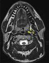
Mild inflammatory hypertrophy of palatine tonsils on axial T2 MRI in a young adult. The lymphoid tissue yields T2 hyperintense, striated signal (arrow) with preserved hypointense constrictor muscle and posterior tonsillar pillar (dotted arrows).
Figure 14.
Figure 14.
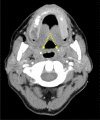
Contrast enhanced CT neck in a 44 year old trauma patient. Bilateral calcific foci in the lateral oropharynx are due to incidental palatine tonsilloliths (dotted arrows).
Figure 15.
Figure 15.
FDG PET-CT for a 72 years old male, performed for staging of a newly diagnosed bronchogenic carcinoma (right). Incidental asymmetric marked FDG uptake identified at the left glossotonsillar groove (left). Associated FDG avid cervical lymph node was also seen in level II (dotted arrow). Tongue base biopsy = Squamous Cell Carcinoma.
Figure 16.
Figure 16.
Axial T2 brain image showing signal loss in right skull base marrow due to normal variant petrous apex pneumatisation (dotted arrow).
Figure 17.
Figure 17.
CT brain (left) in a 34 year old man with headache, facial pain and hearing loss. Unilateral middle ear and mastoid fluid (dotted arrows) indicating Eustachian tube obstruction. Subsequent axial T2 MRI (right) shows asymmetric nasopharyngeal soft tissue mass (asterisk). Biopsy confirmed nasopharyngeal carcinoma.

Similar articles

Cited by

References

    1. Lincango-Naranjo E, Solis-Pazmino P, El Kawkgi O, Salazar-Vega J, Garcia C, Ledesma T, et al. . Triggers of thyroid cancer diagnosis: a systematic review and meta-analysis. Endocrine 2021; 72: 644–59. doi: 10.1007/s12020-020-02588-8 - DOI - PubMed
    1. Drake T, Gravely A, Westanmo A, Billington C. Prevalence of thyroid incidentalomas from 1995 to 2016: a single-center, retrospective cohort study. J Endocr Soc 2020; 4: bvz027. doi: 10.1210/jendso/bvz027 - DOI - PMC - PubMed
    1. Dean DS, Gharib H. Epidemiology of thyroid nodules. Best Pract Res Clin Endocrinol Metab 2008; 22: 901–11. doi: 10.1016/j.beem.2008.09.019 - DOI - PubMed
    1. Grogan RH, Aschebrook-Kilfoy B, White MG, Kaplan EL, Angelos P. Thyroid incidentalomas and the overdiagnosis conundrum. International Journal of Endocrine Oncology 2016; 3: 193–96. doi: 10.2217/ije-2016-0008 - DOI
    1. Russ G, Leboulleux S, Leenhardt L, Hegedüs L. Thyroid incidentalomas: epidemiology, risk stratification with ultrasound and workup. Eur Thyroid J 2014; 3: 154–63. doi: 10.1159/000365289 - DOI - PMC - PubMed

MeSH terms