. 2022 Nov 10;126(44):8957-8969.
doi: 10.1021/acs.jpcb.2c05954. Epub 2022 Nov 1.

Unraveling Complex Local Protein Environments with 4-Cyano-l-phenylalanine

Affiliations

Unraveling Complex Local Protein Environments with 4-Cyano-l-phenylalanine

ByungUk Lee et al. J Phys Chem B. .

Abstract

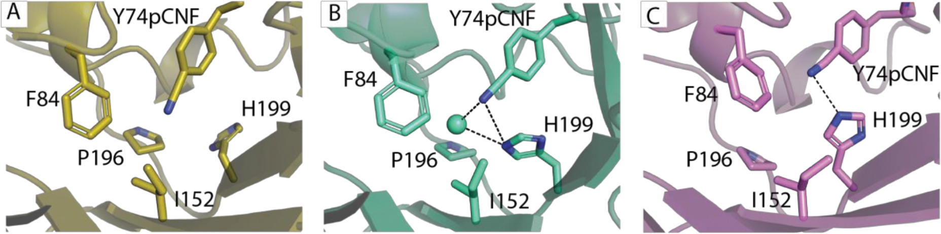
We present a multifaceted approach to effectively probe complex local protein environments utilizing the vibrational reporter unnatural amino acid (UAA) 4-cyano-l-phenylalanine (pCNPhe) in the model system superfolder green fluorescent protein (sfGFP). This approach combines temperature-dependent infrared (IR) spectroscopy, X-ray crystallography, and molecular dynamics (MD) simulations to provide a molecular interpretation of the local environment of the nitrile group in the protein. Specifically, a two-step enantioselective synthesis was developed that provided an 87% overall yield of pCNPhe in high purity without the need for chromatography. It was then genetically incorporated individually at three unique sites (74, 133, and 149) in sfGFP to probe these local protein environments. The incorporation of the UAA site-specifically in sfGFP utilized an engineered, orthogonal tRNA synthetase in E. coli using the Amber codon suppression protocol, and the resulting UAA-containing sfGFP constructs were then explored with this approach. This methodology was effectively utilized to further probe the local environments of two surface sites (sites 133 and 149) that we previously explored with room temperature IR spectroscopy and X-ray crystallography and a new interior site (site 74) featuring a complex local environment around the nitrile group of pCNPhe. Site 133 was found to be solvent-exposed, while site 149 was partially buried. Site 74 was found to consist of three distinct local environments around the nitrile group including nonspecific van der Waals interactions, hydrogen-bonding to a structural water, and hydrogen-bonding to a histidine side chain.


Figures

Figure 1.
Figure 1.
A. Structure of wildtype superfolder green fluorescent protein (PDB ID 2B3P) where three sites studied using pCNPhe are shown in sticks with Asp133 in periwinkle, Asn149 in orange, and Tyr74 in magenta. B. Temperature-dependence of the nitrile symmetric stretching frequency of Boc-pCNPhe (open squares) in THF and pCNPhe (open circles) in an aqueous buffer solution fit to a straight line (solid line). The decreased temperature range utilized for THF compared to water is the result of the lower boiling point of THF. C. Temperature-dependence of the nitrile symmetric stretching frequency of sfGFP-Asn149pCNPhe (open squares) and sfGFP-Asp133pCNPhe (open circles) in an aqueous buffer solution fit to a straight line (solid line). The frequency shifts for Panels B and C were referenced to the nitrile stretching frequency measured at 11.4 °C for each solvent or protein construct, respectively.
Figure 2.
Figure 2.
Time series of normalized solvent accessible surface area (SASA) of pCNPhe side chain atoms from MD simulations of sfGFP-Asp133pCNPhe (blue) and sfGFP-Asn149pCNPhe (red).
Figure 3.
Figure 3.
Room temperature FTIR spectra of pCNPhe (open circles), pC15NPhe (open squares), or p13CNPhe (open triangles) incorporated at site 74 in sfGFP dissolved in an aqueous buffer (20 mM Hepes, pH 7.5) at a concentration of ~1 mM. The spectra were intensity normalized, baseline corrected, and fit with a linear combination of Gaussian and Lorentzian functions. The overall fit is shown as a solid curve while the three subcomponents for each spectrum are shown as dashed curves.
Figure 4.
Figure 4.
Temperature-dependence of the nitrile symmetric stretching frequency of the three subcomponents of the nitrile IR absorbance band for sfGFP-Tyr74pCNPhe in an aqueous buffer solution fit to a straight line (solid lines). The temperature dependence of the low (open circles), intermediate (open squares) and high (open triangles) frequency subcomponents are shown. The frequency shifts were referenced to the nitrile stretching frequency of the three subcomponents measured at 11.4 °C.
Figure 5.
Figure 5.
X-ray crystal structure of sfGFP-Tyr74pCNPhe (orange) aligned with wt-sfGFP (green). A. Overall alignment with wt-sfGFP with 0.23 Å RMSD for 187 Cɑ atoms; B. aligned local environment around site 74 with nearby residues shown in sticks and water molecules < 4 Å from the sidechain at site 74 shown in spheres.
Figure 6.
Figure 6.
Time series of heavy atom distances to pCNPhe nitrile group nitrogen atom from MD simulations of sfGFP-Tyr74pCNPhe. A. For the simulation started with crystallographic waters present at site 74, distances are shown to one of the crystallographic water molecules (blue), the closest atom of the Phe84 side chain (green), and the Nε atom of the His199 side chain (purple). B. For the simulation started with crystallographic waters absent from site 74, distances are shown to the closest bulk water molecule (orange), the closest atom of the Phe84 side chain (green), and the Nε atom of the His199 side chain (purple). The data have been downsampled by a factor of five for clarity.
Figure 7.
Figure 7.
Room temperature FTIR spectra of sfGFP-Tyr74pCNPhe (open square) and sfGFP-Tyr74pCNPhe_His199Leu (open circles) dissolved in an aqueous buffer (20 mM Hepes, pH 7.5) at a concentration of ~1 mM. The spectra were intensity normalized, baseline corrected, and fit with a linear combination of Gaussian and Lorentzian functions. The overall fit is shown as a solid curve while the subcomponents for each spectrum are shown as dashed curves.
Figure 8.
Figure 8.
Time series of heavy atom distances to pCNPhe nitrile group nitrogen atom from MD simulations of sfGFP-Tyr74pCNPhe_His199Leu. A. For the simulation started with crystallographic waters present at site 74, distances are shown to one of the crystallographic water molecules (blue), the closest bulk water molecule (orange), and the closest atoms of the Phe84 side chain (green) and Leu199 side chain (purple). B. For the simulation started with crystallographic waters absent from site 74, distances are shown to the closest atom of the Phe84 side chain (green) and the Leu199 side chain (purple). The data have been downsampled by a factor of five for clarity.
Figure 9.
Figure 9.
Three representative structures of the 74pCNPhe site from the MD simulations of sfGFP-Tyr74pCNPhe. A. The gold structure illustrates a state where the pCNPhe is not involved in a hydrogen bond with a nearby atom and His199 is rotated away from the orientation where it is able to hydrogen bond with the nitrile or stabilize a water molecule hydrogen bonding with the nitrile. B. The violet structure illustrates a state where the pCNPhe is hydrogen bonding to Histidine 199 (d(NCN-NHis) is 3.1 Å). C. The cyan structure illustrates a state where the pCNPhe is hydrogen bonded to a water molecule (d(NCN–OWAT) is 2.8 Å), which is also stabilized by His199 (d(OWAT-NHis) is 3.2 Å and d(NCN-NHis) is 3.5 Å).
Scheme 1
Scheme 1

References

    1. Getahun Z; Huang C-Y; Wang T; De León B; DeGrado WF; Gai F J. Am. Chem. Soc. 2003, 125, 405. - PubMed
    1. Glasscock JM; Zhu Y; Chowdhury P; Tang J; Gai F Biochemistry 2008, 47, 11070. - PMC - PubMed
    1. Weeks CL; Polishchuk A; Getahun Z; Degrado WF; Spiro TG J. Raman Spectrosc. 2008, 39, 1606. - PMC - PubMed
    1. Waegele MM; Culik RM; Gai F J. Phys. Chem. Lett. 2011, 2, 2598. - PMC - PubMed
    1. Fafarman AT; Boxer SG J. Phys. Chem. B 2010, 114, 13536. - PMC - PubMed

Publication types