. 2023 Jan 6;51(D1):D428-D437.
doi: 10.1093/nar/gkac965.

The MHC Motif Atlas: a database of MHC binding specificities and ligands

Affiliations

The MHC Motif Atlas: a database of MHC binding specificities and ligands

Daniel M Tadros et al. Nucleic Acids Res. .

Abstract

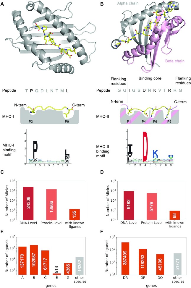
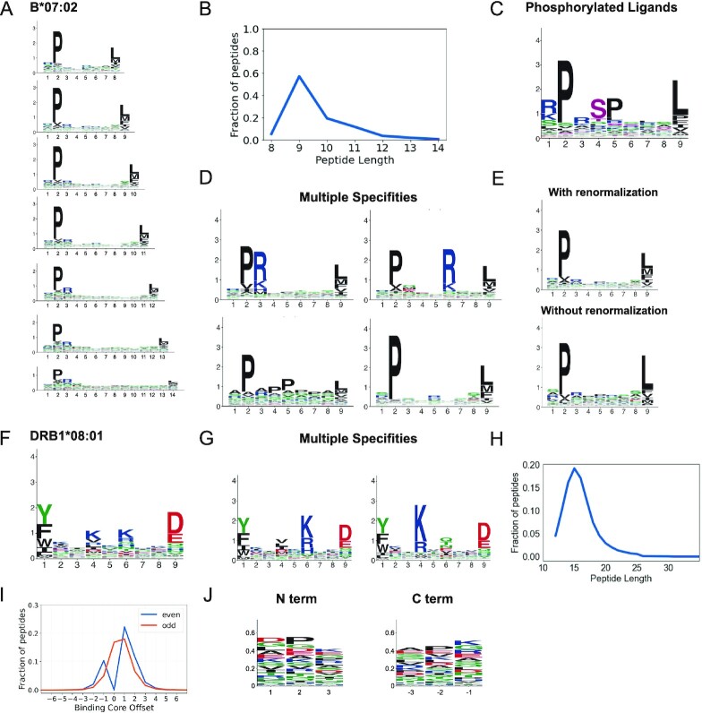
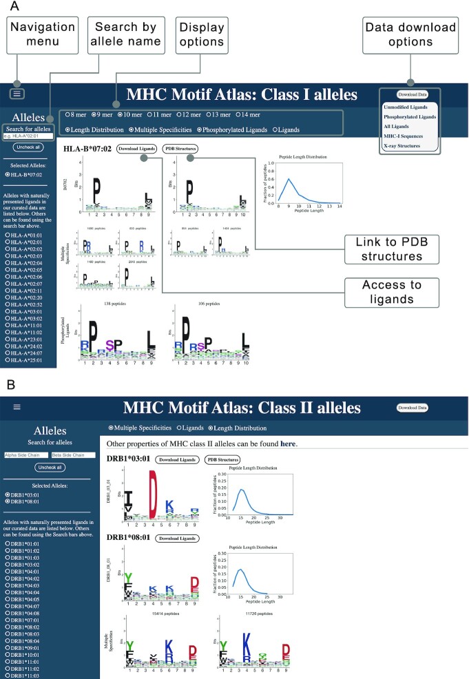
The highly polymorphic Major Histocompatibility Complex (MHC) genes are responsible for the binding and cell surface presentation of pathogen or cancer specific T-cell epitopes. This process is fundamental for eliciting T-cell recognition of infected or malignant cells. Epitopes displayed on MHC molecules further provide therapeutic targets for personalized cancer vaccines or adoptive T-cell therapy. To help visualizing, analyzing and comparing the different binding specificities of MHC molecules, we developed the MHC Motif Atlas (http://mhcmotifatlas.org/). This database contains information about thousands of class I and class II MHC molecules, including binding motifs, peptide length distributions, motifs of phosphorylated ligands, multiple specificities or links to X-ray crystallography structures. The database further enables users to download curated datasets of MHC ligands. By combining intuitive visualization of the main binding properties of MHC molecules together with access to more than a million ligands, the MHC Motif Atlas provides a central resource to analyze and interpret the binding specificities of MHC molecules.


Figures

Figure 1.
Figure 1.
Properties of MHC class I and class II molecules. (A) Description of the peptide binding properties of MHC-I molecules. The upper part shows a crystal structure (PDB:4U1H) (48), with the peptide (TPQDLNTML) in yellow and the MHC-I (HLA-B*07:02) in grey. The middle part shows a schematic view of the binding site, with the two main anchor residues at the second and last position (P2 and P9 for 9-mers). The bottom part shows the motif of HLA-B*07:02 for 9-mers. (B) Description of the peptide binding properties of MHC-II molecules. The upper part shows a crystal structure (PDB:7N19) (49), with the peptide (GGIGSDNKVTRRG) in yellow and the MHC-II in grey (alpha chain, HLA-DRA1*01:01) and pink (beta chain, HLA-DRB1*03:01). The middle part shows a schematic view of the binding site, with the main anchor residues (P1, P4, P6 and P9 of the binding core) and flanking residues on both sides of the core. The binding motif is shown in the lower part and was built based on the binding core of the ligands of this allele. (C) Number of documented MHC-I alleles both at the DNA and protein level in IMGT database (50) (data from https://www.ebi.ac.uk/ipd/imgt/hla/about/statistics/, as of July 2022). The third bar shows the number of MHC-I alleles with known naturally presented ligands. (D) Number of documented MHC-II alleles. The third bar shows the number of MHC-II dimers with known naturally presented ligands. (E) Number of known MHC-I ligands for each gene in human and in other species. (F) Number of known MHC-II ligands for each gene in human and in other species.
Figure 2.
Figure 2.
Binding specificities of MHC molecules. (A) MHC-I binding motifs for different peptide lengths. (B) Peptide length distribution. (C) Motifs for phosphorylated ligands. (D) MHC-I multiple specificities, including mutual exclusivity of charged amino acids at P3 and P6. (E) Illustration of the difference between motifs with and without background frequency renormalization. (F) MHC-II binding motifs. (G) MHC-II multiple specificities capturing a mutual exclusivity of positively charged amino acids at P4 and P6 (see (13)). (H) Average peptide length distribution for MHC-II ligands. (I) Distribution of peptide binding core offsets for MHC-II ligands of even and odd lengths (0 corresponds to a binding core at the middle of peptides with an odd length, and is not defined for peptides with an even length). (J) Motifs in the first and last three N- and C-terminal residues of MHC-II ligands. Panels A–E are built from HLA-B*07:02 ligands. Panels F–G are built from HLA-DRB1*08:01 ligands. Panels H–J are built from all MHC-II ligands (see (13)).
Figure 3.
Figure 3.
Predicting the binding properties of MHC-I molecules. (A) Machine learning framework for the prediction of binding motifs for MHC-I alleles. Distinct neural networks (NN) were built for each peptide length and each position, and the final motif for a given peptide length is built by combining all position. The example illustrates the neural networks for predicting 9-mer binding motifs. (B) Leave-one-allele-out, leave-ligands-out and leave-30-alleles-out cross validation of the predictions of binding motifs for the 30 MHC-I alleles that are not part of the training set of NetMHCpan. Predicted motifs from our method and from NetMHCpan4.1 (at percentile ranks smaller than 0.5% or 2%) were compared to the experimental ones using the Euclidean distance. (C) Architecture of the neural network for the predictions of peptide length distributions for MHC-I alleles without experimental ligands. (D) Leave-one-allele-out, leave-ligands-out and leave-30-alleles-out cross validation of the predictions of peptide length distributions for the 30 MHC-I alleles that are not part of the training set of NetMHCpan. Predicted peptide length distributions from our method and from NetMHCpan4.1 (at percentile ranks smaller than 0.5% or 2%) were compared to the experimental ones using the Euclidean distance. Boxplots indicate the median, upper and lower quartiles. P-values were computed with the paired two-sided Mann–Whitney U-test.
Figure 4.
Figure 4.
MHC Motif Atlas interface of MHC-I and MHC-II. (A) MHC Motif Atlas interface for MHC-I alleles, including visualization of binding motifs, peptide length distributions, multiple specificities and motifs of phosphorylated ligands. The Search field on the top left part enables users to type a part of an allele's name, and all the corresponding alleles will automatically be listed below. By default the alleles listed on the left correspond to those with experimental ligands. The Download Data button allows to download complete lists of MHC-I ligands, as well as MHC-I sequences and X-ray structures PDB identifiers. (B) MHC Motif Atlas interface for MHC-II alleles.

References

    1. Neefjes J., Jongsma M.L.M., Paul P., Bakke O.. Towards a systems understanding of MHC class i and MHC class II antigen presentation. Nat. Rev. Immunol. 2011; 11:823–836. - PubMed
    1. Cobbold M., De La Peña H., Norris A., Polefrone J.M., Qian J., English A.M., Cummings K.L., Penny S., Turner J.E., Cottine J.et al. .. MHC class I-associated phosphopeptides are the targets of memory-like immunity in leukemia. Sci. Transl. Med. 2013; 5:203ra125. - PMC - PubMed
    1. Solleder M., Guillaume P., Racle J., Michaux J., Pak H.-S., Müller M., Coukos G., Bassani-Sternberg M., Gfeller D.. Mass spectrometry based immunopeptidomics leads to robust predictions of phosphorylated HLA class I ligands. Mol. Cell. Proteomics. 2020; 19:390–404. - PMC - PubMed
    1. Ott P.A., Hu Z., Keskin D.B., Shukla S.A., Sun J., Bozym D.J., Zhang W., Luoma A., Giobbie-Hurder A., Peter L.et al. .. An immunogenic personal neoantigen vaccine for patients with melanoma. Nature. 2017; 547:217–221. - PMC - PubMed
    1. Sahin U., Derhovanessian E., Miller M., Kloke B.-P., Simon P., Löwer M., Bukur V., Tadmor A.D., Luxemburger U., Schrörs B.et al. .. Personalized RNA mutanome vaccines mobilize poly-specific therapeutic immunity against cancer. Nature. 2017; 547:222–226. - PubMed

Publication types