. 2022 Nov 11;50(20):11509-11528.
doi: 10.1093/nar/gkac953.

KLF9 and KLF13 transcription factors boost myelin gene expression in oligodendrocytes as partners of SOX10 and MYRF

Affiliations

KLF9 and KLF13 transcription factors boost myelin gene expression in oligodendrocytes as partners of SOX10 and MYRF

Celine Bernhardt et al. Nucleic Acids Res. .

Abstract

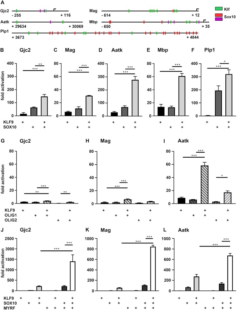
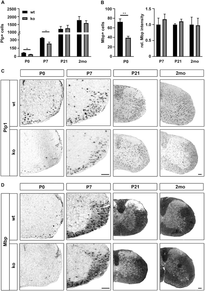
Differentiated oligodendrocytes produce myelin and thereby ensure rapid nerve impulse conduction and efficient information processing in the vertebrate central nervous system. The Krüppel-like transcription factor KLF9 enhances oligodendrocyte differentiation in culture, but appears dispensable in vivo. Its mode of action and role within the oligodendroglial gene regulatory network are unclear. Here we show that KLF9 shares its expression in differentiating oligodendrocytes with the closely related KLF13 protein. Both KLF9 and KLF13 bind to regulatory regions of genes that are important for oligodendrocyte differentiation and equally recognized by the central differentiation promoting transcription factors SOX10 and MYRF. KLF9 and KLF13 physically interact and synergistically activate oligodendrocyte-specific regulatory regions with SOX10 and MYRF. Similar to KLF9, KLF13 promotes differentiation and myelination in primary oligodendroglial cultures. Oligodendrocyte differentiation is also altered in KLF13-deficient mice as demonstrated by a transiently reduced myelin gene expression during the first postnatal week. Considering mouse phenotypes, the similarities in expression pattern and genomic binding and the behaviour in functional assays, KLF9 and KLF13 are important and largely redundant components of the gene regulatory network in charge of oligodendrocyte differentiation and myelination.


Figures

Graphical Abstract
Graphical Abstract
Proposed model for the redundant function of Klf9 and Klf13 transcription factors and synergistic interaction with Sox10 and Myrf in oligodendrocyte differentiation.
Figure 1.
Figure 1.
Expression of BTEB-like Klf factors in oligodendroglial cells. (A–D) Transcript levels for Klf9, Klf13, Klf14 and Klf16 in neuroectodermal cell types (A, astrocyte; N, neuron; OL, newly formed oligodendrocyte) after immunopanning at the time of birth (expressed as FPKM) according to GSE 52564 (30). (E) Transcript levels for Klf9, Klf13, Klf14 and Klf16 in oligodendroglial cells from spinal cord at P6 (expressed as read counts) according to GSE119127 (27). (F) Relative transcript levels for Klf9, Klf13, Klf14, Klf16, Mbp and Pdgfra in primary rat oligodendroglial cultures kept under proliferative conditions (prol) or undergoing differentiation for 3 or 6 days (3d, 6d) as determined by qRT-PCR. Normalized transcript levels for Klf9, Mbp and Pdgfra under proliferating conditions were set to 1, and levels in differentiating oligodendrocytes expressed relative to it (n = 3). (GH) Protein amounts of KLF9, KLF13, MBP and PDGFRA in whole cell extracts from rat oligodendroglial cells kept under proliferating or differentiating conditions as determined by Western blot (G). Numbers on the right of the panels represent approximate size in kDa. For quantification (H), band intensities in proliferating cells were set to 1 after normalization to GAPDH levels, and values for differentiating cells expressed relative to it (n = 3). Statistical significance was determined by two-tailed Student's t test (F) and one way Anova (H) (*P ≤ 0.05; **P ≤ 0.01; ***P ≤ 0.001).
Figure 2.
Figure 2.
Occurrence and localization of KLF9 and KLF13 during oligodendroglial development. (A–D) Detection and quantification of KLF9 (A, B) and KLF13 (C, D) in oligodendroglial cells at P0, P7 and P21 following co-immunohistochemistry of Klf-specific antibodies (in green) with antibodies against SOX10, PDGFRA and MYRF (all in red). Exemplary pictures (A, C) were taken from the ventrolateral white matter of spinal cord tissue at P7. Quantification of the fraction of SOX10-positive oligodendroglial cells and MYRF-positive oligodendrocytes in the white matter that co-expressed KLF9 (B) or KLF13 (D). (E) Subcellular localization of KLF9 and KLF13 proteins by microscopy following co-immunohistochemistry of KLF-specific antibodies (in green) with antibodies against PDGFRA (red) and MBP (yellow), combined with a DAPI nuclear counterstain (magenta) in primary oligodendroglial cells cultured under proliferating (upper row) or differentiating (lower row) conditions. Scale bar: 50 μm (A, C), 25 μm (E).
Figure 3.
Figure 3.
Influence of T3 and SOX10 on oligodendroglial Klf9 and Klf13 expression. (A) Determination of Sox10, Klf9 and Klf13 transcript levels in primary rat oligodendroglial cultures kept under differentiating conditions in the absence (–T3) or presence (+T3) of thyroid hormone by qRT-PCR. Expression levels for each gene in the absence of T3 were set to 1 ± SEM (n = 3). (B) Relative expression of Sox10, Klf9 and Klf13 in the Oln93 cell line (contr) as compared to genome-edited clonal derivatives with Sox10 gene inactivation (SOX10 ko) according to GSE136659 (13). Expression levels for each gene in control Oln93 cells were set to 1 ± SEM (n = 3). (C) qRT-PCR determining Sox10, Klf9 and Klf13 expression in primary oligodendroglial cells from Sox10fl/fl mice (contr) as compared to cells treated with TAT-Cre (SOX10 ko) prior to a 3-day cultivation under differentiating conditions. Expression levels for each gene in untreated cells were set to 1 ± SEM (n = 3). (D, E) qRT-PCR determining Sox10, Klf9 and Klf13 expression in rat oligodendroglial cells transduced with GFP (contr in D,E), GFP and SOX10 (SOX10 oe in D) or GFP and KLF9 (KLF9 oe in E) co-expressing retrovirus. Expression levels for each gene in GFP-transduced cells were set to 1 ± SEM (n = 3).
Figure 4.
Figure 4.
Transcriptional activity of KLF9 on regulatory regions from differentiating oligodendrocytes. (A) Localization of predicted Klf binding motifs (green boxes) within the oligodendroglial regulatory regions from the Gjc2, Mag, Aatk, Mbp and Plp1 genes relative to predicted (red boxes) and validated (magenta boxes) SOX10 binding sites. For each regulatory region, first and last positions relative to the transcriptional start site (arrow) are given. (B–L) Luciferase assays in N2a cells transiently transfected with reporter genes under control of regulatory regions from the Gjc2 (B, G, J), Mag (C, H, K), Aatk (D, I, L), Mbp (E) and Plp1 (F) genes in the presence (+) of various combinations of KLF9 (B–L), SOX10 (B–F, J–L), OLIG1 (G–I), OLIG2 (G–I) and MYRF (J–L) as indicated below the bars. Effector-dependent activation rates are presented as fold inductions ± SEM with transfections in the absence of effectors arbitrarily set to 1 for each reporter construct (n = 3). Differences were statistically significant as determined by one way Anova with Bonferroni correction (*P ≤ 0.05; **P ≤ 0.01; ***P ≤ 0.001).
Figure 5.
Figure 5.
Transcriptional activity of KLF13 on regulatory regions from differentiating oligodendrocytes. (A–K) Luciferase assays in N2a cells transiently transfected with reporter genes under control of regulatory regions from the Gjc2 (A, F, I), Mag (B, G, J), Aatk (C, H, K), Mbp (D) and Plp1 (E) genes in the presence (+) of various combinations of KLF13 (A–K), SOX10 (A–E, I–K), OLIG1 (F–H), OLIG2 (F–H) and MYRF (I–K) as indicated below the bars. Effector-dependent activation rates are presented as fold inductions ± SEM with transfections in the absence of effectors arbitrarily set to 1 for each reporter construct (n = 3). Differences were statistically significant as determined by one way Anova with Bonferroni correction (*P ≤ 0.05; **P ≤ 0.01; ***P ≤ 0.001).
Figure 6.
Figure 6.
Physical interaction of Klf proteins with SOX10 and MYRF. (AB) Co-immunoprecipitation of KLF9 (A) or KLF13 (B) with SOX10 from extracts of transfected HEK293 cells. The Western blot on the left shows the input. The middle panel is from a representative experiment in which HEK293 extracts containing either the Klf protein, SOX10 or a combination of both underwent precipitation after incubation with anti-SOX10 (αSOX10) antibodies. The right panel shows the results from precipitation with anti-myc (αKLF9) or anti-KLF13 (αKLF13) antibodies. (CD) Immunoprecipitation of SOX10 from extracts of primary oligodendroglial cells kept for at least 3 days under differentiating conditions using antisera directed against SOX10 (αSOX10), KLF9 (αKLF9, C) or KLF13 (αKLF13, D) as well as the respective pre-immune sera (PI). The Western blots show SOX10 input and precipitates. (E) Domain structure of Sox10: DIM/HMG, dimerization and HMG-domain; K2, central protein-protein interaction and transactivation domain; TA, carboxy-terminal transactivation domain. (F) Coomassie stain of polyacrylamide-SDS-gel showing GST (–, marked by arrowhead) and fusions between GST and functional SOX10 domains (23) (marked by asterisk) after expression in bacteria, purification and binding to glutathione sepharose beads. (G) Bead-bound KLF9 (upper blot) and KLF13 (lower blot) were visualized after GST pulldown from HEK293 extracts (input) by Western blot with anti-myc antibodies for KLF9 or anti-KLF13 antibodies. (H) Luciferase assays in N2a cells transiently transfected with the Gjc2 luciferase reporter in the presence (+) of various combinations of KLF9, SOX10 and the SOX10 aa1 mutant as indicated below the bars. Effector-dependent activation rates are presented as fold inductions ± SEM with transfections in the absence of effectors arbitrarily set to 1 (n = 3). Differences were statistically significant as determined by one way Anova with Bonferroni correction (*P ≤ 0.05; **P ≤ 0.01; ***P ≤ 0.001). (I, J) Co-immunoprecipitation of MYRF with KLF9 (I) or KLF13 (J) from extracts of transfected HEK293 cells containing either the Klf protein, MYRF or a combination of both. The Western blot on the left shows the input. The blot on the right shows the results from precipitation with antisera against KLF9 (αKLF9) or KLF13 (αKLF13). Numbers on the right side of blots indicate the approximate molecular weight of proteins in kDa.
Figure 7.
Figure 7.
Oligodendroglial development in KLF13-deficient mice. (A–D) Quantification of the number of SOX10-positive (A) and OLIG2-positive (B) oligodendroglial cells, PDGFRA-positive OPCs (C) and MYRF-positive premyelinating and myelinating oligodendrocytes (D) in the spinal cord (forelimb level) of KLF13-deficient mice (ko, gray bars) and age-matched controls (wt, black bars) following immunohistochemistry with respective antibodies at P0, P7, P21 and 2 months (2mo). Three sections per animal and 3 animals per genotype were quantified. Mean numbers per section ± SEM are shown. No statistical significance was detected when ko and wt were compared at a particular time point by Student's t test. (E, F) Representative immunohistochemical stainings of spinal cord sections (right half) at P0, P7, P21 and 2 months for SOX10 (E) and MYRF (F). Scale bars: 100 μm.
Figure 8.
Figure 8.
Myelin gene expression in KLF13-deficient mice. (A–D) Quantification of Plp1- (A) and Mbp- (B) expressing oligodendrocytes in the spinal cord (forelimb level) of KLF13-deficient mice (ko, gray bars) and age-matched controls (wt, black bars) following in situ hybridization with antisense probes directed against Plp1 (C) and Mbp (D) at P0, P7, P21 and 2 months (2mo). For Plp1 at all time points and for Mbp at P0, positive cells were counted on three sections per animal and three animals per genotype. Mean numbers per section ± SEM are shown. For Mbp at P7, P21 and 2 months (2mo) signal intensities were measured. Statistical significance between ko and wt was determined at each time point by Student's t test (*P ≤ 0.05). For representative in situ hybridizations (C, D), the ventral right half of the spinal cord is shown at P0 and P7 and the complete right half for P21 and 2mo. Scale bar: 100 μm.
Figure 9.
Figure 9.
Myelination parameters in KLF13-deficient mice. (A–C) Quantitative RT-PCR on RNA from spinal cord tissue to determine Klf13, Mbp, Mag, Plp1, Acss2, Mboat1 and Lss transcript levels in KLF13-deficient mice (ko, gray bars) and age matched controls (wt, black bars) at P0 (A), P7 (B) and P21 (C). (D, E) Protein amounts of MBP in whole cell extracts from spinal cord of KLF13-deficient mice and age matched controls at P3 as determined by Western blot (D). Numbers on the right of the panels represent approximate size in kDa. For quantification (E), band intensities in wt extracts were set to 1 after normalization to GAPDH levels, and normalized values for ko extracts expressed relative to it (n = 3). Statistical significance between ko and wt was determined by Student's t test (*P ≤ 0.05).
Figure 10.
Figure 10.
Cell-intrinsic role of KLF13 in oligodendroglial differentiation and myelin gene expression. (A) Quantification of the percentage (±SEM) of O4-positive and MBP-positive cells among all oligodendroglial cells from control (wt, black bars) or KLF13-deficient mice (ko, gray bars) after 6 days of culture in differentiating conditions (n = 3). (B–F) Bioinformatic analysis of RNA-seq studies performed on KLF13-deficient (ko, n = 3) and wildtype control (wt, n = 4) oligodendroglial cells cultured in differentiating conditions. (B) Profile of the top 30 differentially expressed genes sorted by their adjusted P-value and depicted in a bi-clustering heatmap by plotting their log2 transformed expression values. (C) Changed expression of genes related to myelination, lipid synthesis and regulation of oligodendroglial differentiation in KLF13-deficient oligodendrocytes. (D) GO analysis of the 278 genes down-regulated with a log2fold change ≥ -1.5 (P-value ≤ 0.05) in KLF13-deficient oligodendrocytes. (E) GO analysis of the 301 genes up-regulated with a log2fold change ≥ 1.5 (P-value ≤ 0.05) in KLF13-deficient oligodendrocytes. (F) GSEA for genes related to ensheathment of neurons, regulation of lipid biosynthetic process and cell cycle in KLF13-deficient oligodendrocytes. NES, normalized enrichment score; FDR, false discovery rate.
Figure 11.
Figure 11.
Functional redundancy of KLF9 and KLF13 in oligodendroglial differentiation and myelin gene expression. (A) Quantification of the percentage (± SEM) of MBP-positive cells among all oligodendroglial cells from wildtype (wt, left panel) or KLF13-deficient mice (Klf13 ko, right panel) after 6 days of culture in differentiating conditions following transfection of siRNA pools against Klf9 (siKlf9) or Klf13 (siKlf13) and a control siRNA pool (siContr) as indicated below the bars (n = 3). (BC) Chromatin immunoprecipitation on rat oligodendroglial cells cultured for 6 days under differentiating conditions (B) or kept under proliferating conditions (C) with anti-KLF9 antiserum, anti-KLF13 antiserum or corresponding pre-immune sera (PI). Enrichment of Gjc2, Mag, Mbp, Plp1 and Aatk regulatory regions or control genomic regions (Contr_1, Contr_2) in the immunoprecipitates was determined by qPCR relative to preimmune sera that were arbitrarily set to 1 (red dotted line). Experiments were performed three times with each PCR in triplicate. (D–F) Consequences of KLF9 or KLF13 overexpression (oe) on the differentiation of retrovirally transduced wildtype rat (D,E) and KLF13-deficient mouse (F) oligodendroglial cells as analysed by immunocytochemical stainings. (D) Representative staining of differentiating rat cultures after transduction with GFP-, GFP- and KLF9-, or GFP- and KLF13-expressing retroviruses using antibodies directed against GFP and MBP. Scale bar: 50 μm (E, F). Quantification of the percentage (± SEM) of MBP-expressing cells among all oligodendroglial cells transduced with the various retroviruses (n = 3, D). Statistical significance (*P ≤ 0.05; **P ≤ 0.01; ***P ≤ 0.001) was determined by one way Anova with Bonferroni correction (A, E, F) or Student's t test (B, C).

References

    1. Emery B. Regulation of oligodendrocyte differentiation and myelination. Science. 2010; 330:779–782. - PubMed
    1. Emery B., Lu Q.R.. Transcriptional and epigenetic regulation of oligodendrocyte development and myelination in the central nervous system. Cold Spring Harb. Perspect. Biol. 2015; 7:a020461. - PMC - PubMed
    1. Sock E., Wegner M.. Transcriptional control of myelination and remyelination. Glia. 2019; 67:2153–2165. - PubMed
    1. Sock E., Wegner M.. Using the lineage determinants olig2 and sox10 to explore transcriptional regulation of oligodendrocyte development. Dev. Neurobiol. 2021; 81:892–901. - PubMed
    1. Nemer M., Horb M.E.. The KLF family of transcriptional regulators in cardiomyocyte proliferation and differentiation. Cell Cycle. 2007; 6:117–121. - PubMed

Publication types

MeSH terms

Substances