

Prostate-Specific Membrane Antigen Targeted Deep Tumor Penetration of Polymer Nanocarriers
- PMID: 36318757
- PMCID: PMC9673064
- DOI: 10.1021/acsami.2c15095
Prostate-Specific Membrane Antigen Targeted Deep Tumor Penetration of Polymer Nanocarriers
Abstract
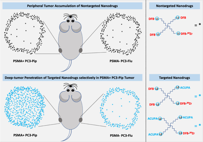
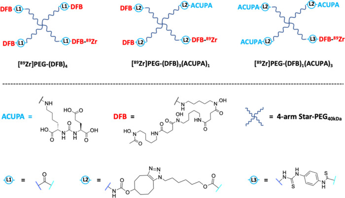
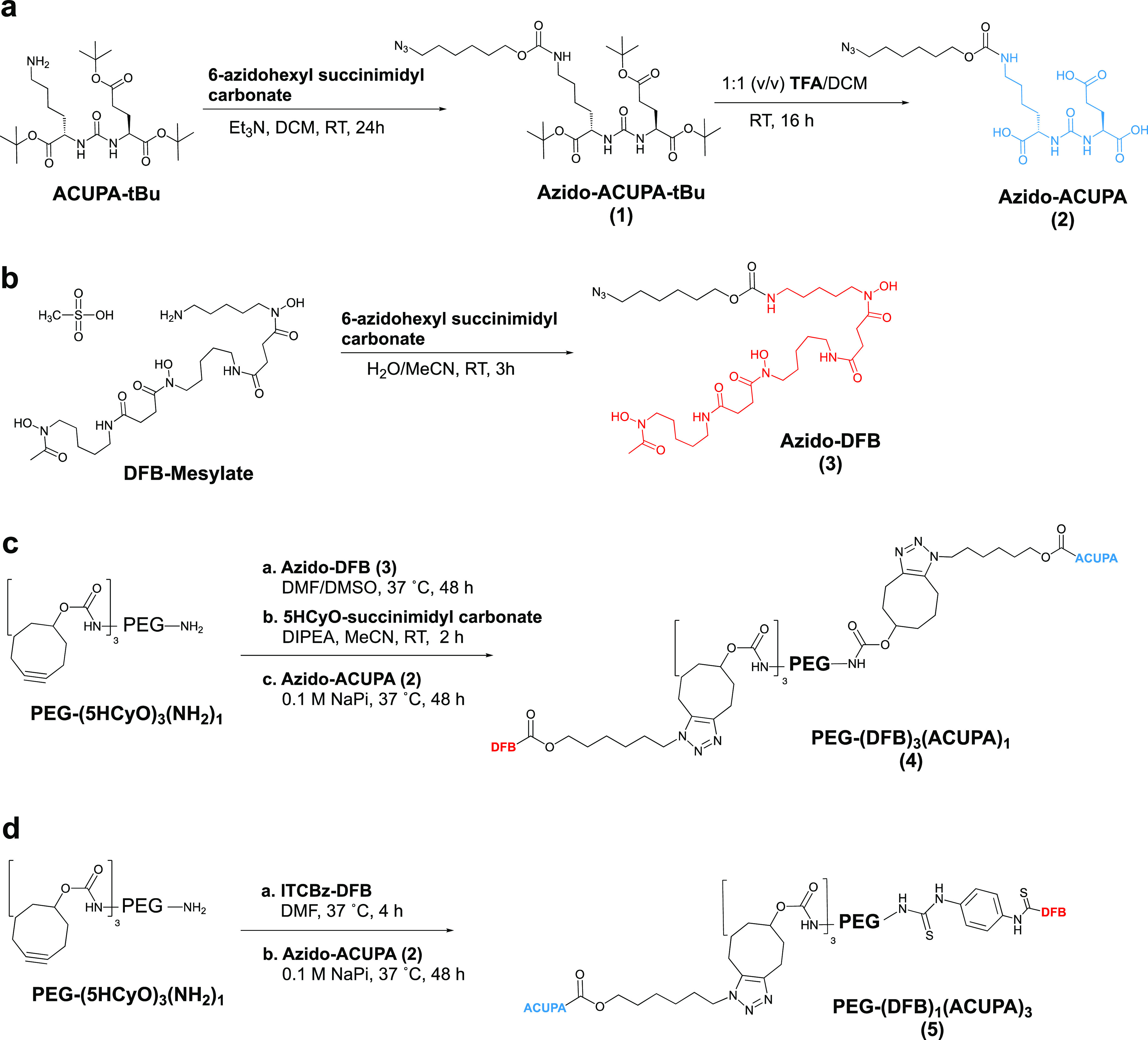
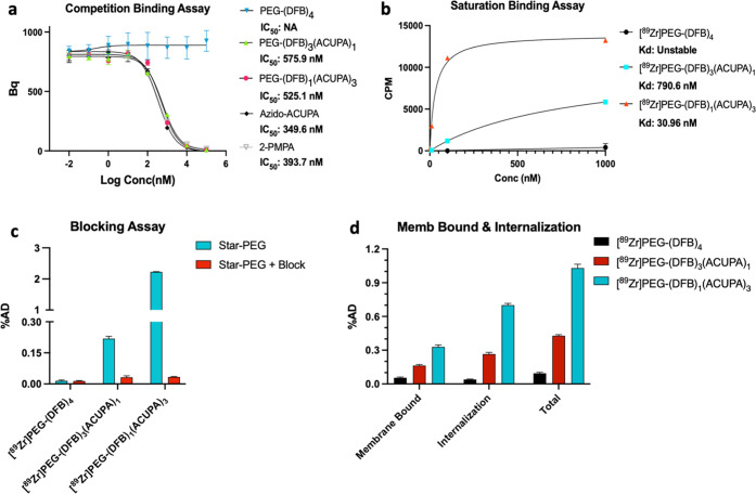
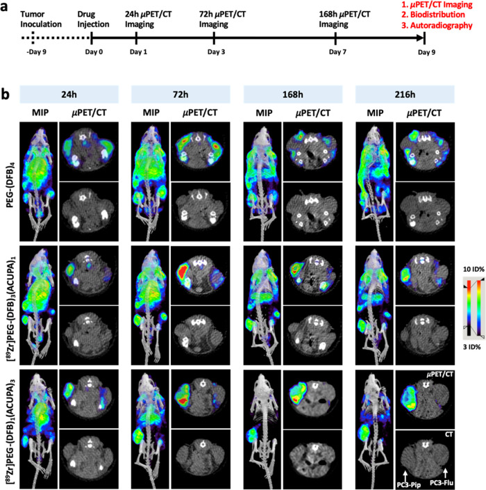
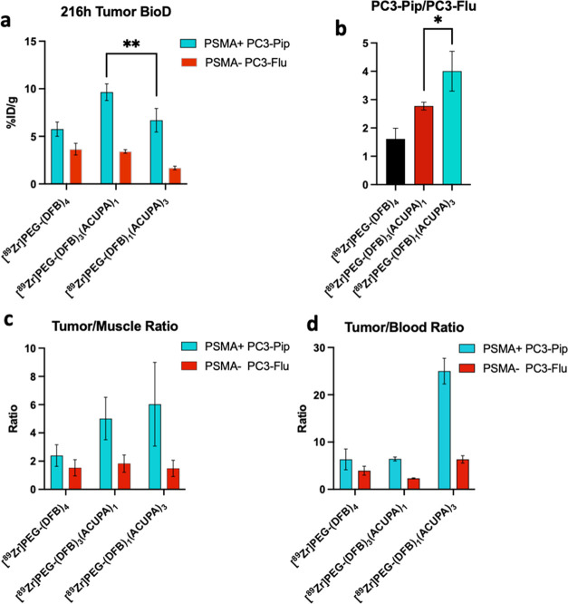
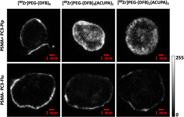
Tumoral uptake of large-size nanoparticles is mediated by the enhanced permeability and retention (EPR) effect, with variable accumulation and heterogenous tumor tissue penetration depending on the tumor phenotype. The performance of nanocarriers via specific targeting has the potential to improve imaging contrast and therapeutic efficacy in vivo with increased deep tissue penetration. To address this hypothesis, we designed and synthesized prostate cancer-targeting starPEG nanocarriers (40 kDa, 15 nm), [89Zr]PEG-(DFB)3(ACUPA)1 and [89Zr]PEG-(DFB)1(ACUPA)3, with one or three prostate-specific membrane antigen (PSMA)-targeting ACUPA ligands. The in vitro PSMA binding affinity and in vivo pharmacokinetics of the targeted nanocarriers were compared with a nontargeted starPEG, [89Zr]PEG-(DFB)4, in PSMA+ PC3-Pip and PSMA- PC3-Flu cells, and xenografts. Increasing the number of ACUPA ligands improved the in vitro binding affinity of PEG-derived polymers to PC3-Pip cells. While both PSMA-targeted nanocarriers significantly improved tissue penetration in PC3-Pip tumors, the multivalent [89Zr]PEG-(DFB)1(ACUPA)3 showed a remarkably higher PC3-Pip/blood ratio and background clearance. In contrast, the nontargeted [89Zr]PEG-(DFB)4 showed low EPR-mediated accumulation with poor tumor tissue penetration. Overall, ACUPA conjugated targeted starPEGs significantly improve tumor retention with deep tumor tissue penetration in low EPR PC3-Pip xenografts. These data suggest that PSMA targeting with multivalent ACUPA ligands may be a generally applicable strategy to increase nanocarrier delivery to prostate cancer. These targeted multivalent nanocarriers with high tumor binding and low healthy tissue retention could be employed in imaging and therapeutic applications.
Keywords: deep tumor penetration; enhanced permeability and retention (EPR) effect; polymer nanocarriers; positron emission tomography (PET) imaging; prostate-specific membrane antigen (PSMA).
Conflict of interest statement
The authors declare the following competing financial interest(s): D.V.S., G.W.A., and S.D.F. are employees of Prolynx Inc. The remaining authors declare no competing financial interest.
Figures
Similar articles
-
Prostate-Specific Membrane Antigen Targeted StarPEG Nanocarrier for Imaging and Therapy of Prostate Cancer.Adv Healthc Mater. 2024 Jul;13(19):e2304618. doi: 10.1002/adhm.202304618. Epub 2024 May 17. Adv Healthc Mater. 2024. PMID: 38700450 Free PMC article.
-
Synthesis and Preliminary Biological Assessment of Carborane-Loaded Theranostic Nanoparticles to Target Prostate-Specific Membrane Antigen.ACS Appl Mater Interfaces. 2021 Nov 24;13(46):54739-54752. doi: 10.1021/acsami.1c16383. Epub 2021 Nov 9. ACS Appl Mater Interfaces. 2021. PMID: 34752058
-
Tumor Uptake of Triazine Dendrimers Decorated with Four, Sixteen, and Sixty-Four PSMA-Targeted Ligands: Passive versus Active Tumor Targeting.Biomolecules. 2019 Aug 28;9(9):421. doi: 10.3390/biom9090421. Biomolecules. 2019. PMID: 31466360 Free PMC article.
-
Targeting Nanomedicines to Prostate Cancer: Evaluation of Specificity of Ligands to Two Different Receptors In Vivo.Pharm Res. 2016 Oct;33(10):2388-99. doi: 10.1007/s11095-016-1945-x. Epub 2016 May 25. Pharm Res. 2016. PMID: 27225496 Review.
-
Prostate specific membrane antigen (PSMA) ligands for diagnosis and therapy of prostate cancer.Expert Rev Mol Diagn. 2016 Nov;16(11):1177-1188. doi: 10.1080/14737159.2016.1243057. Epub 2016 Oct 14. Expert Rev Mol Diagn. 2016. PMID: 27679869 Review.
Cited by
-
Developments in radionanotheranostic strategies for precision diagnosis and treatment of prostate cancer.EJNMMI Radiopharm Chem. 2024 Aug 24;9(1):62. doi: 10.1186/s41181-024-00295-7. EJNMMI Radiopharm Chem. 2024. PMID: 39180599 Free PMC article. Review.
-
PSMA-Targeted Nanotheranostics for Imaging and Radiotherapy of Prostate Cancer.Pharmaceuticals (Basel). 2023 Feb 17;16(2):315. doi: 10.3390/ph16020315. Pharmaceuticals (Basel). 2023. PMID: 37259457 Free PMC article. Review.
-
Enhanced NIR-II Nanoparticle Probe for PSMA-Targeted Molecular Imaging and Prostate Cancer Diagnosis.Int J Nanomedicine. 2025 Aug 9;20:9807-9823. doi: 10.2147/IJN.S532080. eCollection 2025. Int J Nanomedicine. 2025. PMID: 40808713 Free PMC article.
-
Enhanced Prostate-specific Membrane Antigen Targeting by Precision Control of DNA Scaffolded Nanoparticle Ligand Presentation.ACS Nano. 2024 Jul 2;18(26):16674-16683. doi: 10.1021/acsnano.4c01640. Epub 2024 Jun 22. ACS Nano. 2024. PMID: 38907991 Free PMC article.
-
State-of-the-art application of nanoparticles in radiotherapy: a platform for synergistic effects in cancer treatment.Strahlenther Onkol. 2025 Jun;201(6):577-588. doi: 10.1007/s00066-024-02301-y. Epub 2024 Oct 4. Strahlenther Onkol. 2025. PMID: 39367110 Free PMC article. Review.
References
-
- Giesel F.; Knorr K.; Spohn F.; Will L.; Maurer T.; Flechsig P.; Neels O.; Schiller K.; Amaral H.; Weber W.; et al. Detection Efficacy of F-18-PSMA-1007 PET/CT in 251 Patients with Biochemical Recurrence of Prostate Cancer After Radical Prostatectomy. J. Nucl. Med. 2019, 60, 362–368. 10.2967/jnumed.118.212233. - DOI - PMC - PubMed
-
- Morris M.; Rowe S.; Gorin M.; Saperstein L.; Pouliot F.; Josephson D.; Wong J.; Pantel A.; Cho S.; Gage K.; et al. Diagnostic Performance of F-18-DCFPyL-PET/CT in Men with Biochemically Recurrent Prostate Cancer: Results from the CONDOR Phase III, Multicenter Study. Clin. Cancer Res. 2021, 27, 3674–3682. 10.1158/1078-0432.CCR-20-4573. - DOI - PMC - PubMed
MeSH terms
Substances
Grants and funding
LinkOut - more resources
Full Text Sources
Medical
Miscellaneous
