. 2023 Mar;49(2):201-208.
doi: 10.1055/s-0042-1757896. Epub 2022 Nov 1.

Resonant Acoustic Rheometry to Measure Coagulation Kinetics in Hemophilia A and Healthy Plasma: A Novel Viscoelastic Method

Affiliations

Resonant Acoustic Rheometry to Measure Coagulation Kinetics in Hemophilia A and Healthy Plasma: A Novel Viscoelastic Method

Weiping Li et al. Semin Thromb Hemost. 2023 Mar.

Abstract

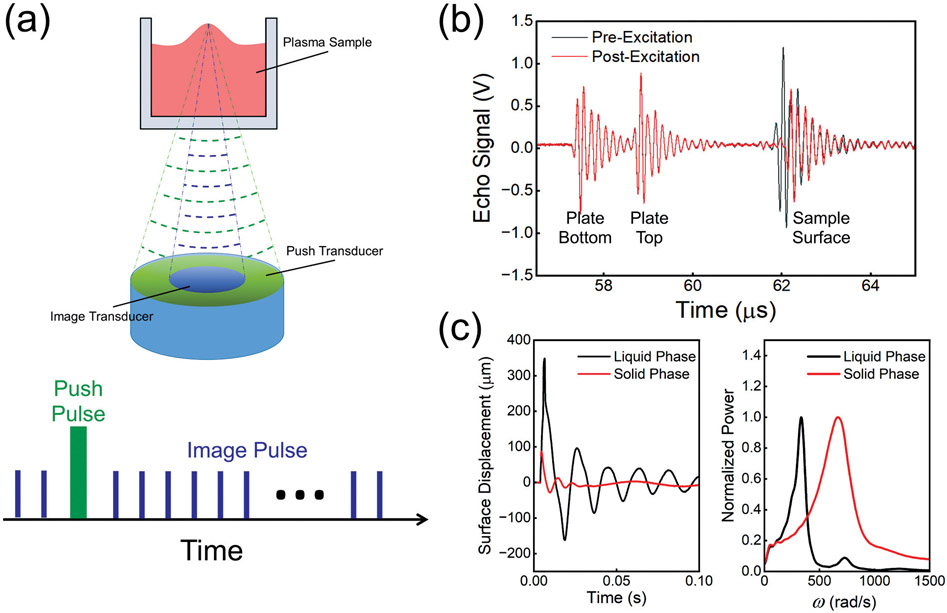
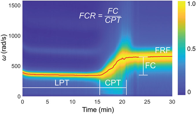
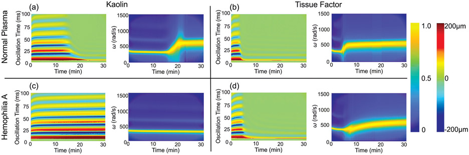
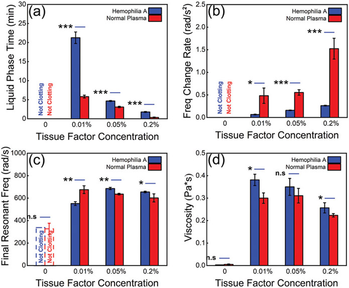
Compared with conventional coagulation tests and factor-specific assays, viscoelastic hemostatic assays (VHAs) can provide a more thorough evaluation of clot formation and lysis but have several limitations including clot deformation. In this proof-of-concept study, we test a noncontact technique, termed resonant acoustic rheometry (RAR), for measuring the kinetics of human plasma coagulation. Specifically, RAR utilizes a dual-mode ultrasound technique to induce and detect surface oscillation of blood samples without direct physical contact and measures the resonant frequency of the surface oscillation over time, which is reflective of the viscoelasticity of the sample. Analysis of RAR results of normal plasma allowed defining a set of parameters for quantifying coagulation. RAR detected a flat-line tracing of resonant frequency in hemophilia A plasma that was corrected with the addition of tissue factor. Our RAR results captured the kinetics of plasma coagulation and the newly defined RAR parameters correlated with increasing tissue factor concentration in both healthy and hemophilia A plasma. These findings demonstrate the feasibility of RAR as a novel approach for VHA, providing the foundation for future studies to compare RAR parameters to conventional coagulation tests, factor-specific assays, and VHA parameters.


Conflict of interest statement

M.M.W. has received honoraria from Alexion Pharmaceuticals.

Figures

Fig. 1
Fig. 1
(a) Schematic diagram of the RAR system with two co-linear focused ultrasound transducers aligned beneath a 96-well tissue culture plate (top). Schematic of pulse signals for detection (imaging) and excitation of sample surface movements (bottom). (b) Echo signal of pre-excitation and post-excitation. (c) Surface displacement of plasma sample as a function of time in the liquid phase and solid phase (left), and normalized power spectra of surface displacement of plasma in the liquid phase and solid phase (right) showing the peak angular frequencies of the surface displacement. RAR, resonant acoustic rheometry; ω, angular frequency.
Fig. 2
Fig. 2
Spectrogram of normal human plasma coagulation triggered by 4% kaolin measured using RAR and definition of clotting parameters. The resonant angular frequency of the surface oscillation stayed constant initially when the sample was in the liquid phase before a distinct increase was detected at t = 15 minutes, indicating a phase change occurring in the sample as it began to transition from the liquid to a state with higher elasticity. The resonant frequency continued to increase rapidly until t = 22 minutes before leveling off, indicating the coagulation process in the sample that changed from the liquid phase to a solid phase. Liquid phase time (LPT) refers to the amount of time duration of the liquid phase before the rapid rise in resonant frequency. Clotting phase time (CPT) refers to the period of time when the rapid rise of frequency occurred. Frequency change rate (FCR), or the slope of the frequency during the rapid clotting phase, is defined as the frequency change (FC) divided by CPT. Final resonant frequency (FRF) refers to the resonant angular frequency measured at the end of the run. CPT, clotting phase time; FC, frequency change; FCR, frequency change rate; FRF, final resonant frequency; LPT, liquid phase time; RAR, resonant acoustic rheometry.
Fig. 3
Fig. 3
(a) Example of the surface displacement heat map (left) and spectrogram (right) for normal plasma samples with the addition of kaolin (4%). The horizontal axis in these images represents the elapsed time of coagulation process during which RAR measurements were performed. The vertical axis in the displacement heat map indicates the oscillation time of the surface oscillation in one single RAR measurement. The vertical axis in the spectrogram is the angular frequency (ω) of the surface displacement. The color represents the normalized power of angular frequency and displacement amplitude, respectively. The peak angular frequency shows the resonant frequency of the surface oscillation, i.e., the frequency of the resonant mode of the surface waves in the sample. (b) Example of the surface displacement heat map (left) and spectrogram (right) for normal plasma samples with the addition of tissue factor. (c, d) Displacement heat map (left) and spectrogram (right) of hemophilia A plasma samples with addition of kaolin (c) and tissue factor (d). Tissue factor concentrations for these examples were at 0.05%. The peak angular frequency shows the resonant frequency of the surface oscillation, i.e., the frequency of the resonant mode of the surface waves in the sample. To the right of the figure, the legend bar ranging from blue (0) to yellow (1.0) indicates the measured ω of the sample during a given time point in the run for the spectrograms. The legend bar ranging from dark blue (−200 μm) to dark red (200 μm) represents the surface displacement in the heat maps. RAR, resonant acoustic rheometry; ω, angular frequency.
Fig. 4
Fig. 4
(a) Liquid phase time for normal plasma and hemophilia A plasma without and with the addition of tissue factor at 0.01, 0.05, and 0.2%. (b) Frequency change rate of normal plasma and hemophilia A plasma without and with tissue factor at 0.01, 0.05, and 0.2%. (c) Final resonant frequency measured in normal and hemophilia A plasma without and with 0.01, 0.05, and 0.2% tissue factor. (d) Viscosity measured in normal and hemophilia A plasma without and with 0.01, 0.05, and 0.2% tissue factor. Unpaired t-test p-values: * < 0.05, ** < 0.01, *** < 0.001, and n.s, not significant.

References

    1. Ramiz S, Hartmann J, Young G, Escobar MA, Chitlur M. Clinical utility of viscoelastic testing (TEG and ROTEM analyzers) in the management of old and new therapies for hemophilia. Am J Hematol 2019;94(02):249–256 - PubMed
    1. Sørensen B, Ingerslev J. Whole blood clot formation phenotypes in hemophilia A and rare coagulation disorders. Patterns of response to recombinant factor VIIa.J Thromb Haemost 2004;2(01):102–110 - PubMed
    1. Bolton-Maggs PH. The rare inherited coagulation disorders. Pediatr Blood Cancer 2013;60(Suppl 1):S37–S40 - PubMed
    1. Aghighi S, Riddell A, Lee CA, Brown SA, Tuddenham E, Chowdary P. Global coagulation assays in hemophilia A: a comparison to conventional assays. Res Pract Thromb Haemost 2019;4(02):298–308 - PMC - PubMed
    1. Nogami K. The utility of thromboelastography in inherited and acquired bleeding disorders. Br J Haematol 2016;174(04):503–514 - PubMed