

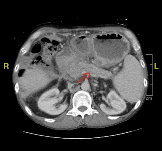
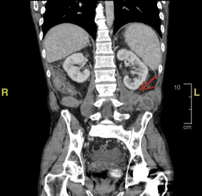
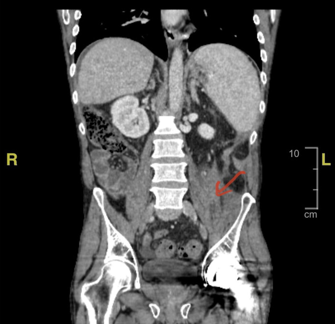
Rare extension of pancreatic pseudocyst with Mycobacterium abscessus into the iliopsoas muscle
- PMID: 36319038
- PMCID: PMC9628508
- DOI: 10.1136/bcr-2022-252777
Rare extension of pancreatic pseudocyst with Mycobacterium abscessus into the iliopsoas muscle
Abstract
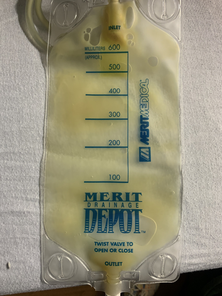
Pancreatic pseudocyst is a well-known complication of both acute and chronic pancreatitis. Although extension into other anatomical sites is common, extension into the retrofascial space causing an iliopsoas abscess is exceedingly rare. Although its low incidence creates a diagnostic challenge for clinicians, early diagnosis is essential to prevent significant complications and poor patient outcomes. We present a case of iliopsoas abscess with unusual culture fluid growth in the setting of acute on chronic pancreatitis secondary to extension of a pancreatic pseudocyst. We also offer a brief review of the literature and pathophysiology of the condition.
Keywords: Gastrointestinal system; Infection (gastroenterology); Infections; Pancreas and biliary tract; Pancreatitis.
© BMJ Publishing Group Limited 2022. No commercial re-use. See rights and permissions. Published by BMJ.
Conflict of interest statement
Competing interests: None declared.
Figures
References
Publication types
MeSH terms
LinkOut - more resources
Full Text Sources