Review
. 2022 Nov 1;21(1):207.
doi: 10.1186/s12943-022-01671-0.

Exosome biogenesis: machinery, regulation, and therapeutic implications in cancer

Affiliations
Review

Exosome biogenesis: machinery, regulation, and therapeutic implications in cancer

Qing-Fang Han et al. Mol Cancer. .

Abstract

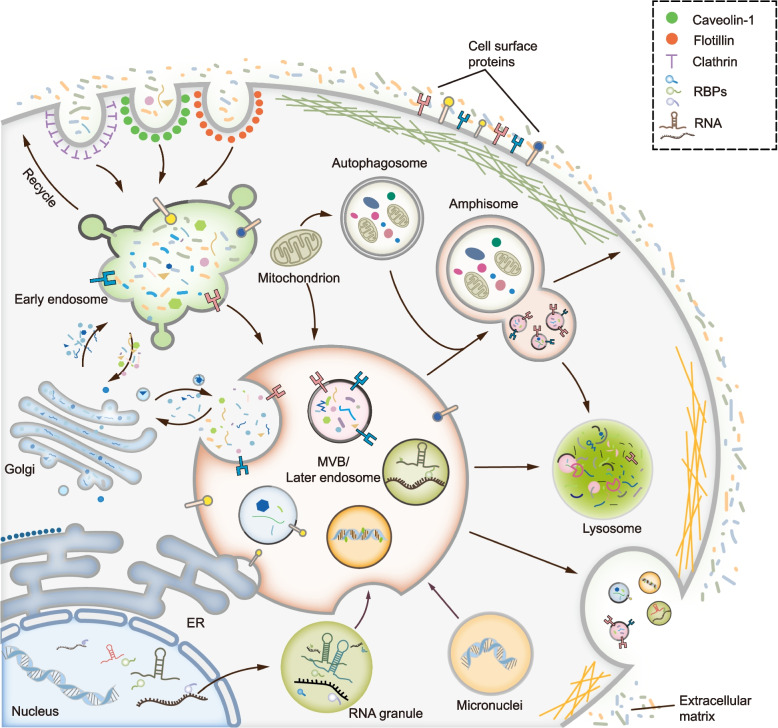
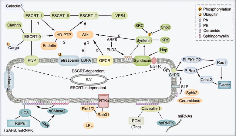
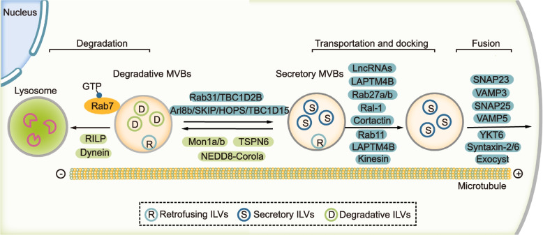
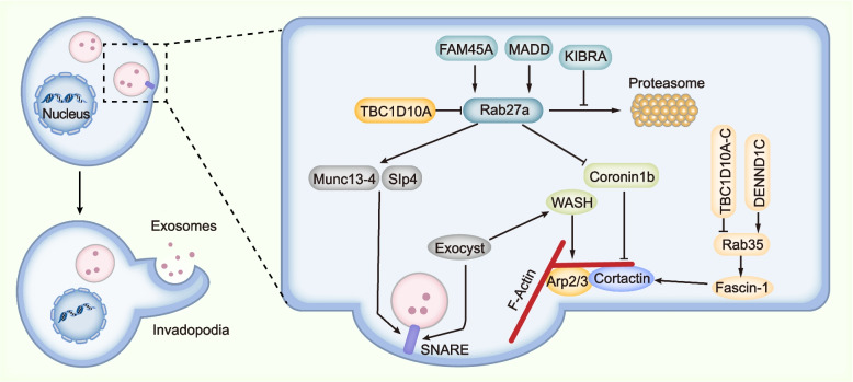
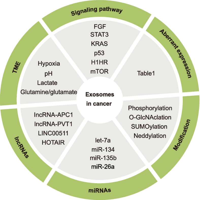
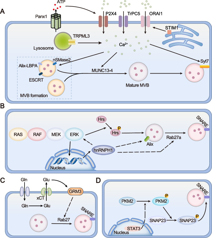
Exosomes are well-known key mediators of intercellular communication and contribute to various physiological and pathological processes. Their biogenesis involves four key steps, including cargo sorting, MVB formation and maturation, transport of MVBs, and MVB fusion with the plasma membrane. Each process is modulated through the competition or coordination of multiple mechanisms, whereby diverse repertoires of molecular cargos are sorted into distinct subpopulations of exosomes, resulting in the high heterogeneity of exosomes. Intriguingly, cancer cells exploit various strategies, such as aberrant gene expression, posttranslational modifications, and altered signaling pathways, to regulate the biogenesis, composition, and eventually functions of exosomes to promote cancer progression. Therefore, exosome biogenesis-targeted therapy is being actively explored. In this review, we systematically summarize recent progress in understanding the machinery of exosome biogenesis and how it is regulated in the context of cancer. In particular, we highlight pharmacological targeting of exosome biogenesis as a promising cancer therapeutic strategy.

Keywords: Biogenesis; Cancer; Exosomes; Implications; Regulation.


Conflict of interest statement

The authors declare that they have no competing interests.

Figures

Fig. 1
Fig. 1
Overview of the process for exosome biogenesis. MVBs take the center of exosome biogenesis. Generally, MVBs are derived from endocytosis, during which different mechanisms mediate the inward budding of the plasma membrane and the formation of early endosomes. MVBs can dynamically communicate with other organelles or compartments including trans-Golgi network (TGN), endoplasmic reticulum (ER), mitochondrion, phagosome, RNA granule and micronuclei, et al. Therefore, different repertoires of cargos such as proteins, RNAs, DNAs or lipids are sorted into MVBs. After the maturation of MVBs, they can either fuse with lysosome to be degraded or fuse with plasma membrane to release ILVs, the so-called exosomes. Of note, MVB can fuse with autophagosome to form amphisome, which can either fuse with lysosome to be degraded or fuse with plasma membrane to secret exosomes
Fig. 2
Fig. 2
Multiple mechanisms regulate the formation of ILVs. MVBs are characterized by containing intralumenal vesicles which can be controlled by multiple mechanisms. Generally, they can be divided into two categories which are ESCRT-dependent pathway and ESCRT-independent pathway. For the classical ESCRT-dependent pathways, ESCRT-0, -I, -II, -III subcomplexes and ATPase VPS4 cooperate in a stepwise manner to mediate the ILV formation (1). For the non-canonical ESCRT-dependent pathways, HD-PTP (2) or Alix (3) can both recognize specific cargos and recruit ESCRT-III and VPS4 to limiting membrane of MVBs, during which other ESCRT subcomplexes are not indispensable. In addition, several other proteins or pathways especially components of lipid rafts paly crucial roles in ESCRT-independent ILV formation. For example, CD63 could promote ILV formation by both ESCRT and ceramide-independent mechanism; nSMase2-ceramide pathway could drive ILV formation and MVB sorting of cargos such as PE-conjugated LC3 and its binding partners in an ESCRT-independent mechanism. Moreover, caveolin-1 or flotillins could drive lipid raft dependent ILV formation, during which process nSMase-ceramide pathway is required in some cell lines. Specially, F-actin formation on the limiting membrane of MVBs that was regulated by S1P signaling promotes ILV sorting of cargos, though the precise role of F-actin formation during the generation of ILVs is still elusive
Fig. 3
Fig. 3
Mechanisms mediating the transport and fate of MVBs. After the maturation of MVBs, they can either fuse with lysosome or fuse with plasma membrane. The activity of Rab7 plays a pivotal role for the fate of MVBs. Mon1a/b and neddylated Coro1a can activate Rab7 and promote dynein-dependent retrograde transport of MVBs towards the minus-end or perinuclear region. On the other hand, Arl8b/SKIP/HOPS/ TBC1D15 cascade or Rab31/TBC1D2B cascade could inactivate RAB7 and promote kinesin dependent antegrade transport of MVBs towards the plus-end or cell periphery. The antegrade transport of MVBs and their docking, tethering and fusion with plasma membrane are controlled by multiple factors including both proteins and lncRNAs. Notably, after the formation of MVBs, the ILVs inside can still retrofuse with the limiting membrane of MVBs and these ILVs are referred as retrofusing ILVs (rILVs). The others are recognized as secretory ILVs (sILVs) or degradative ILVs (dILVs)
Fig. 4
Fig. 4
Proposed model showing the relationship between MVB secretion, actin reorganization and invadopodia formation. These three processes are highly organized and interrelated. Specifically, invadopodia determines the docking and secretion sites of MVBs on the plasma membrane. On the other hand, the fusion of MVBs on the plasma membrane contributes to the formation of invadopodia. And, F-actin formation is vital for both MVB secretion and invadopodia formation. Mechanistically, Rab27a and Rab35 seem to function at the center, the activity of which is regulated by their GAPs or GEFs. Particularly, Rab27a promotes both MVB docking and F-actin formation. Munc13-4 and Slp4 are effectors of Rab27a and function to mediate the docking and fusion of MVBs on the plasma membrane by promoting the formation of SNARE. Concurrently, Rab27a inhibits Coronin1b binding to invadopodia-associated actin and stabilizes Cortactin-mediated branched actin. In addition, actin-binding protein Fascin-1 is an effector of Rab35 and contributes to both invadopodia and exosome secretion. Moreover, exocyst complex binds to WASH, through which to promote Arp2/3 mediated actin polymerization and invadopodia formation. Also, exocyst binds to SNARE and mediates docking and fusion of MVBs on the plasma membrane
Fig. 5
Fig. 5
Categories of mechanisms mediating the dysregulation of exosome biogenesis in cancer
Fig. 6
Fig. 6
Aberrant activation of signaling pathways regulates exosome secretion in cancer. A. Increased Ca2+ in the cytosol that was caused by various stimuli contributes to the activity and function of multiple proteins mediating exosome generation, such as ESCRT, Alix-LBPA interaction, nSMase2, Munc13-4, and Syt7. B. RAS/RAF/MEK/ERK pathway signaling promotes the transcription of hnRNP H1, through which to facilitate exosome biogenesis by upregulating the expression of Alix and Rab27a. Moreover, there is a positive feedback loop between Ras signaling and hnRNP H1. In addition, ERK interacts with and phosphorylates Hrs, thereby promoting exosome secretion. C. Glutamine importer ASCT2 transfers extracellular glutamine (Gln) into the cell, where it can be converted into glutamate (Glu). Subsequently, intracellular glutamate is exported outside the cell by xCT. After binding to its receptor GRM3, GRM3 promotes Rab27-dependent exosome release. D. Activation of STAT3 promotes sequential phosphorylation of PKM2 and SNAP23, thereby accelerating the formation of the SNARE complex and exosome secretion

References

    1. Crescitelli R, Lasser C, Lotvall J. Isolation and characterization of extracellular vesicle subpopulations from tissues. Nat Protoc. 2021;16(3):1548–1580. doi: 10.1038/s41596-020-00466-1. - DOI - PubMed
    1. Harding C. Receptor-mediated endocytosis of transferrin and recycling of the transferrin receptor in rat reticulocytes. J Cell Biol. 1983;97(2):329–339. doi: 10.1083/jcb.97.2.329. - DOI - PMC - PubMed
    1. Johnstone RMAM, Hammond JR, Orr L, Turbide C. Vesicle formation during reticulocyte maturation Association of plasma membrane activities with released vesicles (exosomes) J Biol Chem. 1987;262(19):9412–9420. doi: 10.1016/S0021-9258(18)48095-7. - DOI - PubMed
    1. Kalluri R, LeBleu VS. The biology, function, and biomedical applications of exosomes. Science. 2020;367(6478):eaau6977. doi: 10.1126/science.aau6977. - DOI - PMC - PubMed
    1. Hu W, Liu C, Bi Z-Y, Zhou Q, Zhang H, Li L-L, et al. Comprehensive landscape of extracellular vesicle-derived RNAs in cancer initiation, progression, metastasis and cancer immunology. Molecular Cancer. 2020;19(1):102. doi: 10.1186/s12943-020-01199-1. - DOI - PMC - PubMed

Publication types