. 2022 Oct 14;12(45):29535-29542.
doi: 10.1039/d2ra03796c. eCollection 2022 Oct 11.

Enzyme-linked aptamer-based sandwich assay (ELASA) for detecting Plasmodium falciparum lactate dehydrogenase, a malarial biomarker

Affiliations

Enzyme-linked aptamer-based sandwich assay (ELASA) for detecting Plasmodium falciparum lactate dehydrogenase, a malarial biomarker

Yeon-Jun Kim et al. RSC Adv. .

Abstract

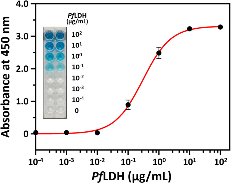
Herein, we report a sensitive and selective enzyme-linked aptamer-based sandwich assay (ELASA) to detect Plasmodium falciparum lactate dehydrogenase (PfLDH), which is an attractive biomarker for malaria diagnosis and antimalarial medication. We performed the sandwich assay with a single aptamer sequence, called 2008s, owing to the structural properties of the PfLDH tetramer instead of using a conventional sandwich assay with two different aptamers (or antibodies) for capturing and probing a target molecule. First, the biotinylated PfLDH aptamer was linked with immobilized streptavidin on a microwell plate for binding flexibility, and then PfLDH was bound to the aptamer. Next, a horseradish peroxidase-conjugated aptamer of the same sequence was used to analyze PfLDH quantitatively. Using this approach, the limit of detection (LOD) of PfLDH with the naked eye was 100 ng mL-1, and the LOD and limit of quantification from the absorbance measurements were 34.9 ng mL-1 and 95.5 ng mL-1, respectively, based on PfLDH spiked blood samples. Our proposed method selectively binds PfLDH, not human lactate dehydrogenase. Therefore, this method may be a valuable tool for diagnosing, monitoring, and quarantining malaria cases easily and rapidly.


Conflict of interest statement

There are no conflicts to declare.

Figures

Fig. 1
Fig. 1. Polyacrylamide gel electrophoresis for Plasmodium falciparum lactate dehydrogenase (PfLDH) and human lactate dehydrogenase (hLDH) proteins. (A) A gel image of each immobilized metal affinity chromatography (IMAC) fraction. Electrophoresis was performed on a 12% polyacrylamide gel under reduced conditions. PL, protein ladder; L, lysate of Escherichia coli BL21(DE3) carrying plasmid pET-21a(+)-PfLDH; F, flow-through fraction after initially loading the BL21(DE3) lysate onto a column packed with nickel–nitrilotriacetic acid agarose beads; W, the washing fraction from the IMAC procedure; E, the elution fraction from the IMAC procedure. (B) Gel images of purified PfLDH (left) and purified hLDH (right). Electrophoresis was performed on a 15% polyacrylamide gel under reduced conditions. (C) Gel images of purified PfLDH (left) and purified hLDH (right). Electrophoresis was performed on a 10% polyacrylamide gel under native (non-reduced) conditions.
Scheme 1
Scheme 1. A schematic illustration of the enzyme-linked aptamer-based sandwich assay for detecting Plasmodium falciparum lactate dehydrogenase (PfLDH). HRP, horseradish peroxidase; TMB, 3,3′,5,5′-tetramethylbenzidine.
Fig. 2
Fig. 2. The principle and the binding affinity results between Plasmodium falciparum lactate dehydrogenase (PfLDH) and the 2008s aptamer. (A) Schematic illustration of the experimental procedures for the dissociation constant (KD) analysis between PfLDH and the 2008s aptamer. (B–D) PfLDH and the 2008s aptamer interaction binding curves using three different buffer conditions for the whole analysis procedures. Phosphate buffered saline (pH 7.4) was used in (B), 25 mM Tris–Cl (pH 7.5), 100 mM NaCl, and 20 mM imidazole was used in (C), and 10 mM Tris (pH 7.4), 10 mM NaCl, and 0.2 mM MgCl2 was used in (D). The circles in (B) to (D) indicate streptavidin (SA, black circles) and bovine serum albumin (BSA, white circles) coated on microplates.
Fig. 3
Fig. 3. Streptavidin and the 2008s aptamer binding. The x-axis represents the horseradish peroxidase (HRP)-conjugated 2008s aptamer concentration, and the y-axis represents the absorbance at 450 nm. A microwell plate was coated with 1 μg mL−1 of streptavidin, and then incubated with various HRP-conjugated 2008s aptamer concentrations (0 to 1000 nM).
Fig. 4
Fig. 4. Plasmodium falciparum lactate dehydrogenase (PfLDH) detection with the enzyme-linked aptamer-based sandwich assay platform. The x-axis represents the PfLDH concentrations (0, 10−4, 10−3, 10−2, 10−1, 100, 101, and 102 μg mL−1), and the y-axis represents the absorbance at 450 nm. The inset image shows the color development after adding the color substrate but before adding the stop solution.
Fig. 5
Fig. 5. Human lactate dehydrogenase (hLDH) detection with the enzyme-linked aptamer-based sandwich assay platform. The x-axis represents the hLDH concentrations (0, 10−4, 10−3, 10−2, 10−1, 100, 101, and 102 μg mL−1), and the y-axis represents the absorbance at 450 nm. The inset image shows the color development after adding the color substrate but before adding the stop solution.
Fig. 6
Fig. 6. A multiple sequence alignment of Plasmodium falciparum lactate dehydrogenase (PfLDH) and human lactate dehydrogenase (hLDH) amino acids using the UniProt database. Each LDH amino acids sequence contains the original sequence without tag. Purple boxes indicate identical amino acids, and yellow boxes indicate aptamer binding sites.
Fig. 7
Fig. 7. Plasmodium falciparum lactate dehydrogenase (PfLDH) detection in a blood sample using the enzyme-linked aptamer-based sandwich assay platform. Mouse blood spiked with different PfLDH concentrations of 0, 10−4, 10−3, 10−2, 10−1, 100, 101, and 102 μg mL−1 (x-axis). The y-axis represents the absorbance at 450 nm. The inset image shows the color development after adding the color substrate but before adding the stop solution.

Similar articles

Cited by

References

    1. World Health Organization (WHO), World Malaria Report 2021, Geneva, 2021
    1. Neves Borgheti-Cardoso L. San Anselmo M. Lantero E. Lancelot A. Serrano J. L. Hernández-Ainsa S. Fernàndez-Busquets X. Sierra T. J. Mater. Chem. B. 2020;8:9428–9448. doi: 10.1039/D0TB01398F. - DOI - PubMed
    1. Phillips M. A. Burrows J. N. Manyando C. van Huijsduijnen R. H. Van Voorhis W. C. Wells T. N. C. Nat. Rev. Dis. Primers. 2017;3:17050. doi: 10.1038/nrdp.2017.50. - DOI - PubMed
    1. Fong Amaris W. M. Martinez C. Cortés-Cortés L. J. Suárez D. R. Malar. J. 2022;21:74. doi: 10.1186/s12936-022-04064-2. - DOI - PMC - PubMed
    1. Eze P. U. Asogwa C. O. Bioengineering. 2021;8:150. doi: 10.3390/bioengineering8110150. - DOI - PMC - PubMed