. 2022 Oct 14;12(45):29469-29481.
doi: 10.1039/d2ra05061g. eCollection 2022 Oct 11.

Amyloid fibrillation of the glaucoma associated myocilin protein is inhibited by epicatechin gallate (ECG)

Affiliations

Amyloid fibrillation of the glaucoma associated myocilin protein is inhibited by epicatechin gallate (ECG)

Ritika Sharma et al. RSC Adv. .

Erratum in

Abstract

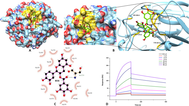
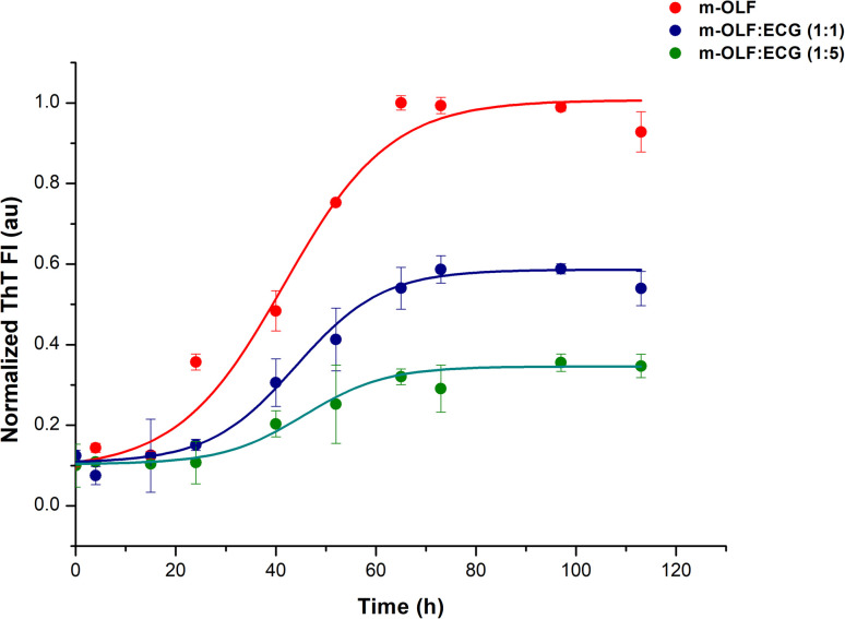
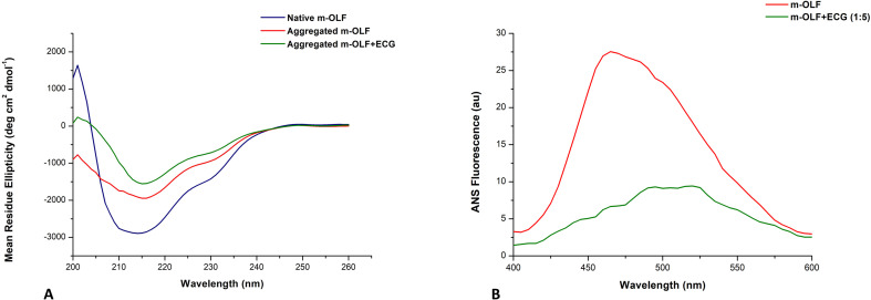
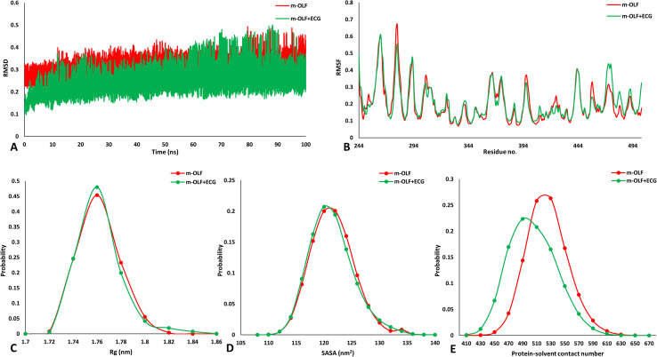
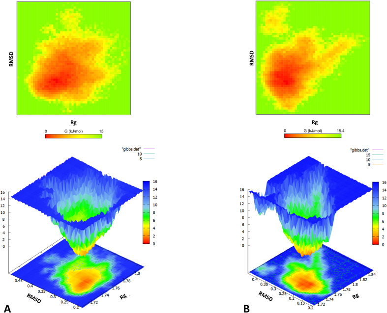
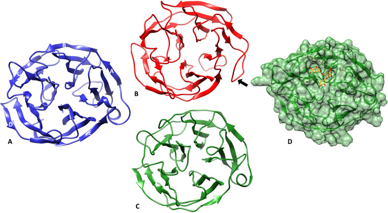
Inherited glaucoma is a recent addition to the inventory of diseases arising due to protein misfolding. Mutations in the olfactomedin (OLF) domain of myocilin are the most common genetic cause behind this disease. Disease associated variants of m-OLF are predisposed to misfold and aggregate in the trabecular meshwork (TM) tissue of the eye. In recent years, the nature of these aggregates was revealed to exhibit the hallmarks of amyloids. Amyloid aggregates are highly stable structures that are formed, often with toxic consequences in a number of debilitating diseases. In spite of its clinical relevance the amyloidogenic nature of m-OLF has not been studied adequately. Here we have studied the amyloid fibrillation of m-OLF and report ECG as an inhibitor against it. Using biophysical and biochemical assays, coupled with advanced microscopic evaluations we show that ECG binds and stabilizes native m-OLF and thus prevents its aggregation into amyloid fibrils. Furthermore, we have used REMD simulations to delineate the stabilizing effects of ECG on the structure of m-OLF. Collectively, we report ECG as a molecular scaffold for designing and testing of novel inhibitors against m-OLF amyloid fibrillation.


Conflict of interest statement

The authors declare that they have no known competing interests.

Figures

Fig. 1
Fig. 1. Evaluation of the binding interactions through molecular docking and SPR. (A) Representation of the m-OLF + ECG docked complex with the ligand occupying the surface groove of the m-OLF structure. Zoomed representation of the binding interactions that stabilize m-OLF + ECG complex in 3D (B) and 2D details (C). (D) Real time binding affinity measurements of ECG using SPR, represented as sensorgrams obtained on injecting ECG at different concentrations over a CM5 chip immobilized with m-OLF.
Fig. 2
Fig. 2. Effect of ECG on the kinetics of m-OLF fibrillation. Variation in the levels of ThT fluorescence plotted as a function of time, depicting the fibrillation of m-OLF alone (red traces) and in the presence of increasing concentrations of ECG (blue and green traces). The kinetics of m-OLF aggregation exhibits a sigmoidal transition and a dose-dependent decline in the levels of ThT fluorescence is observed for m-OLF in the presence of ECG.
Fig. 3
Fig. 3. Morphological characterization of the m-OLF aggregates formed in the presence and absence of ECG. Negatively stained amyloid fibrils formed by m-OLF in the absence of ECG as observed using TEM (A) and AFM (B; where arrows in white pinpoint the straight fibrils). Amorphous aggregates formed by m-OLF samples in the presence of ECG as visualized using TEM (C) and AFM (D). Scale bars represent 100 nm and 1 μM for TEM and AFM images respectively. Colour bars in (B) and (D) provide a gradation for the height of the observed aggregates, structures in bright yellow have the maximum height.
Fig. 4
Fig. 4. Secondary structural changes and hydrophobicity assessment of the aggregates formed by m-OLF. (A) Far UV CD spectra for aggregated m-OLF in the absence and presence of ECG exhibited a decrease in the CD signal at 215 nm, and a slight shift of the spectral minimum towards 220 nm was observed for m-OLF in the absence of ECG. (B) ECG binding results in formation of m-OLF aggregates with reduced surface hydrophobicity.
Fig. 5
Fig. 5. Evaluation of the REMD simulation trajectories for studying conformational variations. (A) Backbone RMSD variations of m-OLF alone (red traces) and m-OLF + ECG complex (green traces) during the complete simulation. (B) Computed RMSF depicting the structural fluctuations by each amino acid residue for the stable time frame. Probability distribution plots of Rg (C), SASA (D) and protein–solvent contacts (E) compared for both the systems.
Fig. 6
Fig. 6. Free energy landscape of m-OLF. 2D and 3D Gibbs free energy landscapes (kcal mol−1) of m-OLF projected as a function of the backbone Rg and RMSD values in the absence (A) and presence of ECG (B).
Fig. 7
Fig. 7. Comparison of the structural variation in m-OLF protein at the end of the simulation. (A) Native structure of the monomeric m-OLF domain, which comprises of a β propeller structure having five blades numbered A–E. Each blade is composed of four antiparallel β sheets which are joined by intermediate loops with two helical turns and a short α-helix in between the outer strands of the A blade. (B) Structure of m-OLF when simulated alone for 100 ns, the arrow highlights an unfolded α-helix. (C) m-OLF simulated in the presence of ECG with the side helix intact at the end of the simulation. (D) Surface representation of the docked complex of m-OLF + ECG at the end of the simulation.

Similar articles

Cited by

References

    1. Leite M. T. Sakata L. M. Medeiros F. A. Arquivos Brasileiros de Oftalmologia. 2011;74:83–84. - PMC - PubMed
    1. Quigley H. A. Broman A. T. Br. J. Ophthalmol. 2006;90:262–267. - PMC - PubMed
    1. Tham Y.-C. Li X. Wong T. Y. Quigley H. A. Aung T. Cheng C.-Y. Ophthalmology. 2014;121:2081–2090. - PubMed
    1. Kapetanakis V. V. Chan M. P. Y. Foster P. J. Cook D. G. Owen C. G. Rudnicka A. R. Br. J. Ophthalmol. 2016;100:86–93. - PMC - PubMed
    1. Fingert J. H. Héon E. Liebmann J. M. Yamamoto T. Craig J. E. Rait J. Kawase K. Hoh S.-T. Buys Y. M. Dickinson J. Hum. Mol. Genet. 1999;8:899–905. - PubMed