. 2022 Sep 21;12(41):26789-26799.
doi: 10.1039/d2ra03285f. eCollection 2022 Sep 16.

Hydroxyapatite coating on an aluminum/bioplastic scaffold for bone tissue engineering

Affiliations

Hydroxyapatite coating on an aluminum/bioplastic scaffold for bone tissue engineering

Oratai Jongprateep et al. RSC Adv. .

Abstract

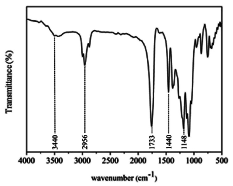
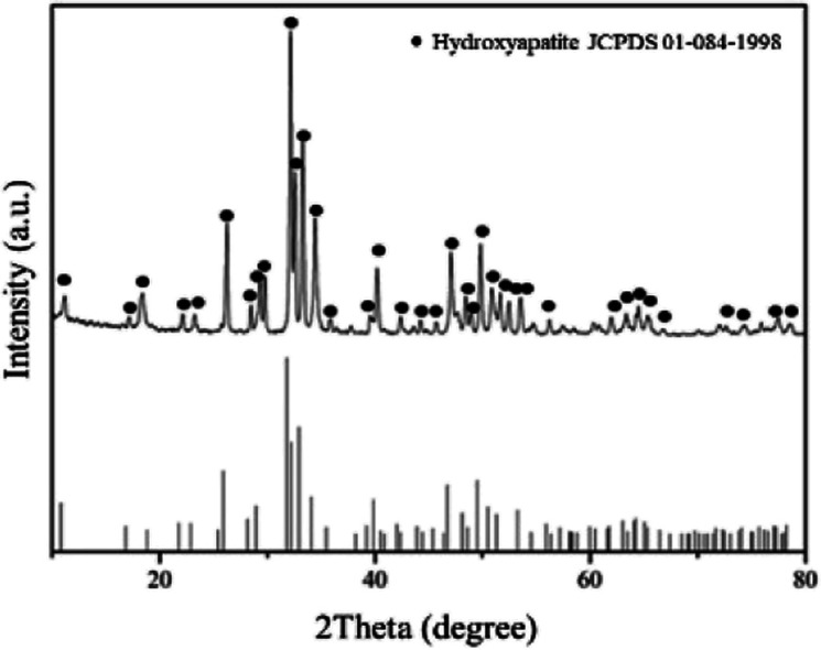
Three-dimensional printing can produce scaffolds with shapes and dimensions tailored for practical clinical applications. Enhanced osteoconductivity of such scaffolds is generally desired. Hydroxyapatite (HA) is an inorganic ceramic that can be used to coat such scaffolds and to accelerate healing during the bone restoration process. In this study, HA-coated aluminum/bioplastic scaffolds were fabricated, and their structural characteristics and osteoconductivity were evaluated. Aluminum/bioplastic scaffolds were fabricated by three-dimensional printing, and HA slurries with solids loadings of 10-20 vol% were used for coating. As solids loadings increased, the thickness of the coating layers slightly increased, whereas pore sizes decreased. The average compressive strength was comparable to that of cancellous bone. Potential osteoconductivity was tested by simulated body fluid immersion for 28 days, and the formation of the HA phase on the surface along with a weight increase indicates the potential bioactivity of the samples.


Conflict of interest statement

There are no conflicts to declare.

Figures

Fig. 1
Fig. 1. 3D-printed scaffold.
Fig. 2
Fig. 2. 3D printer and filament used to fabricate the scaffold.
Fig. 3
Fig. 3. HA synthesis by solution combustion.
Fig. 4
Fig. 4. Scanning electron micrograph of the scaffold microstructure.
Fig. 5
Fig. 5. XRD pattern of the scaffold.
Fig. 6
Fig. 6. Elemental mapping of the scaffold.
Fig. 7
Fig. 7. Fourier transform infrared spectrum of the scaffold.
Fig. 8
Fig. 8. Thermal analysis of the bone scaffold: (a) TGA curve and (b) DSC curve.
Fig. 9
Fig. 9. X-ray diffraction pattern of HA powder synthesized by solution combustion.
Fig. 10
Fig. 10. (a) and (b) Scanning electron micrographs showing the morphology of HA particle. (c) Scanning electron micrographs showing agglomeration of HA particles into clusters.
Fig. 11
Fig. 11. Particle size distributions of HA particles.
Fig. 12
Fig. 12. Scanning electron micrographs showing the microstructure of scaffolds coated by (a) 10, (b) 15, and (c) 20 vol% HA.
Fig. 13
Fig. 13. Scanning electron micrographs showing the pore structure of scaffolds coated by (a) 10, (b) 15, and (c) 20 vol% HA.
Fig. 14
Fig. 14. Average pore sizes of the coated scaffolds analyzed by ImageJ.
Fig. 15
Fig. 15. Average porosity of scaffolds coated by HA analyzed by ImageJ and Origin Pro8.5.
Fig. 16
Fig. 16. Scanning electron micrographs, EDS spectrum, and elemental mapping of the scaffolds coated by (a) 10, (b) 15, and (c) 20 vol% HA.
Fig. 17
Fig. 17. Scanning electron micrographs showing thickness of the (a) uncoated sample, coated with (b) 10 vol% solids loading, (c) 15 vol% solids loading, and (d) 20 vol% solids loading.
Fig. 18
Fig. 18. Thickness of the coating layer on the scaffold according to solids loading.
Fig. 19
Fig. 19. Compressive strength of uncoated and coated scaffolds.
Fig. 20
Fig. 20. Scanning electron micrographs showing the sample coated with (a) 10 vol% solids loading, (b) 15 vol% solids loading, and (c) 20 vol% solids loading.
Fig. 21
Fig. 21. Weibull plots for the compressive strength of scaffolds: (a) control, (b) coated with 10 vol% HA, (c) coated with 15 vol% HA, and (d) coated with 20 vol% HA.
Fig. 22
Fig. 22. Scanning electron micrographs and elemental mapping of the scaffolds coated by hydroxyapatite slurries with 10 vol% (a) before immersion in SBF, and (b) after immersion in SBF 28 days.

References

    1. Tariverdian T., Sefat F., Gelinsky M. and Mozafari M., Handbook of Tissue Engineering Scaffolds: Volume One, Elsevier, 2019, pp. 189–209
    1. Ghassemi T. Shahroodi A. Ebrahimzadeh M. H. Mousavian A. Movaffagh J. Moradi A. Arch. Bone Jt. Surg. 2018;6:90–99. - PMC - PubMed
    1. De Witte T. M. Fratila-Apachitei L. E. Zadpoor A. A. Peppas N. A. Regener. Biomater. 2018;5:197–211. - PMC - PubMed
    1. Brown B. N. Valentin J. E. Stewart-Akers A. M. McCabe G. P. Badylak S. F. Biomaterials. 2009;30:1482–1491. - PMC - PubMed
    1. Hutmacher D. W. Biomaterials. 2000;21:2529–2543. - PubMed