Review
. 2023 Nov 9;43(Suppl 1):S19-S31.
doi: 10.1093/asj/sjac267.

Integrative Assessment for Optimizing Aesthetic Outcomes When Treating Glabellar Lines With Botulinum Toxin Type A: An Appreciation of the Role of the Frontalis

Review

Integrative Assessment for Optimizing Aesthetic Outcomes When Treating Glabellar Lines With Botulinum Toxin Type A: An Appreciation of the Role of the Frontalis

Vince Bertucci et al. Aesthet Surg J. .

Abstract

Despite the perception that treatment of glabellar lines with botulinum toxin A is straightforward, the reality is that the glabellar region contains a number of interrelated muscles. To avoid adverse outcomes, practitioners need to appreciate how treatment of 1 facial muscle group influences the relative dominance of others. In particular, practitioners need to understand the independent role of the frontalis in eyebrow outcomes and the potential for negative outcomes if the lower frontalis is unintentionally weakened by botulinum toxin A treatment. In addition, practitioners must recognize how inter-individual variation in the depth, shape, and muscle fiber orientation among the upper facial muscles can affect outcomes. For optimal results, treatment of the glabellar complex requires a systematic and individualized approach based on anatomical principles of opposing muscle actions rather than a one-size-fits-all approach. This review provides the anatomical justification for the importance of an integrated assessment of the upper facial muscles and eyebrow position prior to glabellar treatment. In addition, a systematic and broad evaluation system is provided that can be employed by practitioners to more comprehensively assess the glabellar region in order to optimize outcomes and avoid negatively impacting resting brow position and dynamic brow movement.


Figures

Figure 1.
Figure 1.
Role of the frontalis muscle in eyelid elevation. (A) In this 52-year-old female, the upper eyelid is located at the upper margin of the iris at rest. (B) At maximum lift, the frontalis muscle raises the eyelid 1 to 2 mm above the upper margin of the iris. (C) Lateral frontalis lines above the left eyebrow at rest suggest involuntary frontalis activity is compensating for levator weakness in this 47-year-old female.
Figure 2.
Figure 2.
Eyelid ptosis severities. (A) No eyelid ptosis. In this 47-year-old female, the upper eyelids are located just slightly below the margin of the iris. However, lateral frontalis lines above the left eyebrow are suggestive of left eyelid ptosis masked by compensatory frontalis activity (marginal reflex distance = 4.5 mm for the upper right eyelid and 5 mm for the upper left eyelid). (B) Mild bilateral ptosis. In this 56-year-old female, the upper lid is located below the upper margin of the iris at rest, and approximately one-third of the upper iris is obscured (marginal reflex distance = 3 mm for the upper right eyelid and 4 mm for the upper left eyelid). (C) Moderate bilateral eyelid ptosis. In this 44-year-old man, the upper lid bisects the pupil in the right and left eyes at rest (marginal reflex distance = 0 mm for both upper eyelids), and 50% of the iris is obscured.
Figure 3.
Figure 3.
Eyebrow ptosis. (A) In this 52-year-old female, right eyebrow ptosis is present at rest and (B) is indicated by eyebrow asymmetry between the relative positions of the medial and lateral brows.
Figure 4.
Figure 4.
Forehead line patterns as indicators of frontalis muscle activity. (A) Straight central forehead lines at maximum lift are suggestive of a strong central pillar of frontalis muscle in this 41-year-old female. (B) Wavy-shaped central forehead lines at maximum lift are suggestive of a weaker central pillar of frontalis muscle in this 48-year-old female. (C) No central forehead lines at maximum lift are suggestive of a weaker central pillar of frontalis muscle in this 41-year-old female.
Figure 5.
Figure 5.
Muscle activity that contributes to glabellar line formation (A) is shown in a representative image of this 54-year-old female at maximum frown (B). The location of muscle activity and glabellar lines are shown by lines (A) and arrows (B), respectively, where blue denotes the procerus, yellow denotes the medial region of the corrugator, green denotes the lateral region of the corrugator, and purple denotes the orbicularis oculi.
Figure 6.
Figure 6.
Balancing act of eyebrow position. (A) Eyebrow position is determined by a balance between the caudal pull of the depressor complex (procerus, depressor supercilii, orbicularis oculi, and corrugator supercilii) and the upward pull of the frontalis muscle. (B) Weakening of the suspensory activity of the lower frontalis (eg, by exposure to botulinum toxin) creates an imbalance in the forces holding the brow in position, resulting in suboptimal outcomes. This can occur through inactivation of the medial region of the lower frontalis, causing a drop in the medial region of the brow and elevation of the lateral region of the brow or (C) through inactivation of the medial and lateral regions of the lower frontalis, causing a drop across all regions of the brow. (D) Weakening of the downward pull of the depressor complex can elevate the medial region of the eyebrow, resulting in aesthetically pleasing outcomes.
Figure 7.
Figure 7.
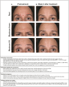
Eyebrow outcomes following glabellar line treatment for patients with a weaker central frontalis in (A) this 54-year-old female with a lower central frontalis that was not exposed to botulinum toxin A (BoNTA) and (B) this 36-year-old female with a lower central frontalis inadvertently exposed to botulinum toxin A. In Patient B, injection points to the medial regions of the corrugators were accurately targeting the medial region, but injections to the lateral regions of the corrugators and the procerus exposed the lower frontalis to botulinum toxin A. MRD, marginal reflex distance.
Figure 8.
Figure 8.
Aesthetic outcomes following glabellar line treatment for this 52-year-old female with a strong central frontalis and mild bilateral ptosis prior to treatment (A) and 2 weeks following treatment with botulinum toxin A (B).

References

    1. Kaminer MS, Cox SE, Fagien S, Kaufman J, Lupo MP, Shamban A. Re-examining the optimal use of neuromodulators and the changing landscape: a consensus panel update. J Drugs Dermatol. 2020;19(4):s5–s15. - PubMed
    1. Benedetto AV, Lahti JG. Measurement of the anatomic position of the corrugator supercilii. Dermatol Surg. 2005;31(8 Pt 1):923–927. doi: 10.1097/00042728-200508000-00006 - DOI - PubMed
    1. Lee HJ, Lee KW, Tansatit T, Kim HJ. Three-dimensional territory and depth of the corrugator supercilii: application to botulinum neurotoxin injection. Clin Anat. 2020;33(5):795–803. doi: 10.1002/ca.23507 - DOI - PubMed
    1. Macdonald MR, Spiegel JH, Raven RB, Kabaker SS, Maas CS. An anatomical approach to glabellar rhytids. Arch Otolaryngol Head Neck Surg. 1998;124(12):1315–1320. doi: 10.1001/archotol.124.12.1315 - DOI - PubMed
    1. Cohn JE, Greco TM. Advanced techniques for the use of neurotoxins in non-surgical facial rejuvenation. Aesthetic Plast Surg. 2020;44(5):1788–1799. doi: 10.1007/s00266-020-01691-5 - DOI - PubMed

Substances

Grants and funding