. 2022 Nov 4;8(44):eadc9566.
doi: 10.1126/sciadv.adc9566. Epub 2022 Nov 2.

Lipids mediate supramolecular outer membrane protein assembly in bacteria

Affiliations

Lipids mediate supramolecular outer membrane protein assembly in bacteria

Melissa N Webby et al. Sci Adv. .

Abstract

β Barrel outer membrane proteins (OMPs) cluster into supramolecular assemblies that give function to the outer membrane (OM) of Gram-negative bacteria. How such assemblies form is unknown. Here, through photoactivatable cross-linking into the Escherichia coli OM, coupled with simulations, and biochemical and biophysical analysis, we uncover the basis for OMP clustering in vivo. OMPs are typically surrounded by an annular shell of asymmetric lipids that mediate higher-order complexes with neighboring OMPs. OMP assemblies center on the abundant porins OmpF and OmpC, against which low-abundance monomeric β barrels, such as TonB-dependent transporters, are packed. Our study reveals OMP-lipid-OMP complexes to be the basic unit of supramolecular OMP assembly that, by extending across the entire cell surface, couples the requisite multifunctionality of the OM to its stability and impermeability.


Figures
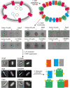
Fig. 1.
Fig. 1.. Strategy for identifying OMP near-neighbors by BPA-mediated cross-linking into the E. coli OM.
Schematic outlining the key steps of the protocol for UV-activated OMPBPA cross-linking, including OmpF (green), BtuB (blue), and FepA (orange). (1) A plasmid (blue and green) encoding the omp gene of interest with an amber stop codon (TAG) at a single site was transformed into a knockout cell line devoid of the omp of interest, in conjunction with a plasmid for expressing the tRNase (pEVOL-pBpF, brown) required for BPA incorporation. Cells were then grown in the presence of BPA (brown star; shown as a yellow star following UV activation) and omp gene expression induced with 0.15% arabinose. (2) A colicin-based cytotoxic assay was used to test for OMP functionality in the OM. The colicins used in the study each require the OMP of interest as a receptor before import and cell killing. Hence, colicin cytotoxicity is a simple readout of appropriate expression/insertion into the OM. Examples of successful killing of E. coli cells expressing BPA-incorporated OMPs are shown for each protein target. (3) Cells were exposed to UV light (λ365) for 90 min to activate BPA cross-linking. (4) The cellular distribution of the OMP of interest was subsequently reanalyzed, to ascertain whether it was similar to that of the wild-type protein, using fluorescent colicin labels as in figs. S3 to S5. Scale bar, 1 μm. (5) Cells were lysed, the OM was extracted, and OMPs were solubilized with detergent (1% β-OG) and purified by chromatography for further characterization (see Materials and Methods for details).
Fig. 2.
Fig. 2.. BtuBBPA and FepABPA cross-linking into the OM enhances associations with other OMPs and lipids.
(A) Denaturing SDS-polyacrylamide gel of BtuBBPA variants extracted from the OM of E. coli RK5016 cells following UV exposure and stained with Coomassie blue for protein (top) and emerald green for LPS (bottom). Several variants cross-link to LPS, as indicated by a gel shift and fluorescence with LPS stain, which is not observed in wild-type BtuB or a no-UV BtuB W164BPA control. Small amounts of OmpA are observed in BtuB variants that contain BPA. The noncovalent recruitment of BamA to BtuB W164BPA (black asterisk) was confirmed by peptide fingerprinting and Western blot. (B) Top view of BtuB β barrel showing BPA incorporation sites (gray spheres) where BPA cross-linking into the membrane results in covalent attachment of LPS. (C) Denaturing SDS-polyacrylamide gel of FepABPA variants extracted from the OM of E. coli BW25113 ΔFepA cells following UV exposure and stained with Coomassie blue for protein (top) and emerald green for LPS (bottom). FepABPA mutants that have been exposed to UV copurify with the same complement of additional OMPs, OmpF/C, FhuE, and LptD, but their yields vary between BPA incorporation sites. In the absence of UV exposure, there is a notable reduction in the copurification of OMPs. Although the number of LPS cross-links appear minimal (only two are detected on the gel), it is likely that the increased copurification of OmpF/C for FepABPA mutants arises from lipid-mediated interactions with cross-linked PL and/or with LPS associated noncovalently with porins. (D) Top view of FepA β barrel showing BPA incorporation sites (gray spheres) where cross-linking into the membrane results in covalent attachment of LPS.
Fig. 3.
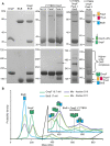
Fig. 3.. OmpFBPA cross-linking to the asymmetric lipids of the OM stabilizes promiscuous associations with TBDTs.
(A) Representative UV280 absorbance recording from the final anion exchange purification step showing wild-type OmpF eluting in a major peak (peak 1) with a trailing edge. Peak 1 persists following exposure of OmpFBPA mutants to UV and a second peak (peak 2) appears eluting later in the salt gradient; representative profile of OmpF V177BPA is shown. (B) OmpFBPA peak 1 samples were analyzed by denaturing SDS-PAGE and stained with Coomassie blue for protein (top) and ProQ emerald green for LPS (bottom). In some cases, exposure of OmpFBPA mutants to UV results in elevated levels of copurified FepA. (C) OmpFBPA peak 2 samples were analyzed by denaturing SDS-PAGE and stained with Coomassie blue for protein (top) and ProQ emerald green for LPS (bottom). Cross-links between OmpFBPA mutants and LPS, as indicated by fluorescence following emerald green staining of SDS-PAGE gels, result in enhanced copurification of the TBDTs FepA and FhuA. (D) Native-MS spectrum of UV-activated OmpF V177BPA peak 1 shows OmpF trimer cross-linked to one to three PL molecules. Similar lipid cross-linking profiles are observed for peak 1 of other OmpFBPA mutants (figs. S10 and S11). Inset: a zoomed view of 21+ charge state. Observed masses are listed in table S4. Gel inset of the same sample stained with Coomassie and emerald green confirms the absence of LPS staining for OmpF V177BPA peak 1. (E) Native-MS spectrum for OmpF V177BPA from peak 2. Peak 2 corresponds to OmpF cross-linked to LPS with or without PLs and copurified FepA and FhuA (apo- and LPS-bound forms). Insets: a zoomed view of OmpF trimer 21+ charge state and SDS-PAGE gel of sample confirm the presence of LPS in the cross-linked sample.
Fig. 4.
Fig. 4.. OM lipids mediate promiscuous higher-order complexes between OMPs in vitro.
(A) Denaturing SDS-PAGE (top) and blue-native PAGE (bottom) of detergent-solubilized OmpF and BtuB confirm that no high–molecular weight complexes are detected in single protein controls and in a 1:1 mixture (20 μM protein concentration). OmpF V177BPA without UV exposure resembles wild-type OmpF in both SDS-PAGE and native PAGE. Peak 1 and peak 2 of UV-exposed OmpF V177BPA cross-linking to PL and LPS, respectively, copurify with monomeric OMPs FhuA and/or FepA. The abundance of FepA and FhuA in the peak 2 (LPS bound) sample is sufficient to detect a higher–molecular weight complex in blue-native PAGE demonstrating that cross-linked LPS promotes a promiscuous complex between trimeric OmpF and the copurified TBDTs (arrow). Addition of purified BtuB (15 μM, with LPS bound noncovalently; fig. S7) to OmpF V177BPA peak 1 and peak 2 samples results in the appearance of higher-order complexes that are absent in the single protein controls. (B) Mass photometry data were collected following passage of OMPs through a size exclusion column (SEC). The average plot for the pooled elution peak fractions is shown for OmpF V177BPA peak 1 (green, 17 nM) and BtuB (blue, 17 nM). Two discrete SEC fractions (D10 and D11) of the BtuB-OmpF V177BPA mixture (pale green and purple) are also plotted. BtuB control data show that most of the species are monomeric with some dimer formation. Similarly, the OmpF V177BPA peak 1 control data show some self-association of OmpF trimers. Comparison of these individual protein controls with data for BtuB and OmpF V177BPA mixture reveals an increased abundance of high–molecular mass species relative to the control samples. The inset shows the assignment of OMP complexes within the sample to corresponding high mass peaks.
Fig. 5.
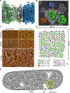
Fig. 5.. OMP-lipid-OMP complexes are the functional units of supramolecular OMP assemblies that stretch across the entire E. coli OM.
(A) Snapshot of MD simulation for the OmpF-LPS/PL-BtuB complex showing the mutual sharing of asymmetric lipids. OmpF mid-barrel residues L259 and I273 (purple spheres) are highlighted for reference. (B) Snapshot of molecular dynamics (MD) simulation for a heterologous lipid-mediated complex formed between trimeric OmpF (green) and neighboring OMPs. LPS is shown in dark gray with molecules surrounding the central OmpF in white. (C) AFM imaging the OM of a live E. coli MG1655 cell labeled with FepA-binding ColB-mCherry (marked as pink balls). As in previous work (30), phase images provide the highest contrast for detecting the trimeric porin network (purple dots). The simultaneously recorded height image shows the positions of ColB-mCherry fluorescent labels as local height maxima. The overlay of FepA positions with the trimeric porins demonstrates that FepA is embedded within the porin network. In addition, there are regions where OMPs do not appear, previously identified as LPS-enriched domains (30). Scale bars, 50 nm. Color (phase/height) scales are 1.1°/2.5 nm and 1.1°, respectively. (D) Model of the simulated OMP island (SOI) in which OmpF (green) hosts heterologous OMPs within its hexagonal arrays. Monomeric β barrels and associated proteins incorporated into the island (detailed in key) were all identified in the present work. A total of 102 OmpF trimers are presented in the model, forming 18 hexagons. n, number of specific OMP in simulation. (E) Cartoon of an E. coli cell depicting the OMP-LPS-OMP network across the OM. The high relative abundance of OmpF results in the porin being spread over much of the cell surface. OMP islands (highlighted by colored OMP island) immersed within a black and white OMP background are embedded within the OmpF network.

References

    1. Murray C. J., Ikuta K. S., Sharara F., Swetschinski L., Robles Aguilar G., Gray A., Han C., Bisignano C., Rao P., Wool E., Johnson S. C., Browne A. J., Chipeta M. G., Fell F., Hackett S., Haines-Woodhouse G., Kashef Hamadani B. H., Kumaran E. A. P., McManigal B., Agarwal R., Akech S., Albertson S., Amuasi J., Andrews J., Aravkin A., Ashley E., Bailey F., Baker S., Basnyat B., Bekker A., Bender R., Bethou A., Bielicki J., Boonkasidecha S., Bukosia J., Carvalheiro C., Castañeda-Orjuela C., Chansamouth V., Chaurasia S., Chiurchiù S., Chowdhury F., Cook A. J., Cooper B., Cressey T. R., Criollo-Mora E., Cunningham M., Darboe S., Day N. P. J., de Luca M., Dokova K., Dramowski A., Dunachie S. J., Eckmanns T., Eibach D., Emami A., Feasey N., Fisher-Pearson N., Forrest K., Garrett D., Gastmeier P., Giref A. Z., Greer R. C., Gupta V., Haller S., Haselbeck A., Hay S. I., Holm M., Hopkins S., Iregbu K. C., Jacobs J., Jarovsky D., Javanmardi F., Khorana M., Kissoon N., Kobeissi E., Kostyanev T., Krapp F., Krumkamp R., Kumar A., Kyu H. H., Lim C., Limmathurotsakul D., Loftus M. J., Lunn M., Ma J., Mturi N., Munera-Huertas T., Musicha P., Mussi-Pinhata M. M., Nakamura T., Nanavati R., Nangia S., Newton P., Ngoun C., Novotney A., Nwakanma D., Obiero C. W., Olivas-Martinez A., Olliaro P., Ooko E., Ortiz-Brizuela E., Peleg A. Y., Perrone C., Plakkal N., Ponce-de-Leon A., Raad M., Ramdin T., Riddell A., Roberts T., Robotham J. V., Roca A., Rudd K. E., Russell N., Schnall J., Scott J. A. G., Shivamallappa M., Sifuentes-Osornio J., Steenkeste N., Stewardson A. J., Stoeva T., Tasak N., Thaiprakong A., Thwaites G., Turner C., Turner P., van Doorn H. R., Velaphi S., Vongpradith A., Vu H., Walsh T., Waner S., Wangrangsimakul T., Wozniak T., Zheng P., Sartorius B., Lopez A. D., Stergachis A., Moore C., Dolecek C., Naghavi M., Global burden of bacterial antimicrobial resistance in 2019: A systematic analysis. Lancet 399, 629–655 (2022). - PMC - PubMed
    1. ECDC/EMEA Joint Working Group, The Bacterial Challenge: Time to React: A Call to Narrow the Gap Between Multidrug-Resistant Bacteria in the EU and the Development of New Antibacterial Agents (EUR-OP, 2009).
    1. Nikaido H., Molecular basis of bacterial outer membrane permeability revisited. Microbiol. Mol. Biol. Rev. 67, 593–656 (2003). - PMC - PubMed
    1. Delcour A. H., Outer membrane permeability and antibiotic resistance. Biochim. Biophys. Acta 1794, 808–816 (2009). - PMC - PubMed
    1. Wimley W. C., The versatile beta-barrel membrane protein. Curr. Opin. Struct. Biol. 13, 404–411 (2003). - PubMed

MeSH terms

LinkOut - more resources