Review
. 2022 Nov 4;8(44):eadd4920.
doi: 10.1126/sciadv.add4920. Epub 2022 Nov 2.

Untangling the roles of TOP2A and TOP2B in transcription and cancer

Affiliations
Review

Untangling the roles of TOP2A and TOP2B in transcription and cancer

Liis Uusküla-Reimand et al. Sci Adv. .

Abstract

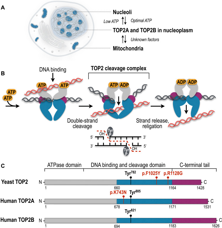
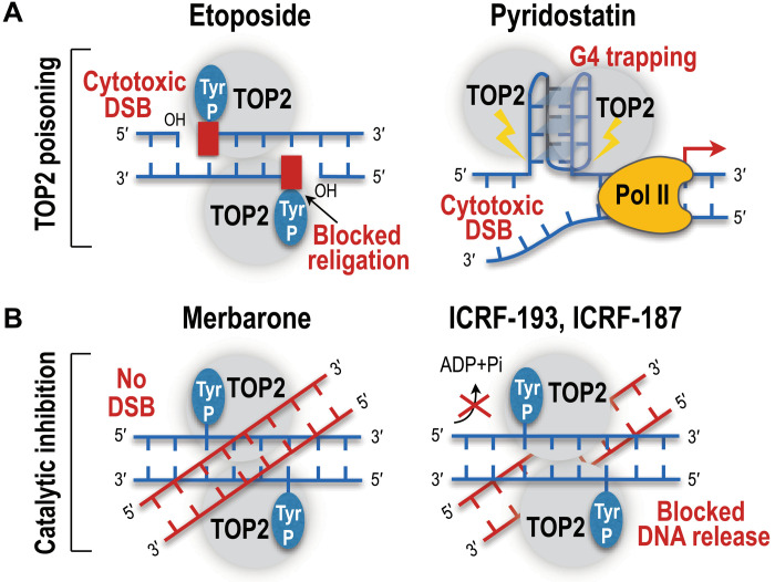
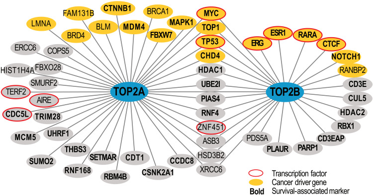
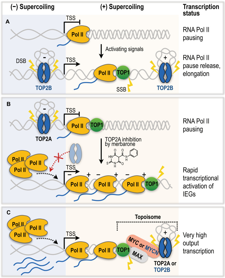
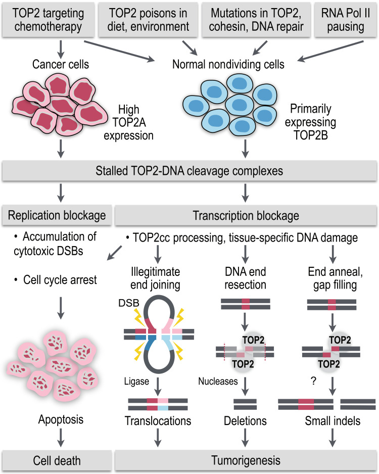
Type II topoisomerases (TOP2) are conserved regulators of chromatin topology that catalyze reversible DNA double-strand breaks (DSBs) and are essential for maintaining genomic integrity in diverse dynamic processes such as transcription, replication, and cell division. While controlled TOP2-mediated DSBs are an elegant solution to topological constraints of DNA, DSBs also contribute to the emergence of chromosomal translocations and mutations that drive cancer. The central importance of TOP2 enzymes as frontline chemotherapeutic targets is well known; however, their precise biological functions and impact in cancer development are still poorly understood. In this review, we provide an updated overview of TOP2A and TOP2B in the regulation of chromatin topology and transcription, and discuss the recent discoveries linking TOP2 activities with cancer pathogenesis.


Figures

Fig. 1.
Fig. 1.. Cellular localization, catalytic cycle, and evolutionary conservation of TOP2 enzymes.
(A) TOP2A and TOP2B are detected in the nucleoplasm, nucleoli, and mitochondria in the cell. (B) Eukaryotic TOP2 binds helix-helix crossovers of DNA as a homodimer that cleaves both strands of duplex DNA with 5′-four base overhangs and forms the TOP2cc by covalently attaching the catalytic tyrosine (Tyr) to the 5′-ends of the cleaved DNA. TOP2cc allows the passage of the uncleaved helix and the subsequent seamless religation of the DNA breaks. (C) Protein domains of yeast and human TOP2 enzymes. Colors show homologous regions of the enzymes. The N-terminal domain is responsible for ATP binding and hydrolysis. The central domain harbors tyrosine (Tyr) active sites to form covalent bonds with DNA and cleave and religate the DNA helix. The C-terminal domain shows the largest extent of evolutionary diversity and is involved in the association and dissociation of TOP2 with the DNA. TOP2 mutations associated with the self-poisoning phenotype and short insertions and deletions are shown in red.
Fig. 2.
Fig. 2.. Mechanism of action of TOP2 poisons and inhibitors in chemotherapy.
(A) TOP2 poisons such as etoposide trap TOP2 to 5′ termini of DNA DSBs, block religation of breaks, and cause accumulation of TOP2cc-blocked DSBs. In contrast, the small-molecule ligand pyridostatin traps TOP2A at the G-quadruplex (G4) structures and induces DNA damage and cell cycle arrest. (B) Merbarone inhibits TOP2 catalytically, stabilizes noncovalent TOP2-DNA complexes, and inhibits the formation of enzyme-mediated DSBs. The TOP2 inhibitors ICRF-193 and ICRF-187 decrease the ATPase activity of TOP2 and lock TOP2 in a closed clamp conformation that holds the cleaved and uncleaved DNA duplexes.
Fig. 3.
Fig. 3.. High-confidence protein-protein interactions of TOP2A and TOP2B.
Known protein-protein interactions were retrieved from the BioGRID database (v4.4.205) and the recent studies (75, 86, 130). Interactions were filtered to only include interactions detected in at least two independent studies or experimental approaches. Orange nodes indicate known cancer driver genes (from COSMIC Cancer Gene Census database), red circles highlight human transcription factors [from (156)], and cancer patient survival–associated markers are shown in bold [data from The Cancer Genome Atlas (TCGA)].
Fig. 4.
Fig. 4.. The roles of TOP2A, TOP2B, and TOP1 in RNA Pol II transcription regulation.
(A) The breaking model. TOP2B catalyzes DNA DSBs to promote permissive chromatin structures and facilitate RNA Pol II pause release for rapid activation of transcription. TOP1 removes DNA supercoils of elongating RNA Pol II via single-strand breaks (SSBs). (B) The nonbreaking model of transcriptional activation of immediate early genes (IEGs). Catalytic inhibition of TOP2A induces RNA Pol II promoter-proximal pause release independently of formation of DNA breaks. Negative supercoiling at TSS facilitates separation of DNA molecules, allowing tandem recruitment and transcription of multiple RNA Pol II complexes. Topological feedback loop of transcribing polymerases cancels negative and positive supercoiling. (C) Topoisomes assembled by MYC or MYCN transcription factors unite TOP1 and TOP2A or TOP2B, respectively. Topoisomes stimulate enzymatic activities of topoisomerases to overcome topological challenges resulting from high-output transcription of MYC/MYCN-regulated genes.
Fig. 5.
Fig. 5.. TOP2 role in tumorigenesis.
Normally short-lived TOP2-DNA cleavage intermediates become stalled and trapped to the chromatin by (i) chemotherapeutic, dietary, and environmental TOP2 poisons, (ii) mutations in TOP2, cohesin, and DNA repair enzymes, and (iii) endogenous cellular processes that prolong TOP2-DNA interactions, such as RNA Pol II pausing. In proliferating cancer cells, trapped TOP2ccs block the essential steps in DNA replication and cell division, which overwhelms the DNA damage repair mechanisms and leads to accumulation of cytotoxic DNA lesions and apoptosis of the cancer cell. However, encounter of the stalled TOP2ccs with the transcription machinery in the normal nondividing cells and rapid processing of TOP2-DNA intermediates into protein-free DSBs provide the means for emergence of tissue-specific DNA damage and mutagenesis, including tumorigenic translocations, deletions, and short indels.

References

    1. Champoux J. J., DNA topoisomerases: Structure, function, and mechanism. Annu. Rev. Biochem. 70, 369–413 (2001). - PubMed
    1. Pommier Y., Sun Y., Huang S.-Y. N., Nitiss J. L., Roles of eukaryotic topoisomerases in transcription, replication and genomic stability. Nat. Rev. Mol. Cell Biol. 17, 703–721 (2016). - PMC - PubMed
    1. Ashour M. E., Atteya R., El-Khamisy S. F., Topoisomerase-mediated chromosomal break repair: An emerging player in many games. Nat. Rev. Cancer 15, 137–151 (2015). - PubMed
    1. Forterre P., Gribaldo S., Gadelle D., Serre M.-C., Origin and evolution of DNA topoisomerases. Biochimie 89, 427–446 (2007). - PubMed
    1. Corbett K. D., Berger J. M., Structure, molecular mechanisms, and evolutionary relationships in DNA topoisomerases. Annu. Rev. Biophys. Biomol. Struct. 33, 95–118 (2004). - PubMed

MeSH terms