. 2023 Feb 2;141(5):490-502.
doi: 10.1182/blood.2022017097.

Therapeutic activity of GARP:TGF-β1 blockade in murine primary myelofibrosis

Affiliations

Therapeutic activity of GARP:TGF-β1 blockade in murine primary myelofibrosis

Sara Lecomte et al. Blood. .

Abstract

Primary myelofibrosis (PMF) is a myeloproliferative neoplasm characterized by the clonal expansion of myeloid cells, notably megakaryocytes (MKs), and an aberrant cytokine production leading to bone marrow (BM) fibrosis and insufficiency. Current treatment options are limited. TGF-β1, a profibrotic and immunosuppressive cytokine, is involved in PMF pathogenesis. While all cell types secrete inactive, latent TGF-β1, only a few activate the cytokine via cell type-specific mechanisms. The cellular source of the active TGF-β1 implicated in PMF is not known. Transmembrane protein GARP binds and activates latent TGF-β1 on the surface of regulatory T lymphocytes (Tregs) and MKs or platelets. Here, we found an increased expression of GARP in the BM and spleen of mice with PMF and tested the therapeutic potential of a monoclonal antibody (mAb) that blocks TGF-β1 activation by GARP-expressing cells. GARP:TGF-β1 blockade reduced not only fibrosis but also the clonal expansion of transformed cells. Using mice carrying a genetic deletion of Garp in either Tregs or MKs, we found that the therapeutic effects of GARP:TGF-β1 blockade in PMF imply targeting GARP on Tregs. These therapeutic effects, accompanied by increased IFN-γ signals in the spleen, were lost upon CD8 T-cell depletion. Our results suggest that the selective blockade of TGF-β1 activation by GARP-expressing Tregs increases a CD8 T-cell-mediated immune reaction that limits transformed cell expansion, providing a novel approach that could be tested to treat patients with myeloproliferative neoplasms.


Conflict of interest statement

Conflict-of-interest disclosure: Patents pertaining to blocking antibodies against human GARP:TGF-β1 have been filed under the Patent Cooperation Treaty (International application Number PCT/IB2019/053753) with S. Lucas and P.G.C. as inventors and UCLouvain as applicant. The anti–mouse GARP:TGF-β1 antibody (clone 58A2) used herein is a surrogate for these antibodies. The laboratory of S. Lucas received research funding from AbbVie which licensed the anti-human GARP:TGF-β1 antibodies. The remaining authors declare no competing financial interests.

Figures

None
Graphical abstract
Figure 1.
Figure 1.
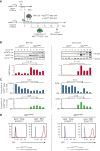
GARP protein is increased in murine PMF and is expressed exclusively on MKs and Tregs. (A) Schematic representation of the experimental design. BM cells were isolated from 5-FU-treated donor mice, transduced with a retrovirus encoding MPLW508A or MPLWT and transplanted into lethally irradiated recipient mice (n = 5 mice/group). BM cells and splenocytes were isolated from MPLW508A or MPLWT control mice 34 days after transplantation. (B) Western blot analyses of BM cells and splenocytes with antibodies against GARP, pSTAT5, or β-actin. Bar graphs show quantification of enhanced chemiluminescence signals. P values were calculated using a two-tailed Student t test. (C) Proportions of GARP+ Tregs and GARP+ MKs in live single cells, as determined by flow cytometry. Fold differences between mean proportions in MPLW508A and MPLWT mice are indicated above bar graphs. (D) Histograms showing GFP expression on Tregs and MKs in the BM and spleen from 1 representative MPLW508A mouse. Cells from a C57BL/6 mouse that did not undergo transplantation were used as negative controls. The experiment is representative of at least 3 other independent experiments.
Figure 2.
Figure 2.
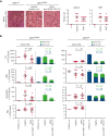
Blocking anti-GARP:TGF-β1 mAbs reduces fibrosis and tumor burden in murine PMF. MPLW508A and MPLWT control mice, generated as indicated in Figure 1, were injected IP weekly with the indicated mAbs, starting 1 day before transplantation of the transduced BM cells (n = 8-10 mice per group). C57BL/6 mice that did not undergo transplantation served as additional controls (ctrl). Blood was taken on day 28 and mice were euthanized on day 34 to collect femurs and spleens. (A) Histological assessment of fibrosis in sections of paraffin-embedded spleens and femurs after silver staining. Representative sections are shown on the left. Fibrosis scores, corresponding to reticulin staining grades expressed on a modified Bauermeister scale, are indicated on the right. Data points: scores in individual mice; horizontal bars: mean + SEM per group. (B) Blood cell counts and GFP expression were determined using a hematological analyzer and flow cytometry. Data points on the left: blood cell counts in individual mice; horizontal bars: mean + SEM per group. Stacked bar graphs on the right: mean + SEM of GFP+ and GFP- cells. Data from 1 of at least 4 independent experiments. P values calculated using a two-tailed Student t test. SEM, standard error of the mean.
Figure 3.
Figure 3.
Reduced tumor burden and fibrosis in PMF do not require effector functions of anti-GARP:TGF-β1 mAbs. (A) MPLW508A mice were treated with wild-type or Fc-dead mAbs as indicated in Figure 2 (n = 8-10 mice per group). Blood was taken on day 21 and mice were euthanized on day 32 to collect femurs and spleens. Blood cell counts, GFP expression, and spleen fibrosis were measured as indicated in Figure 2. Data points: values in individual mice; horizontal lines: mean ± SEM per group; stacked bars: mean + SEM. P values were calculated using a two-tailed Student t test. (B) 32D cells were transduced with MplWT-IRES-GFP or MplW508A-IRES-GFP RV and GFP+ cells were sorted by flow cytometry. Parental 32D cells, sorted MPLWT/GFP+, or MPLW508A/GFP+ bulk cell populations, or clones (supplemental Figure 6) were cultured in complete medium, in the absence or presence of recombinant IL-3, TPO, or TGF-β1, as indicated in the graphical legend. Proliferation was measured in 3H-thymidine incorporation assays. Maximum growth inhibition (%) in the presence of rTGF-β1 is indicated in colored text on the right and was calculated as follows: (cpm w/o rTGF-β1 – cpm at the highest concentration of rTGF-β1) ÷ (cpm w/o rTGF-β1). Data points: mean cpm ± SD (triplicates). Results are representative of 2 independent experiments. cpm w/o rTGF-β1, counts per minute in the absence of rTGF-β1; SD, standard deviation.
Figure 4.
Figure 4.
Absence of GARP:TGF-β1 complexes on Tregs, but not on MKs, reduces fibrosis and tumor burden in PMF mouse models. (A) BM cells were isolated from 5-FU-treated Foxp3Cre × Garpwt/wt mice or Foxp3Cre × Garpfl/fl mice, transduced with a retrovirus encoding MPLW508A, and transplanted into irradiated recipient mice of the same genotype as the donor mice (n = 8-9 mice per group). C57BL/6 mice that did not undergo transplantation were used as controls. Blood was taken on day 28 and mice were euthanized on day 34 to collect femurs and spleens. Blood cell counts, GFP expression, and spleen fibrosis were measured as indicated in Figure 2. Data points: values in individual mice; horizontal lines: mean ± SEM per group; stacked bars: mean + SEM. P values were calculated using a two-tailed Student t test. (B) Same as in (A), except that BM cells were isolated from Garpfl/fl or Tg(Pf4Cre) × Garpfl/fl mice and transplanted into irradiated Garpfl/fl recipients (n = 4-6 mice per group).
Figure 5.
Figure 5.
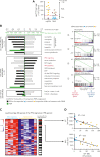
Anti-GARP:TGF-β1 mAbs induce an IFNγ signature in murine PMF. (A) MPLW508A/GFP+ WBC counts on day 34 in mice from experiment in Figure 2. Data points: individual mice; horizontal lines: median + SEM per group. MPLW508A mice responding to 58A2 (n = 5, blue symbols) are defined as mice with a low tumor burden (cut-off: median MPLW508A/GFP+ WBC counts in the 58A2 group). Controls correspond to nontreated (mIgG2a) MPLW508A mice with a high tumor burden (n = 7, orange symbols). (B) GSEA of RNAseq data from spleens collected from MPLW508A mice on days 13, 20, or 34. White bars: ES of the indicated signatures in the list of transcripts ordered by mean fold-change between day 20 and 13 in nontreated controls. Black bars: ES in transcripts ordered by mean fold-change between 58A2 responders and non-treated controls. Green bars: FDR obtained by calculating ES for 1000-gene-set permutations. Right panel shows representative GSEA plots, with black vertical bars indicating position of genes from various gene sets in the ordered transcript list for the responder vs control comparison. EMT, epithelial mesenchymal transition. (C) Heatmap representation of the expression of genes in the leading edge of the IFNγ response signature. Each row represents a gene and each column a mouse. Black boxes on the right indicate genes that are also present in IFNα response signature. (D) Correlation between expression of IFN-induced genes by RNAseq analysis and number of MPLW508A/GFP+ WBCs in all mice.
Figure 6.
Figure 6.
Therapeutic efficacy of anti-GARP:TGF-β1 mAb in murine PMF requires T cell function. (A) Recipient mice were transplanted with transformed MPLW508A/GFP+ cells and injected IP weekly with 58A2 and/or anti-CD8α mAbs at the time points indicated. Blood samples were collected weekly starting on day 14 for blood cell count and flow cytometry. Mice were euthanized on day 35. (B) Counts of GFP+ blood neutrophiles during PMF progression. Data points: mean values per group (n = 6-8 mice per group). P values were calculated with a repeated measure analysis of variance.

References

    1. Szuber N, Mudireddy M, Nicolosi M, et al. 3023 Mayo Clinic patients with myeloproliferative neoplasms: risk-stratified comparison of survival and outcomes data among disease subgroups. Mayo Clin Proc. 2019;94(4):599–610. - PubMed
    1. Cahu X, Constantinescu SN. Oncogenic drivers in myeloproliferative neoplasms: from JAK2 to calreticulin mutations. Curr Hematol Malig Rep. 2015;10(4):335–343. - PubMed
    1. Deininger M, Radich J, Burn TC, Huber R, Paranagama D, Verstovsek S. The effect of long-term ruxolitinib treatment on JAK2p.V617F allele burden in patients with myelofibrosis. Blood. 2015;126(13):1551–1554. - PMC - PubMed
    1. Harrison C, Kiladjian JJ, Al-Ali HK, et al. JAK inhibition with ruxolitinib versus best available therapy for myelofibrosis. N Engl J Med. 2012;366(9):787–798. - PubMed
    1. Vannucchi AM, Kantarjian HM, Kiladjian JJ, et al. A pooled analysis of overall survival in COMFORT-I and COMFORT-II, 2 randomized phase III trials of ruxolitinib for the treatment of myelofibrosis. Haematologica. 2015;100(9):1139–1145. - PMC - PubMed

Publication types

MeSH terms