. 2023 Feb 2;141(5):534-549.
doi: 10.1182/blood.2022018221.

Germ line DDX41 mutations define a unique subtype of myeloid neoplasms

Affiliations

Germ line DDX41 mutations define a unique subtype of myeloid neoplasms

Hideki Makishima et al. Blood. .

Erratum in

Abstract

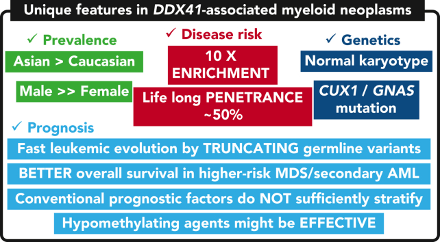
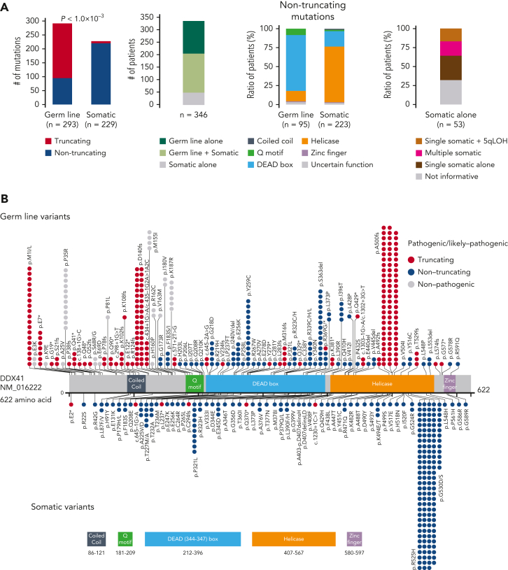
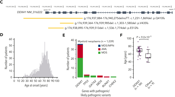
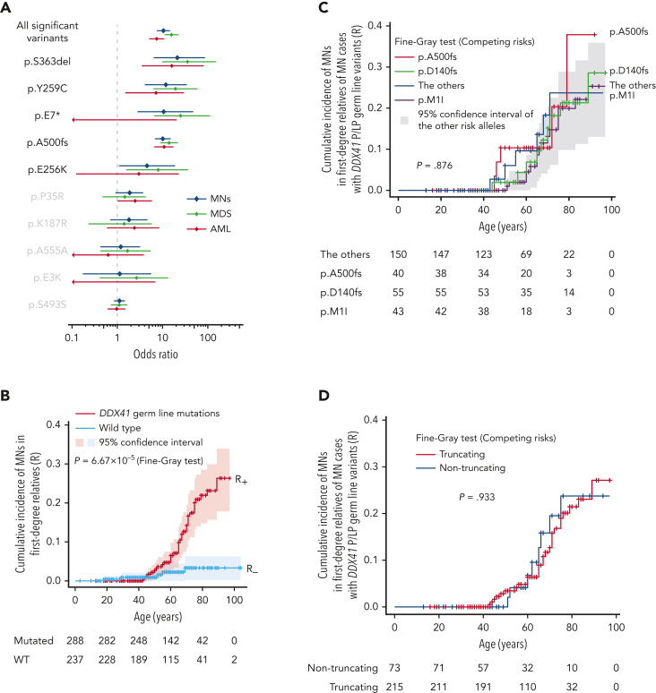
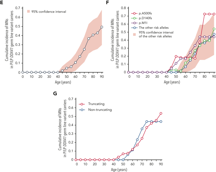
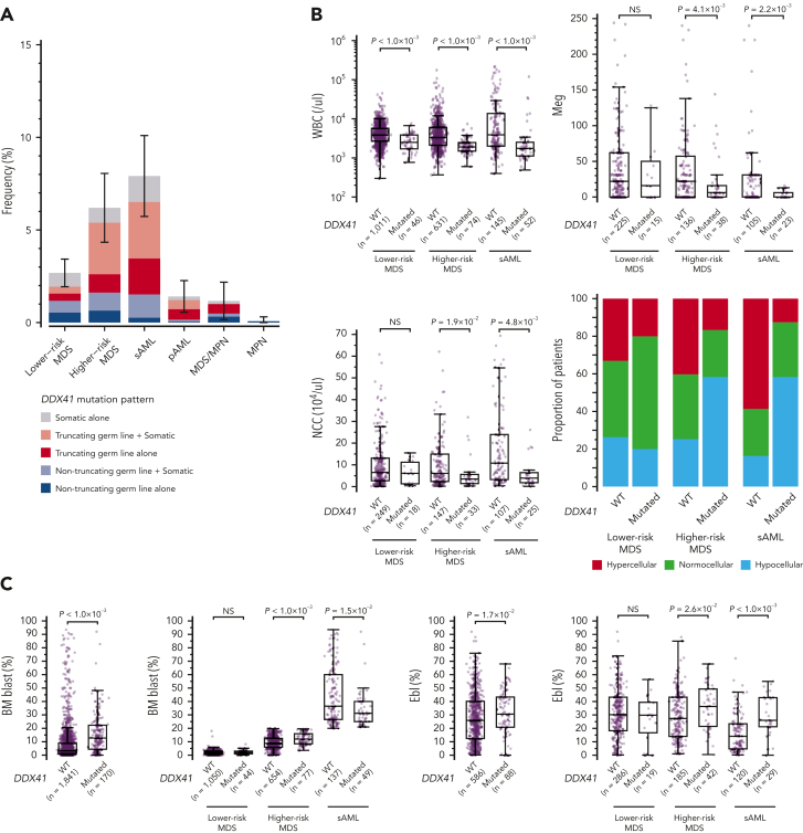
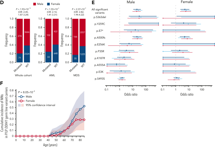
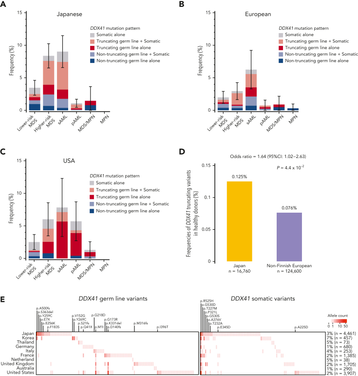
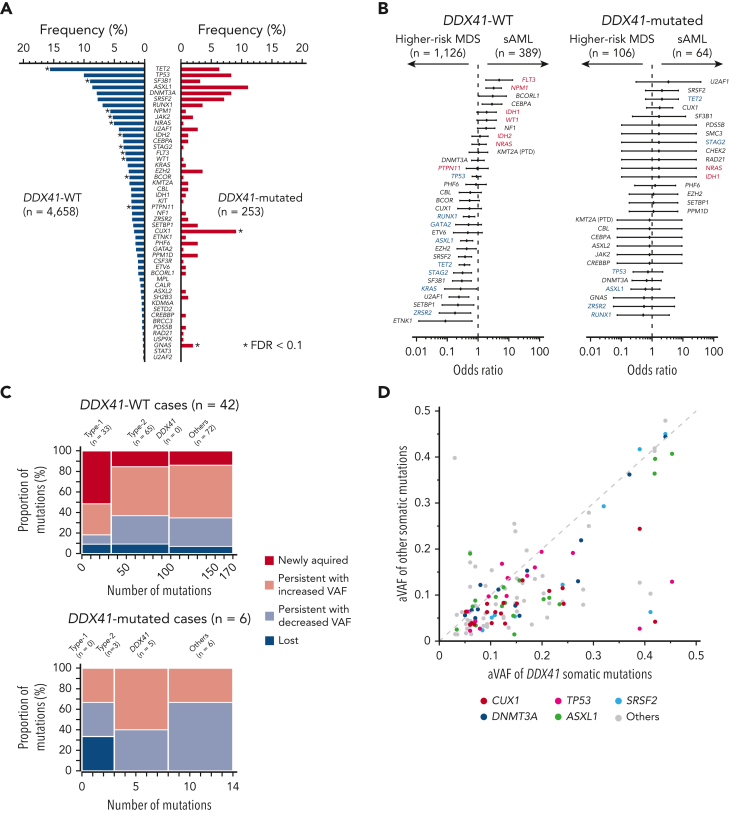
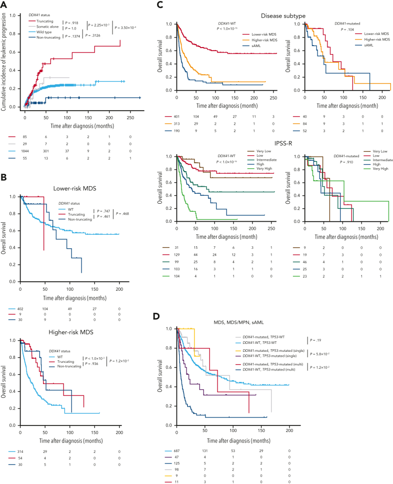
Germ line DDX41 variants have been implicated in late-onset myeloid neoplasms (MNs). Despite an increasing number of publications, many important features of DDX41-mutated MNs remain to be elucidated. Here we performed a comprehensive characterization of DDX41-mutated MNs, enrolling a total of 346 patients with DDX41 pathogenic/likely-pathogenic (P/LP) germ line variants and/or somatic mutations from 9082 MN patients, together with 525 first-degree relatives of DDX41-mutated and wild-type (WT) patients. P/LP DDX41 germ line variants explained ∼80% of known germ line predisposition to MNs in adults. These risk variants were 10-fold more enriched in Japanese MN cases (n = 4461) compared with the general population of Japan (n = 20 238). This enrichment of DDX41 risk alleles was much more prominent in male than female (20.7 vs 5.0). P/LP DDX41 variants conferred a large risk of developing MNs, which was negligible until 40 years of age but rapidly increased to 49% by 90 years of age. Patients with myelodysplastic syndromes (MDS) along with a DDX41-mutation rapidly progressed to acute myeloid leukemia (AML), which was however, confined to those having truncating variants. Comutation patterns at diagnosis and at progression to AML were substantially different between DDX41-mutated and WT cases, in which none of the comutations affected clinical outcomes. Even TP53 mutations made no exceptions and their dismal effect, including multihit allelic status, on survival was almost completely mitigated by the presence of DDX41 mutations. Finally, outcomes were not affected by the conventional risk stratifications including the revised/molecular International Prognostic Scoring System. Our findings establish that MDS with DDX41-mutation defines a unique subtype of MNs that is distinct from other MNs.


Conflict of interest statement

Conflict-of-interest disclosure: S.O.: Leadership position/advisory role for: KAN Research Institute, Inc, ChordiaTherapeutics, Inc. Stockholder in: Asahi Genomics Co, Ltd. Grant/Research funding from: KAN Research Institute, Inc, ChordiaTherapeutics, Inc, Sumitomo Dainippon Pharma Co, Ltd, Otsuka Pharmaceutical Co, Ltd, Eisai Co, Ltd. Accepted a researcher from: ChordiaTherapeutics, Inc. The remaining authors declare no competing financial interests.

Figures

None
Graphical abstract
Figure 1.
Figure 1.
P/LP germ line variants and somatic mutations in DDX41 found in 346 cases with MNs. (A) Frequency of truncating and nontruncating variants within P/LP germ line variants and somatic mutations (left), cases with P/LP germ line and/or somatic variants of DDX41 (middle left), nontruncating variants involving known functional domains (middle right), and cases with somatic DDX41 mutations alone with mono and biallelic involvement (right). (B) Distribution of 325 germ line variants (top) and 229 somatic mutations (bottom). Truncating (red) and nontruncating (blue) P/LP germ line variants were defined based on the American College of Medical Genetics and Genomics criteria. Gray color indicates those classified as germ line variants with undetermined significance. Amino acid locations of relevant functional domains are indicated at the bottom. (C) Germ line variants with deletions including multiple exons identified in 3 cases. Genomic positions of DDX41 (NM_016222) and affected regions were shown according to the GRCh37/hg19 reference. (D) Distributions of age at disease onset in 1039 patients with MN in whom germ line samples were examined. (E) The number of cases with P/LP alleles of DDX41 and other 22 known leukemia predisposing genes in 1039 Japanese cases with MN. The germ line origin of each variant was confirmed in buccal smear samples. (F) Distributions of age at disease onset in those with P/LP germ line variants in DDX41 and the other genes. Statistical significance was tested by 2-sided Wilcoxon test.
Figure 1.
Figure 1.
P/LP germ line variants and somatic mutations in DDX41 found in 346 cases with MNs. (A) Frequency of truncating and nontruncating variants within P/LP germ line variants and somatic mutations (left), cases with P/LP germ line and/or somatic variants of DDX41 (middle left), nontruncating variants involving known functional domains (middle right), and cases with somatic DDX41 mutations alone with mono and biallelic involvement (right). (B) Distribution of 325 germ line variants (top) and 229 somatic mutations (bottom). Truncating (red) and nontruncating (blue) P/LP germ line variants were defined based on the American College of Medical Genetics and Genomics criteria. Gray color indicates those classified as germ line variants with undetermined significance. Amino acid locations of relevant functional domains are indicated at the bottom. (C) Germ line variants with deletions including multiple exons identified in 3 cases. Genomic positions of DDX41 (NM_016222) and affected regions were shown according to the GRCh37/hg19 reference. (D) Distributions of age at disease onset in 1039 patients with MN in whom germ line samples were examined. (E) The number of cases with P/LP alleles of DDX41 and other 22 known leukemia predisposing genes in 1039 Japanese cases with MN. The germ line origin of each variant was confirmed in buccal smear samples. (F) Distributions of age at disease onset in those with P/LP germ line variants in DDX41 and the other genes. Statistical significance was tested by 2-sided Wilcoxon test.
Figure 2.
Figure 2.
DDX41-variant–associated risk of MN development. (A) Enrichment of 10 major germ line DDX41 alleles in 4461 Japanese cases with MNs compared with 20 238 cases from ethnicity-matched controls. ORs are plotted with 95% CIs for each allele. (B-D) Cumulative incidence of MNs calculated by Fine-Gray test of ages at disease onset and death in the first-degree relatives of MN cases with (red) or without (blue) germ line DDX41 mutations are shown in the whole kin cohorts (B), in each P/LP variant (C), and truncating vs nontruncating variants (D). (E-G) Cumulative incidences of MNs in carriers of DDX41 risk alleles estimated by kin-cohort analysis are demonstrated in the whole kin cohorts (E), in each P/LP variant (F), and truncating vs nontruncating variants (G).
Figure 2.
Figure 2.
DDX41-variant–associated risk of MN development. (A) Enrichment of 10 major germ line DDX41 alleles in 4461 Japanese cases with MNs compared with 20 238 cases from ethnicity-matched controls. ORs are plotted with 95% CIs for each allele. (B-D) Cumulative incidence of MNs calculated by Fine-Gray test of ages at disease onset and death in the first-degree relatives of MN cases with (red) or without (blue) germ line DDX41 mutations are shown in the whole kin cohorts (B), in each P/LP variant (C), and truncating vs nontruncating variants (D). (E-G) Cumulative incidences of MNs in carriers of DDX41 risk alleles estimated by kin-cohort analysis are demonstrated in the whole kin cohorts (E), in each P/LP variant (F), and truncating vs nontruncating variants (G).
Figure 3.
Figure 3.
Demographic features of DDX41-mutated patients. (A) Frequency of DDX41-mutated patients in different subtypes of MNs including LR- and HR-MDS, sAML, pAML, MDS/MPN, and MPN with DDX41-mutation status in terms of mono vs biallelic and truncating vs nontruncating variants. (B-C) Comparison of WBC counts, BM NCC, megakaryocyte (Meg) counts, BM cellularity (B), BM blast and erythroblast (Ebl) counts (C) between DDX41-mutated and unmutated cases. P values are provided using the Wilcoxon rank-sum test. (D) Male and female distributions in disease phenotypes. P values and ORs are provided using the Fisher exact test. (E) Enrichment of 10 major germ line DDX41 alleles in Japanese MN cases compared with control Japanese populations were separately shown for male/female subjects. ORs are plotted with 95% CI for each allele. (F) Cumulative incidence of MNs in male vs female carriers of DDX41 risk alleles estimated by kin-cohort analysis. P value was provided using the Wilcoxon signed-rank test. NCC, nucleated cell counts; pAML, primary AML.
Figure 3.
Figure 3.
Demographic features of DDX41-mutated patients. (A) Frequency of DDX41-mutated patients in different subtypes of MNs including LR- and HR-MDS, sAML, pAML, MDS/MPN, and MPN with DDX41-mutation status in terms of mono vs biallelic and truncating vs nontruncating variants. (B-C) Comparison of WBC counts, BM NCC, megakaryocyte (Meg) counts, BM cellularity (B), BM blast and erythroblast (Ebl) counts (C) between DDX41-mutated and unmutated cases. P values are provided using the Wilcoxon rank-sum test. (D) Male and female distributions in disease phenotypes. P values and ORs are provided using the Fisher exact test. (E) Enrichment of 10 major germ line DDX41 alleles in Japanese MN cases compared with control Japanese populations were separately shown for male/female subjects. ORs are plotted with 95% CI for each allele. (F) Cumulative incidence of MNs in male vs female carriers of DDX41 risk alleles estimated by kin-cohort analysis. P value was provided using the Wilcoxon signed-rank test. NCC, nucleated cell counts; pAML, primary AML.
Figure 4.
Figure 4.
Geographic distribution of P/LP germ line variants and somatic mutations in DDX41. (A-C) Disease-specific frequencies of DDX41 variants in different geographical areas including Japan (A), Europe (B), and USA (C). (D) Frequency of truncating germ line variants in DDX41 in the Japanese and European general populations. (E) Geographic distribution of major P/LP germ line DDX41 alleles found in 13 249 cases with MNs (left) is compared with that of somatic DDX41 variants (right). The number of distinct DDX41 alleles/variants (horizontal axis) in each country is shown in color gradient. Highly recurrent major alleles/variants are indicated.
Figure 5.
Figure 5.
Comutation patterns of DDX41 mutated and unmutated cases with MNs. (A) Frequency of co-occurring driver mutations in DDX41-mutated and unmutated cases. Significant difference (q < 0.1) is shown by an asterisk. (B) Comparison of the frequency of driver mutations between HR-MDS and sAML in DDX41-mutated and -unmutated cases in forest plots of ORs and their 95% CI. Type-1 (FLT3, NRAS, WT1, NPM1, IDH1, IDH2, and PTPN11) and Type-2 (GATA2, KRAS, TP53, RUNX1, STAG2, ASXL1, ZRSR2, and TET2) genes are indicated by red and blue characters, respectively. (C) Compositions of Type-1, Type-2, somatic DDX41, and other mutations are shown for each set of mutations that were newly acquired, that persisted with increased or decreased clone size, and that were lost in the second sampling. (D) Diagonal plot comparing variant allele frequencies adjusted by CNAs (aVAF) of somatic DDX41 mutations (the horizontal axis) and co-occurring driver mutations in other genes indicated by colors (the vertical axis).
Figure 6.
Figure 6.
Clinical impacts of DDX41 germ line and somatic variants. (A) Cumulative incidence of leukemic progression in MNs patients with P/LP germ line truncating/nontruncating DDX41 variants, those with somatic alone, and those without any DDX41 mutation. (B) Kaplan-Meier curves of OS in MDS with and without DDX41 mutations (truncating and nontruncating variants). LR- and HR-MDS were separately shown. (C) Kaplan-Meier curves of OS depending on disease subtype and IPSS-R. Patients with or without any DDX41 mutations were separately shown. (D) Kaplan-Meier curves of OS in MDS, MDS/MPN, and sAML depending on the allelic status of TP53 and the presence of DDX41 mutations. Patients not examined by copy-number analysis, for whom the allelic status of TP53 could not be determined, were excluded from the analysis. P values are provided using Gray test in panel A, and the log-rank test in panels B-D.

Comment in

References

    1. Arber DA, Orazi A, Hasserjian R, et al. The 2016 revision to the World Health Organization classification of myeloid neoplasms and acute leukemia. Blood. 2016;127(20):2391–2405. - PubMed
    1. Cazzola M. Myelodysplastic syndromes. N Engl J Med. 2020;383(14):1358–1374. - PubMed
    1. Godley LA, Shimamura A. Genetic predisposition to hematologic malignancies: management and surveillance. Blood. 2017;130(4):424–432. - PMC - PubMed
    1. Churpek JE, Pyrtel K, Kanchi KL, et al. Genomic analysis of germ line and somatic variants in familial myelodysplasia/acute myeloid leukemia. Blood. 2015;126(22):2484–2490. - PMC - PubMed
    1. Ogawa S. Genetics of MDS. Blood. 2019;133(10):1049–1059. - PMC - PubMed

Publication types

Substances