Review
. 2022 Dec:77:102484.
doi: 10.1016/j.sbi.2022.102484. Epub 2022 Oct 28.

Recent advances and current trends in cryo-electron microscopy

Affiliations
Review

Recent advances and current trends in cryo-electron microscopy

Margherita Guaita et al. Curr Opin Struct Biol. 2022 Dec.

Abstract

All steps of cryogenic electron-microscopy (cryo-EM) workflows have rapidly evolved over the last decade. Advances in both single-particle analysis (SPA) cryo-EM and cryo-electron tomography (cryo-ET) have facilitated the determination of high-resolution biomolecular structures that are not tractable with other methods. However, challenges remain. For SPA, these include improved resolution in an additional dimension: time. For cryo-ET, these include accessing difficult-to-image areas of a cell and finding rare molecules. Finally, there is a need for automated and faster workflows, as many projects are limited by throughput. Here, we review current developments in SPA cryo-EM and cryo-ET that push these boundaries. Collectively, these advances are poised to propel our spatial and temporal understanding of macromolecular processes.


Conflict of interest statement

Conflict of interest statement Nothing declared.

Figures
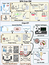
Figure 1.
Figure 1.. Overview of advances in SPA cryo-EM and cryo-ET.
SPA cryo-EM panel, A typical workflow includes sample extraction and purification from a recombinant or native source such as tissue followed by vitrification by plunge freezing. Vitrification sub-panel (A). Alternative vitrification methods such as spraying-mixing (B), mixing-spraying (C), and light time-resolved cryo-EM (D) facilitate the capture of short-lived states. After vitrification, the specimen can be imaged. Cryo-ET panel, Panels on the left outline a general workflow. Cells can either be vitrified by plunge freezing or, like small amounts of tissue, by high-pressure freezing. If the specimen is thin enough and the location of the molecule of interest is known, it can be imaged without further processing. Finally, particles are either identified manually and, potentially, with 3DTM or using 2DTM. Panels on the right outline optional techniques. Live-cell fLM is used to monitor the expression and stage of the sample to determine the optimal timepoint for vitrification. After vitrification, cryo-fLM can be leveraged to identify the locations of fluorescently-tagged molecules of interest to guide subsequent milling using a cryo-FIB for immediate image acquisition.

References

    1. Bai XC, Seeing Atoms by Single-Particle Cryo-EM. Trends Biochem Sci, 2021. 46(4): p. 253–254. - PubMed
    1. Bohning J and Bharat TAM, Towards high-throughput in situ structural biology using electron cryotomography. Prog Biophys Mol Biol, 2021. 160: p. 97–103. - PubMed
    1. Bouvette J, et al., Beam image-shift accelerated data acquisition for near-atomic resolution single-particle cryo-electron tomography. Nat Commun, 2021. 12(1): p. 1957. - PMC - PubMed
    1. Cheng A, et al., High resolution single particle cryo-electron microscopy using beam-image shift. J Struct Biol, 2018. 204(2): p. 270–275. - PMC - PubMed
    1. Turonova B, et al., Benchmarking tomographic acquisition schemes for high-resolution structural biology. Nat Commun, 2020. 11(1): p. 876. - PMC - PubMed

MeSH terms

Substances