Review
. 2022 Nov 1;34(11):1620-1653.
doi: 10.1016/j.cmet.2022.10.008.

Mitochondrial signal transduction

Affiliations
Review

Mitochondrial signal transduction

Martin Picard et al. Cell Metab. .

Abstract

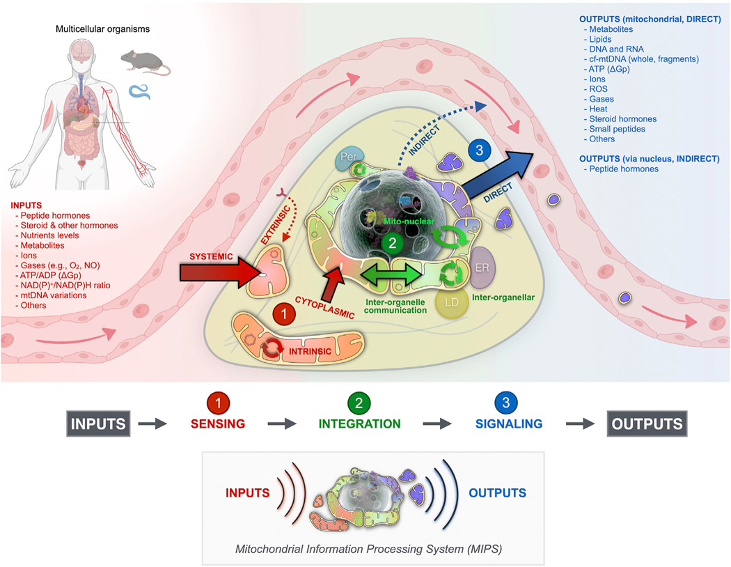
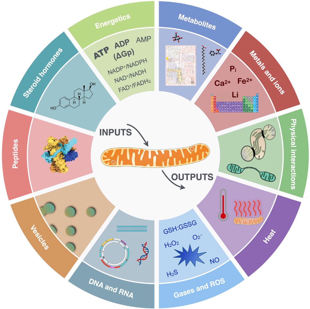
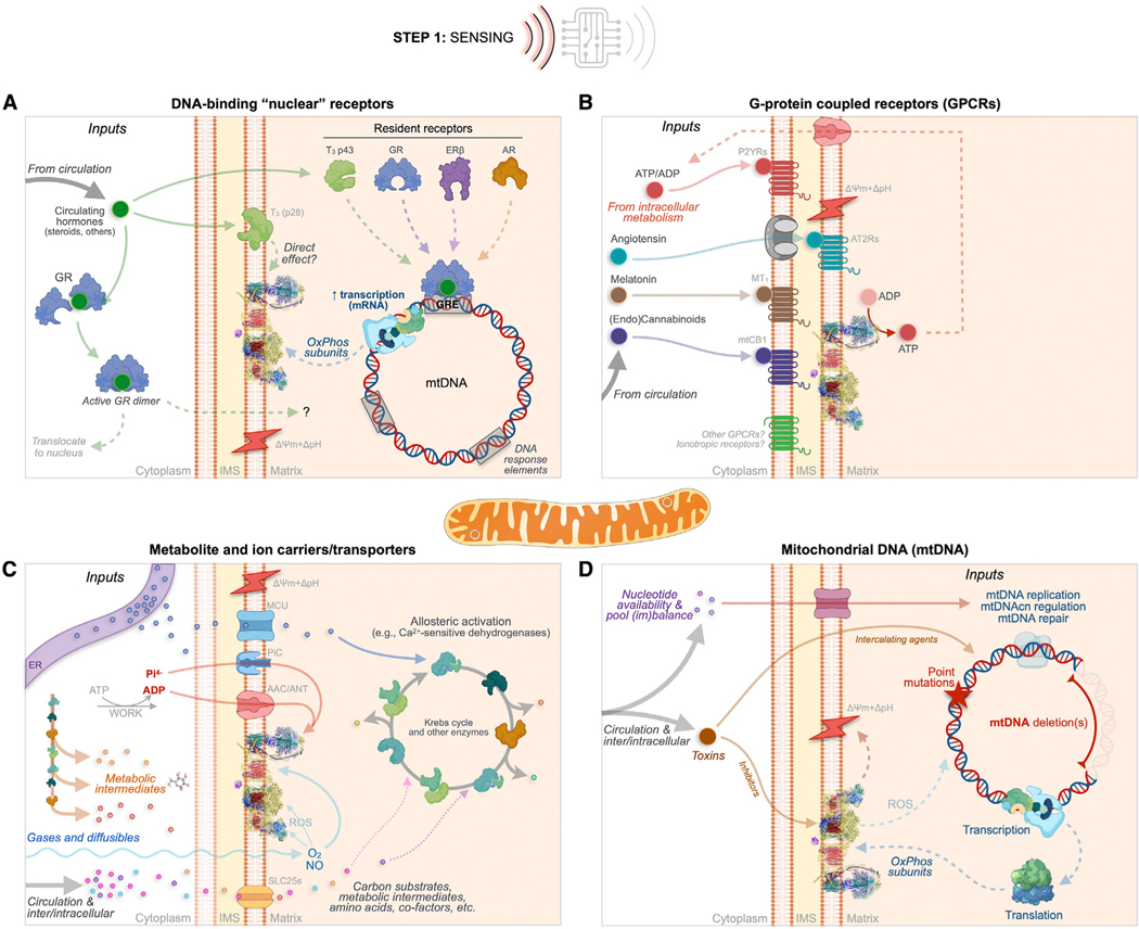
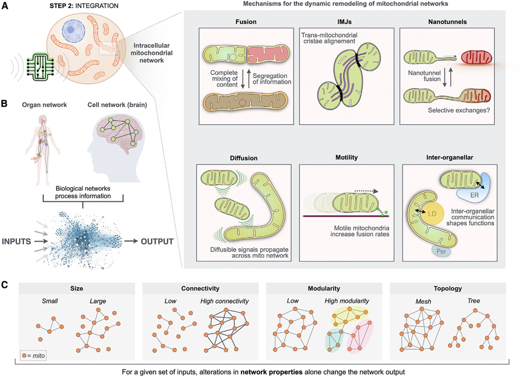
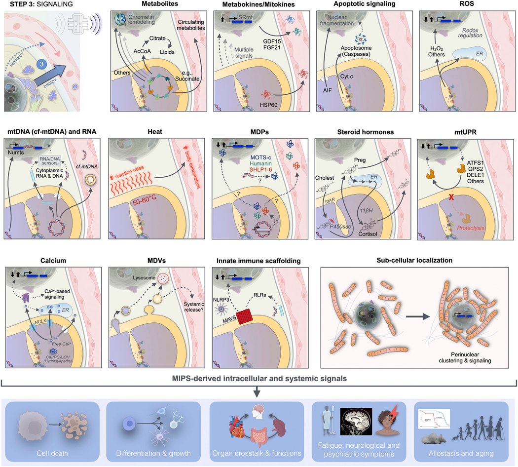
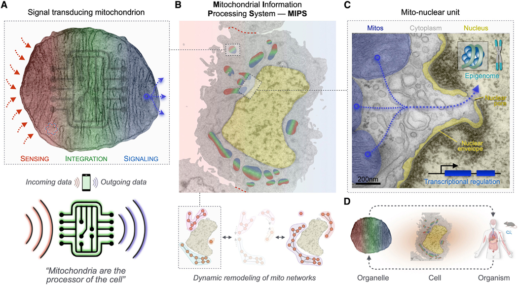
The analogy of mitochondria as powerhouses has expired. Mitochondria are living, dynamic, maternally inherited, energy-transforming, biosynthetic, and signaling organelles that actively transduce biological information. We argue that mitochondria are the processor of the cell, and together with the nucleus and other organelles they constitute the mitochondrial information processing system (MIPS). In a three-step process, mitochondria (1) sense and respond to both endogenous and environmental inputs through morphological and functional remodeling; (2) integrate information through dynamic, network-based physical interactions and diffusion mechanisms; and (3) produce output signals that tune the functions of other organelles and systemically regulate physiology. This input-to-output transformation allows mitochondria to transduce metabolic, biochemical, neuroendocrine, and other local or systemic signals that enhance organismal adaptation. An explicit focus on mitochondrial signal transduction emphasizes the role of communication in mitochondrial biology. This framework also opens new avenues to understand how mitochondria mediate inter-organ processes underlying human health.

Keywords: amplification; communication; energy; evolution; health; membrane potential; metabokines; mito-nuclear signaling; mitochondrial networks; mitokines; mitotypes; receptors; signal transduction; steroid hormones; stress responses; tissue-specific.


Conflict of interest statement

Declaration of interests O.S.S. is a co-founder of Capacity and Inspire Bio.

Figures

Figure 1.
Figure 1.. Modern historical landmarks in mitochondrial research illustrate the need for an integrative view of this multifaceted organelle
Proportion of biomedical publications by organelle, corrected for total published articles across biomedicine. Selected discoveries that challenged prior views about mitochondria are noted, as well as some historical landmarks for context. Figure adapted from Picard et al. with data retrieved from https://pubmed.ncbi.nlm.nih.gov/ on February 12, 2022.
Figure 2.
Figure 2.. Three-step model of mitochondrial signal transduction
As the mitochondrial information processing system (MIPS), mitochondria are input integrators and output generators. Within the cytoplasm, mitochondria are topologically positioned at the interface between incoming signals from the outside extracellular space and the inside compartment of the nucleus where the (epi) genome is stored. In a three-step process, mitochondria receive, integrate, and generate signals that contribute to cellular and organismal adaptation. All mitochondria have the potential to perform sensing, integration, and signaling steps. Here the contributions to signal transduction are color-coded and matched to specific topologies for illustrative purposes. ER, endoplasmic reticulum; LD, lipid droplet; Per, peroxisome; mtDNA, mitochondrial DNA; NO, nitric oxide; ΔGp, phosphorylation potential.
Figure 3.
Figure 3.. The hallmarks of mitochondrial signal transduction
Depicted is the mitochondrial repertoire of mechanisms and substrates through which mitochondria receive, integrate, and transmit intracellular and systemic signals.
Figure 4.
Figure 4.. MIPS step 1: Sensing
As in excitable cells where a broad variety of chemical inputs (e.g., neurotransmitters) converge onto membrane potential variations, extrinsic and intrinsic MIPS inputs trigger molecular changes that converge into morpho-functional mitochondrial states. Mitochondria sense extrinsic and intrinsic information through four main classes of mechanisms. (A) Canonical DNA-binding “nuclear” receptors for steroid hormones including glucocorticoids (GC), estrogen (ER), and androgen (AR) exist in mitochondria or can translocate upon ligand binding. (B) G protein-coupled receptors (GPCRs) embedded within mitochondrial membranes including the angiotensin (AT1R and AT2R), the cannabinoid (mtCB1), melatonin (MT1), and purine (Py2Rs) receptors, and possibly others (e.g., GPR35). (C) Metabolite and ion carriers/transporters such as the ADP/ATP carrier protein (AAC, also adenine nucleotide translocator [ANT]) and the SLC25 family of transporters. Also shown are some gases and ions that either freely diffuse through the IMM or whose import/export is mediated by other carriers/transporters. (D) Acquired sequence variation in the mtDNA sequence, including mutations and deletions that cause functional changes within the OxPhos system. The top path shows nucleotide availability/imbalance, and the bottom path shows exogenous toxins that can interfere with electron transport chain function and secondarily cause mtDNA instability. AAC/ANT, ADP/ATP carrier or adenine nucleotide translocator; AT2Rs, angiotensin receptors; AR, androgen receptor; ERβ, estrogen receptor beta; GR, glucocorticoid receptor; GRE, glucocorticoid response element (used here as an example for other gene regulatory elements); IMS, intermembrane space; MCU, mitochondrial calcium uniporter; mtCB1, mitochondrial cannabinoid receptor; MT1, melatonin 1 receptor; mtDNA, mitochondrial DNA; NO, nitric oxide; O2, molecular oxygen; OxPhos, oxidative phosphorylation; P2YRs, purine receptors; ROS, reactive oxygen species; SLC25s, solute carriers family 25; T3, triiodothyronine; ΔpH+Δψm, mitochondrial proton motive force.
Figure 5.
Figure 5.. MIPS step 2: Signal integration
The physical and functional binding of multiple energized units (mitochondria) into sparsely connected networks naturally gives rise to signal integration. (A) Mechanisms of mitochondrial network remodeling and inter-organellar communication (mito-mito, mito-other organelles) among the MIPS. (B) Conceptual representation of the organism’s organ network and of the brain, where information from one group of units (e.g., neurons) is transmitted to other units, giving rise to computational agents. Information processing is not a private property of brains; it is a generalizable property of all life forms. (C) Four examples of network properties that may be used to define the organization of mitochondrial collectives processing biochemical, metabolic, endocrine, and other inputs into coherent outputs.
Figure 6.
Figure 6.. MIPS step 3: Signaling
Mitochondria synthesize and release signals evolved to influence cellular and organismal functions. Mitochondrial signals arise from various mitochondrial compartments and reach the cytoplasm, nucleus, and other organelles, where they induce cell-autonomous responses. These responses are transmitted to the systemic circulation either directly as mitochondria-derived metabolites and mitokines, or indirectly through transcriptional regulation of nuclear genes encoding metabokines or other hormone-like mediators. Mito-nuclear signaling is a form of signal amplification and integration. The MIPS converts metabolic signals into extracellular proteinaceous, secreted factors, allowing mitochondria to signal their state well beyond the confine of the cell in which they reside. AcCoA, acetyl coenzyme A; AIF, apoptosis inducible factor; ATFS1, activating transcription factor associated with stress-1; cf-mtDNA, cell-free mitochondrial DNA; Cholest, cholesterol; Cyt c, cytochrome c; DELE1, DAP3-binding cell death enhancer 1; ER, endoplasmic reticulum; FGF21, fibroblast growth factor 21; GDF15, growth differentiation factor 15; GPS2, G-protein pathway suppressor 2; HSP60, heat shock protein 60; ISRmt, integrated stress response; MAVS, mitochondrial antiviral signaling; MDVs, mitochondria-derived vesicles; MPDs, mitochondria-derived peptides; NLRP3, NLR family pyrin domain containing 3; Numts, nuclear mitochondrial DNA segments; Preg, pregnenolone; P450ssc, side chain cleavage enzyme cytochrome P450; RLRs, RIG-I-like receptors; StAR, steroidogenic acute regulatory protein; UPRmito, mitochondrial unfolded protein response; 11βH, 11β-hydroxylase (mitochondrial cytochrome P450 11B1).
Figure 7.
Figure 7.. Core MIPS components for mitochondrial signal transduction
(A) All mitochondria contain the machinery and capacity to sense, integrate, and signal information. Based on evidence reviewed above, mitochondria emerge as “the processor of the cell.” (B) Electron micrograph of a cultured cell (human 143B) with mitochondria (blue, green, and red) and nucleus (yellow) highlighted; dotted red lines indicate cell-cell boundaries. The functions of the MIPS are determined by intrinsic properties of individual mitochondria and surrounding organelles, constantly shaped by the dynamic remodeling of organelle networks over minutes to hours. Topologically, the MIPS sits at the interface of the extracellular space harboring endocrine, metabolic, and biochemical signals, and of the (epi)genome in the nucleus. (C) The mito-nuclear unit enhances and amplifies the effects of mitochondrial outputs, tapping into a rich variety of evolved nuclear DNA-encoded (epi)genetic stress response pathways that communicate local mitochondrial states to the organism. This includes the signaling of peripherally derived metabokines on the brain. (D) The framework of mitochondrial signaling from organelle to organism. In multicellular animals, the MIPS is a core regulatory element acting both subcellularly and systemically to optimize adaptation and organismal health.

References

    1. Altman R (1890). Die Elementarorganismen und ihre Beziehungen zu den Zellen (Veit).
    1. Mitchell P. (1961). Coupling of phosphorylation to electron and hydrogen transfer by a chemi-osmotic type of mechanism. Nature 191, 144–148. 10.1038/191144a0. - DOI - PubMed
    1. Chance B, Williams GR, Holmes WF, and Higgins J. (1955). Respiratory enzymes in oxidative phosphorylation. V. A mechanism for oxidative phosphorylation. J. Biol. Chem. 217, 439–451. 10.1016/s0021-9258(19)57193-9. - DOI - PubMed
    1. Siekevitz P. (1957). Powerhouse of the Cell (Scientific American). https://www.scientificamerican.com/article/powerhouse-of-the-cell/.
    1. Giles RE, Blanc H, Cann HM, and Wallace DC (1980). Maternal inheritance of human mitochondrial DNA. Proc. Natl. Acad. Sci. USA 77, 6715–6719. 10.1073/pnas.77.11.6715. - DOI - PMC - PubMed

Publication types

LinkOut - more resources