. 2022 Nov 2;12(1):18500.
doi: 10.1038/s41598-022-22576-4.

Discovery and structural characterization of chicoric acid as a SARS-CoV-2 nucleocapsid protein ligand and RNA binding disruptor

Affiliations

Discovery and structural characterization of chicoric acid as a SARS-CoV-2 nucleocapsid protein ligand and RNA binding disruptor

Gustavo Fernando Mercaldi et al. Sci Rep. .

Abstract

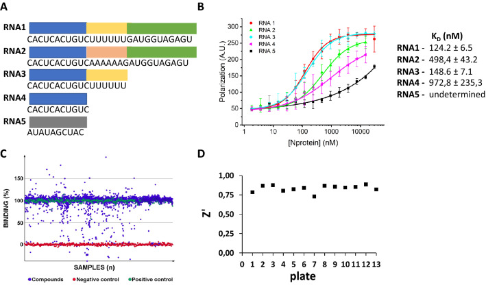
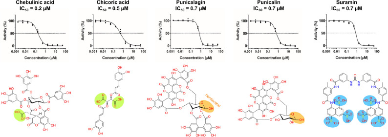
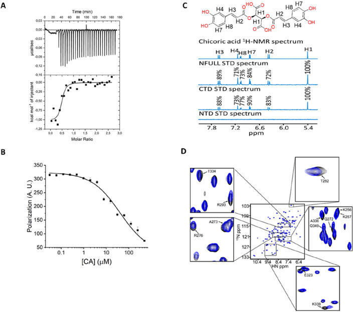
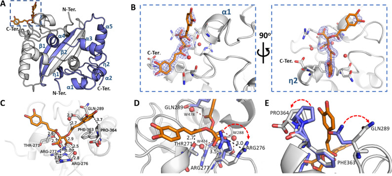
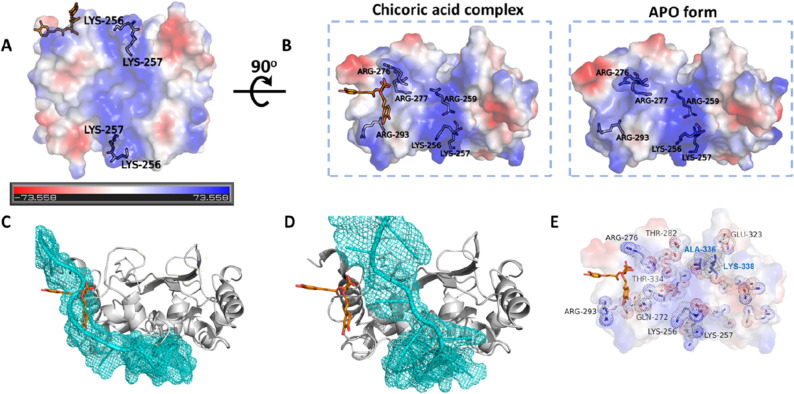
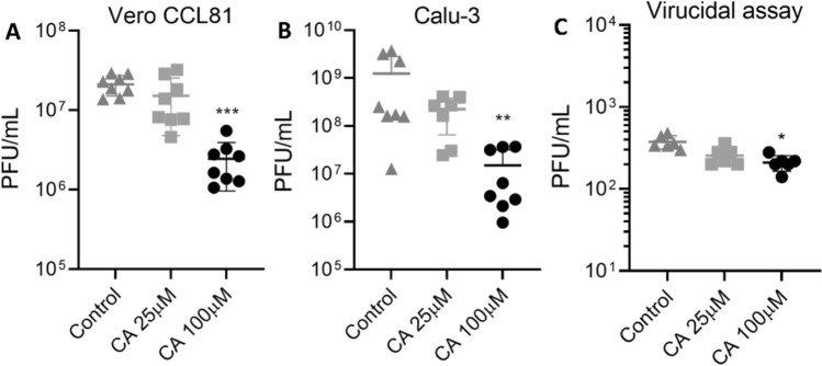
The nucleocapsid (N) protein plays critical roles in coronavirus genome transcription and packaging, representing a key target for the development of novel antivirals, and for which structural information on ligand binding is scarce. We used a novel fluorescence polarization assay to identify small molecules that disrupt the binding of the N protein to a target RNA derived from the SARS-CoV-2 genome packaging signal. Several phenolic compounds, including L-chicoric acid (CA), were identified as high-affinity N-protein ligands. The binding of CA to the N protein was confirmed by isothermal titration calorimetry, 1H-STD and 15N-HSQC NMR, and by the crystal structure of CA bound to the N protein C-terminal domain (CTD), further revealing a new modulatory site in the SARS-CoV-2 N protein. Moreover, CA reduced SARS-CoV-2 replication in cell cultures. These data thus open venues for the development of new antivirals targeting the N protein, an essential and yet underexplored coronavirus target.


Conflict of interest statement

The authors declare no competing interests.

Figures

Figure 1
Figure 1
Development of a biochemical assay for the identification of compounds that disrupt the SARS-CoV-2 N protein-RNA interaction. (A) Nucleotide sequences of the RNA molecules used as N-protein ligands. The blue, yellow and green regions represent the 3’ arm, the UUUUUU motif and the 5’ arm, respectively. (B) FP assay showing the binding affinity curves of the N protein with the FITC-labelled RNA probes 1 to 5 and the corresponding binding equilibrium constants (KD) determined by FP. High-affinity binding was observed with RNAs 1 and 3 derived from the putative SARS-CoV-2 PS sequence. The mutated RNA sequences 2 and 4, and RNA5, used as a negative control, show, in comparison, much lower binding affinities. Error bars represent standard deviation of the means from triplicate experiments. (C) Scatterplot of HTS results showing the binding percentage of each compound in the library calculated using polarization data from negative (RNA alone) and positive (RNA plus protein) controls. Seventy-eight compounds reduced the binding of the N protein to RNA1 to less than 30%, of which 45 were selected for concentration-dependent assays. Compounds that would promote RNA-N complex formation (activators, binding > 100%) were not further considered. (D) Z' values obtained for each assay plate confirming the robustness of the HTS trials.
Figure 2
Figure 2
Concentration–response curves and chemical structures of compounds identified as submicromolar N-protein-RNA1 disruptors in the HTS trials. Curves were measured in triplicates, normalized to positive and negative controls and fitted with the log 4-parameter curve in GraphPad Prism. The main non-phenolic functional groups were highlighted as follows: carboxylic acids (green); hemiacetal—in equilibrium with the aldehyde form—(orange); as well as the sulfonic acids (blue) for the polyamide suramin.
Figure 3
Figure 3
Chicoric acid (CA) is a nanomolar N-protein affinity ligand that binds the CTD and promotes dissociation of the N protein-RNA1 complex. (A) ITC assay showing the thermodynamic parameters of interaction between the N protein and CA. The free N protein (20 µM) was titrated against CA (250 µM). The data were fitted to the One Set of Site model, resulting in the following thermodynamic parameters: association constant (KA) = 2.27E6 1.3E6 M-1 (KD = 0.25 ± 7.9 E-9 µM); binding enthalpy change (ΔH) = − 953.2 ± 104.4 cal/mol and binding entropy change (ΔS) = 27.0 ±1.0 cal/mol/deg. The isotherm is representative of three replicates and the values reported are average and standard deviation of the three independent experiments. (B) Dissociation curve showing that CA promotes the dissociation of previously formed N protein-RNA1 complex with a KD value of 41.1 ± 11.7 µM. (C) One-dimension 1H-STD NMR spectra of CA in the presence of free N protein (full-length), CTD or NTD. The relative degree of saturation of each hydrogen atom of CA is mapped onto each spectrum and normalized by hydrogen H1. (D) 1H-15 N-HSQC spectra of free 15 N-CTD in the absence (black signals) or presence (blue signals) of CA in a 1:1 protein:CA ratio. Assigned peaks were based on the BMRB entry 50,518. Highlighted peaks correspond to signals that changed by more than 0.02 ppm using the deviation equation Δδ(15 N + 1H) = [(Δδ15N/10)2 + (Δδ1H)2]1/2.
Figure 4
Figure 4
The crystal structure of SARS-CoV-2 N protein CTD binding chicoric acid (CA) reveals a network of polar contacts and structural readjustments to accommodate the symmetric ligand in the CA-N protein binding site. (A) Cartoon representation of the N protein CTD dimer crystal structure depicting its secondary structure elements (in blue), including two 310 (η) helices, five α-helices and two antiparallel β-strands and the CA binding site (inset highlighted by the blue dashed square). (B) Detailed CA-binding site from the inset of panel (A). The CA molecule is represented as sticks (orange) with its electron-density map in blue. CA binds to a shallow pocket formed between α-helices 1–2 and η-helix 2, close to the C-terminus (C-Ter). (C) CA atomic interactions with the N protein residues. The CA carboxyl groups are at ideal distances to engage electrostatic interactions and hydrogen bonds with Arg276 side chain (NH1 atom), Arg277 main chain amine and a structural water molecule (W288) stabilized by Arg276 NH2. Thr271 and Gln289 can further position hydrogen bond donors (Thr271Oγ and a structural water molecule, W478, stabilized by the Gln289 carbonyl) to engage a symmetric interaction with the carbonyl groups from both caffeoyl units of CA. One of the catechol motifs of CA is well accommodated near the C-terminal Pro364, showing a well-defined electron density (vide panel B). (D,E) Superposition of the CA-binding site in the native N protein CTD (blue sticks, PDB ID 7UXX) and CA-N protein CTD complex (grey sticks (PDB ID 7UXZ) highlighting structural readjustments induced by CA binding (highlighted by red arrows). Figures were generated with Pymol (Schroedinger Inc.). Polar contacts are indicated by dashed lines with the measured distances in Angstroms. Oxygen atoms are shown in red, nitrogen atoms in blue. Water molecules are represented as red spheres.
Figure 5
Figure 5
Chicoric acid (CA) binding to SARS-CoV-2 N protein CTD changes the topology and electrostatic character of the putative RNA-binding region with implications to RNA binding. (A) N protein CTD electrostatic potential with red and blue colors indicating negative and positive potentials, respectively (please refer to the electrostatic scale bar). The positively charged groove, formed by Lys256, Lys257, Lys259 Lys261 and Arg262, is thought to contribute for RNA binding. (B) The positively charged groove depicted in panel A extends towards the CA-binding pocket through Arg259, Arg276, Arg277 and Arg293. The eletrostacic potentials calculated from the CA-N protein CTD (PDB ID 7UXZ) and native N protein CTD (PDB ID 7UXX) crystal structures are shown. Figures and electrostatic potential were generated with Pymol (Schroedinger Inc.). (C,D) Structural alignment between the CTD-CA complex and a model of the N protein complexed with an RNA. The CTD chain (grey cartoon) complexed with CA (orange sticks) was superposed to two CTD chains from a N protein model complexed with RNA (cyan), which shows two distinct RNA-binding modes. In the first alignment (C), the CA molecule fully occupies the predicted RNA-binding site, whereas in the second alignment (D), the CA site is near the RNA-binding site. In both scenarios CA should interfere in N protein binding to RNA. (E) CTD residues that underwent chemical shift changes (shown as sticks and dots) upon CA binding in the HSQC experiment, here mapped on the CTD crystal structure binding CA. Residues discussed in the text are labelled. Ala-336 and Lys-338 (labelled in blue) are form the second protomer of the CTD dimer.
Figure 6
Figure 6
Anti-SARS-CoV-2 activity of CA in cell assays. In vitro replication assays performed in Vero CCL81 (A) and Calu-3 (B) cells. Cells were infected with SARS-CoV-2 for 1 h and later incubated with fresh media containing CA at 25 µM or 100 µM. The viral load was quantified by the plaque assay (PFU/ml) in the cell supernatants collected 48 h post-infection. (C) Virucidal assays performed in Vero CCL-81 cells, where SARS-CoV-2 particles were incubated with CA at 25 µM or 100 µM for 1 h and then used to infect the cells for 1 h. The viral load was quantified using the plaque assay to assess the infectious viral progeny. DMSO at 0.2% final concentration was used as control. p ≤ 0.05 (*), p ≤ 0.01 (**), p ≤ 0.001 (***). Results are expressed as individual values (n = 8 for replication assay and n = 4 for virucidal assay) with mean +/− standard deviation.

Similar articles

Cited by

References

    1. Nicola M, et al. The socio-economic implications of the coronavirus pandemic (COVID-19): A review. Int. J. Surg. 2020;78:185–193. doi: 10.1016/j.ijsu.2020.04.018. - DOI - PMC - PubMed
    1. Das, S., Wingender, P., Barrett, P., Pugacheva, E. & Magistretti, G. After-effects of the COVID-19 pandemic: Prospects for medium-term economic damage. In IMF Working Papers2021, 1 (2021).
    1. Beigel JH, et al. Remdesivir for the treatment of Covid-19 — final report. N. Engl. J. Med. 2020;383:1813–1826. doi: 10.1056/NEJMoa2007764. - DOI - PMC - PubMed
    1. Jayk Bernal A, et al. Molnupiravir for oral treatment of Covid-19 in Nonhospitalized Patients. N. Engl. J. Med. 2022;386:509–520. doi: 10.1056/NEJMoa2116044. - DOI - PMC - PubMed
    1. Kozlov M. Merck’s COVID pill loses its lustre: What that means for the pandemic. Nature. 2021 doi: 10.1038/d41586-021-03667-0. - DOI - PubMed

Publication types