

Differential expression of immune-regulatory proteins C5AR1, CLEC4A and NLRP3 on peripheral blood mononuclear cells in early-stage non-small cell lung cancer patients
- PMID: 36323738
- PMCID: PMC9630369
- DOI: 10.1038/s41598-022-21891-0
Differential expression of immune-regulatory proteins C5AR1, CLEC4A and NLRP3 on peripheral blood mononuclear cells in early-stage non-small cell lung cancer patients
Abstract
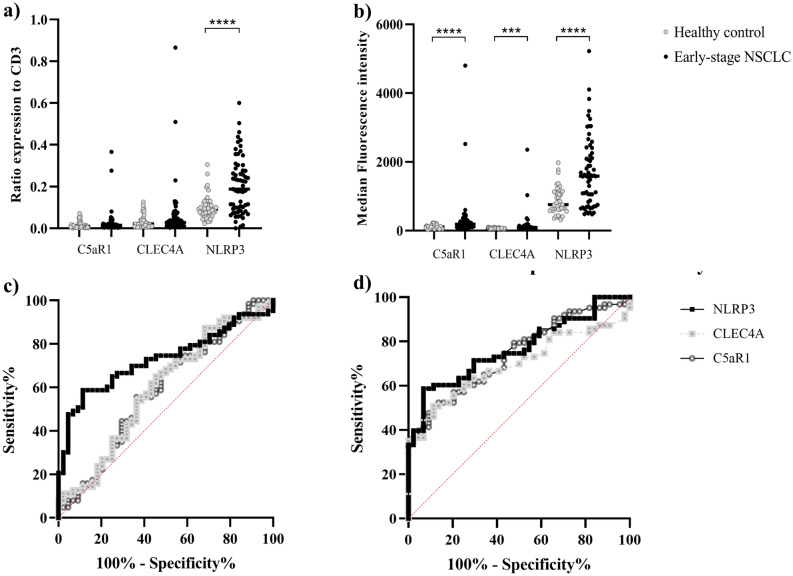
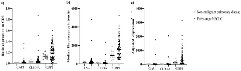
Changes in gene expression profiling of peripheral blood mononuclear cells (PBMC) appear to represent the host's response to the cancer cells via paracrine signaling. We speculated that protein expression on circulating T-lymphocytes represent T-lymphocyte trafficking before infiltration into the tumor microenvironment. The possibility of using protein expression on circulating T-lymphocytes as a biomarker to discriminate early-stage non-small cell lung cancer (NSCLC) was explored. Four independent PBMC gene expression microarray datasets (GSE12771, GSE13255, GSE20189 and GSE3934) were analyzed. We selected C5AR1, CLEC4A and NLRP3 based on their significant protein expression in tumor-infiltrating lymphocytes, but not in normal lymphoid tissue. A validation study using automated flow cytometry was conducted in 141 study participants including 76 treatment-naive early-stage non-small cell lung cancer patients (NSCLC), 12 individuals with non-malignant pulmonary diseases, and 53 healthy individuals. Median ratios of C5AR1, CLEC4A and NLRP3 specific antibody staining to CD3 positive cells in early-stage NSCLC patients compared to healthy controls were 0.014 [0-0.37] vs. 0.01 [0-0.07, p = 0.13], 0.03 [0-0.87] vs. 0.02 [0-0.13, p = 0.10] and 0.19 [0-0.60] vs. 0.09 [0.02-0.31, p < 0.0001], respectively. Median fluorescence intensity (MFI) of CD3+C5AR1+, CD3+CLEC4A+ and CD3+NLRP3+ expression in early-stage NSCLC patients compared to healthy volunteers was 185 [64.2-4801] vs. 107.5 [27-229, p < 0.0001], 91.2 [42.4-2355] vs. 71.25 [46.2-103, p = 0.0005], and 1585 [478-5224] vs. 758.5 [318-1976, p < 0.0001], respectively. NLRP3:CD3 ratio, CD3+C5AR1+, CD3+CLEC4A+ and CD3+NLRP3+ MFI were significantly higher in early-stage NSCLC than healthy volunteers with an area under the ROC curve of 0.69-0.76. The CD3+NLRP3+ MFI provided the most distinguishable expression at 71.5% sensitivity and 70% specificity. Furthermore, CD3+NLRP3+ MFI potentially discriminated between early-stage NSCLC from malignant-mimic inflammation and infection pulmonary disease. Further validation in various pulmonary inflammatory disease might be warranted. Our proof-of-principle findings strengthen the hypothesis that malignancies generate distinctive protein expression fingerprints on circulating T-lymphocytes.
© 2022. The Author(s).
Conflict of interest statement
The authors declare no competing interests.
Figures
References
-
- Sung H, Ferlay J, Siegel RL, Laversanne M, Soerjomataram I, Jemal A, et al. Global Cancer Statistics 2020: GLOBOCAN estimates of incidence and mortality worldwide for 36 cancers in 185 countries. CAs. 2021;71(3):209–49. - PubMed
-
- Siegel RL, Miller KD, Fuchs HE, Jemal A. Cancer statistics, 2021. CA. 2021;71(1):7–33. - PubMed
Publication types
MeSH terms
Substances
Grants and funding
LinkOut - more resources
Full Text Sources
Medical
Molecular Biology Databases
