

Deep Learning for Image Enhancement and Correction in Magnetic Resonance Imaging-State-of-the-Art and Challenges
- PMID: 36323914
- PMCID: PMC9984670
- DOI: 10.1007/s10278-022-00721-9
Deep Learning for Image Enhancement and Correction in Magnetic Resonance Imaging-State-of-the-Art and Challenges
Abstract
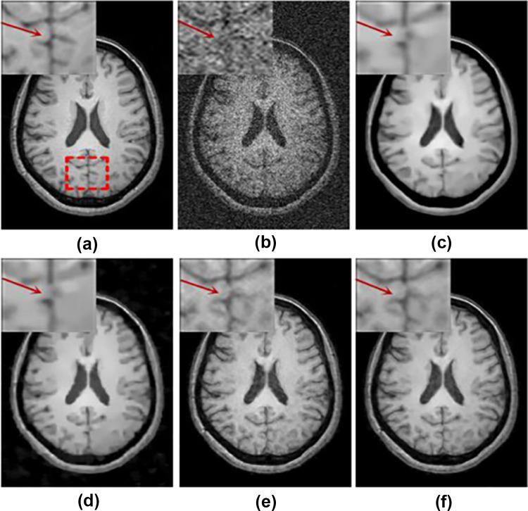
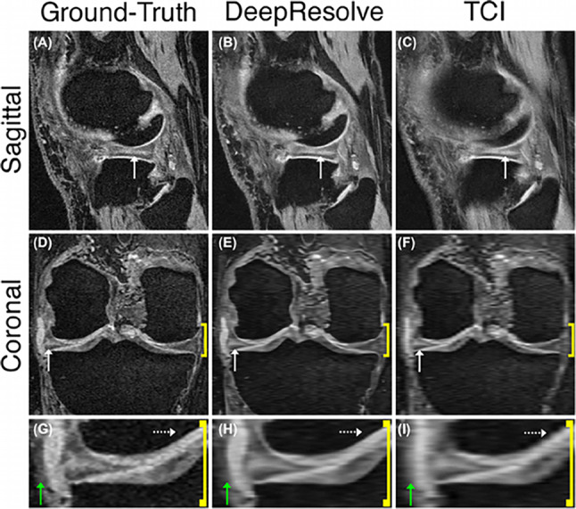
Magnetic resonance imaging (MRI) provides excellent soft-tissue contrast for clinical diagnoses and research which underpin many recent breakthroughs in medicine and biology. The post-processing of reconstructed MR images is often automated for incorporation into MRI scanners by the manufacturers and increasingly plays a critical role in the final image quality for clinical reporting and interpretation. For image enhancement and correction, the post-processing steps include noise reduction, image artefact correction, and image resolution improvements. With the recent success of deep learning in many research fields, there is great potential to apply deep learning for MR image enhancement, and recent publications have demonstrated promising results. Motivated by the rapidly growing literature in this area, in this review paper, we provide a comprehensive overview of deep learning-based methods for post-processing MR images to enhance image quality and correct image artefacts. We aim to provide researchers in MRI or other research fields, including computer vision and image processing, a literature survey of deep learning approaches for MR image enhancement. We discuss the current limitations of the application of artificial intelligence in MRI and highlight possible directions for future developments. In the era of deep learning, we highlight the importance of a critical appraisal of the explanatory information provided and the generalizability of deep learning algorithms in medical imaging.
Keywords: Artefact correction; Image enhancement; Magnetic resonance imaging; Noise; Post-processing; Super-resolution.
© 2022. The Author(s).
Conflict of interest statement
The authors declare no competing interests.
Figures
Similar articles
-
Deep learning methods for 3D magnetic resonance image denoising, bias field and motion artifact correction: a comprehensive review.Phys Med Biol. 2024 Nov 28;69(23). doi: 10.1088/1361-6560/ad94c7. Phys Med Biol. 2024. PMID: 39569887 Review.
-
Stop moving: MR motion correction as an opportunity for artificial intelligence.MAGMA. 2024 Jul;37(3):397-409. doi: 10.1007/s10334-023-01144-5. Epub 2024 Feb 22. MAGMA. 2024. PMID: 38386151 Review.
-
Recent developments in denoising medical images using deep learning: An overview of models, techniques, and challenges.Micron. 2024 May;180:103615. doi: 10.1016/j.micron.2024.103615. Epub 2024 Mar 2. Micron. 2024. PMID: 38471391 Review.
-
Complexities of deep learning-based undersampled MR image reconstruction.Eur Radiol Exp. 2023 Oct 4;7(1):58. doi: 10.1186/s41747-023-00372-7. Eur Radiol Exp. 2023. PMID: 37789241 Free PMC article. Review.
-
Deep learning-based image reconstruction and post-processing methods in positron emission tomography for low-dose imaging and resolution enhancement.Eur J Nucl Med Mol Imaging. 2022 Jul;49(9):3098-3118. doi: 10.1007/s00259-022-05746-4. Epub 2022 Mar 21. Eur J Nucl Med Mol Imaging. 2022. PMID: 35312031 Free PMC article. Review.
Cited by
-
Pilot Lightweight Denoising Algorithm for Multiple Sclerosis on Spine MRI.J Digit Imaging. 2023 Aug;36(4):1877-1884. doi: 10.1007/s10278-023-00816-x. Epub 2023 Apr 17. J Digit Imaging. 2023. PMID: 37069452 Free PMC article.
-
Multidisciplinary quantitative and qualitative assessment of IDH-mutant gliomas with full diagnostic deep learning image reconstruction.Eur J Radiol Open. 2024 Dec 4;13:100617. doi: 10.1016/j.ejro.2024.100617. eCollection 2024 Dec. Eur J Radiol Open. 2024. PMID: 39717474 Free PMC article.
-
Myelin water quantification in multiple sclerosis using short repetition time adiabatic inversion recovery prepared-fast spin echo (STAIR-FSE) imaging.Quant Imaging Med Surg. 2024 Feb 1;14(2):1673-1685. doi: 10.21037/qims-23-1021. Epub 2024 Jan 18. Quant Imaging Med Surg. 2024. PMID: 38415151 Free PMC article.
-
Enhancing repeatability of follicle counting with deep learning reconstruction high-resolution MRI in PCOS patients.Sci Rep. 2025 Jan 7;15(1):1241. doi: 10.1038/s41598-024-84812-3. Sci Rep. 2025. PMID: 39775101 Free PMC article.
-
Association of T1-weighted hyperintensity with timing of peak bilirubin levels in neonates with hyperbilirubinemia.Neuroradiology. 2025 Jun 30. doi: 10.1007/s00234-025-03690-2. Online ahead of print. Neuroradiology. 2025. PMID: 40586872 No abstract available.
References
Publication types
MeSH terms
LinkOut - more resources
Full Text Sources
Other Literature Sources
