

Apolipoprotein E imbalance in the cerebrospinal fluid of Alzheimer's disease patients
- PMID: 36324176
- PMCID: PMC9628034
- DOI: 10.1186/s13195-022-01108-2
Apolipoprotein E imbalance in the cerebrospinal fluid of Alzheimer's disease patients
Abstract
Objective: The purpose of this study was to examine the levels of cerebrospinal fluid (CSF) apolipoprotein E (apoE) species in Alzheimer's disease (AD) patients.
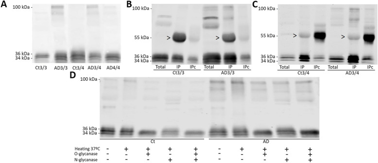
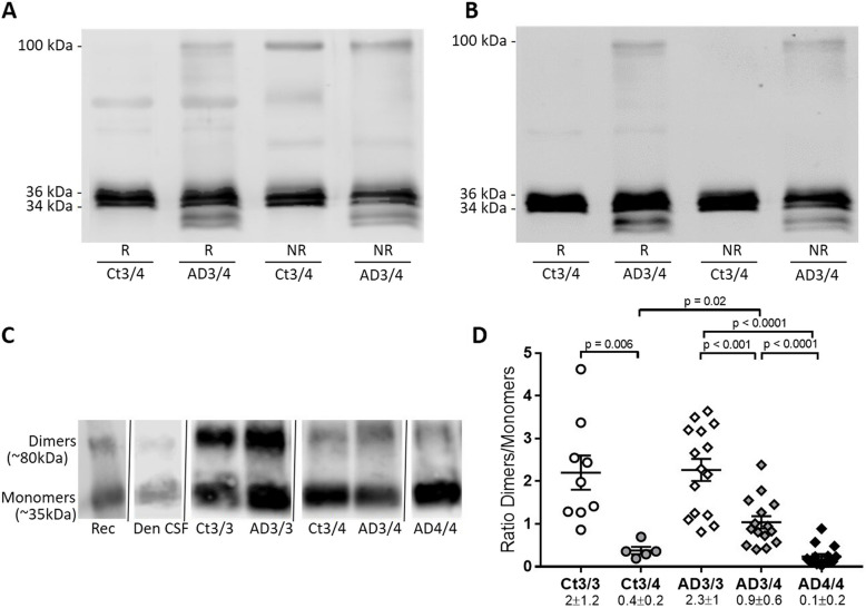
Methods: We analyzed two CSF cohorts of AD and control individuals expressing different APOE genotypes. Moreover, CSF samples from the TgF344-AD rat model were included. Samples were run in native- and SDS-PAGE under reducing or non-reducing conditions (with or without β-mercaptoethanol). Immunoprecipitation combined with mass spectrometry or western blotting analyses served to assess the identity of apoE complexes.
Results: In TgF344-AD rats expressing a unique apoE variant resembling human apoE4, a ~35-kDa apoE monomer was identified, increasing at 16.5 months compared with wild-types. In humans, apoE isoforms form disulfide-linked dimers in CSF, except apoE4, which lacks a cysteine residue. Thus, controls showed a decrease in the apoE dimer/monomer quotient in the APOE ε3/ε4 group compared with ε3/ε3 by native electrophoresis. A major contribution of dimers was found in APOE ε3/ε4 AD cases, and, unexpectedly, dimers were also found in ε4/ε4 AD cases. Under reducing conditions, two apoE monomeric glycoforms at 36 kDa and at 34 kDa were found in all human samples. In AD patients, the amount of the 34-kDa species increased, while the 36-kDa/34-kDa quotient was lower compared with controls. Interestingly, under reducing conditions, a ~100-kDa apoE complex, the identity of which was confirmed by mass spectrometry, also appeared in human AD individuals across all APOE genotypes, suggesting the occurrence of aberrantly resistant apoE aggregates. A second independent cohort of CSF samples validated these results.
Conclusion: These results indicate that despite the increase in total apoE content the apoE protein is altered in AD CSF, suggesting that function may be compromised.
Keywords: Aberrant complexes; Alzheimer’s disease; Biomarker; Cerebrospinal fluid; Glycoform imbalance; apoE.
© 2022. The Author(s).
Conflict of interest statement
HZ has served at scientific advisory boards and/or as a consultant for Abbvie, Alector, Annexon, Artery Therapeutics, AZTherapies, CogRx, Denali, Eisai, Nervgen, Novo Nordisk, Pinteon Therapeutics, Red Abbey Labs, Passage Bio, Roche, Samumed, Siemens Healthineers, Triplet Therapeutics, and Wave; has given lectures in symposia sponsored by Cellectricon, Fujirebio, Alzecure, Biogen, and Roche; and is a co-founder of Brain Biomarker Solutions in Gothenburg AB (BBS), which is a part of the GU Ventures Incubator Program (outside submitted work). KB has served as a consultant, at advisory boards, or at data monitoring committees for Abcam, Axon, Biogen, JOMDD/Shimadzu, Julius Clinical, Lilly, MagQu, Novartis, Prothena, Roche Diagnostics, and Siemens Healthineers and is a co-founder of Brain Biomarker Solutions in Gothenburg AB (BBS), which is a part of the GU Ventures Incubator Program, all unrelated to the work presented in this paper. JF has served as a consultant for Novartis and Lundbeck; has received honoraria for lectures from Roche, NovoNordisk, Nestle, Esteve, and Biogen; and served at advisory boards for AC Immune, Zambon, and Lundbeck. D.A. participated in advisory boards from Fujirebio-Europe and Roche Diagnostics and received speaker honoraria from Fujirebio-Europe, Roche Diagnostics, Nutricia, Krka Farmacéutica S.L., Zambon S.A.U., and Esteve Pharmaceuticals S.A. AL has served at scientific advisory boards from Fujirebio-Europe, Nutricia, Roche-Genentech, Biogen, Grifols, and Roche Diagnostics and has filed a patent application of synaptic markers in neurodegenerative diseases.
Figures


References
-
- Strittmatter WJ, Weisgraber KH, Huang DY, Dong LM, Salvesen GS, Pericak-Vance M, et al. Binding of human apolipoprotein E to synthetic amyloid beta peptide: isoform-specific effects and implications for late-onset Alzheimer disease. Proc Natl Acad Sci U S A. 1993;90:8098–102 Available from: https://pubmed.ncbi.nlm.nih.gov/8367470/. [Cited 2022 Feb 22]. - PMC - PubMed
-
- Husain MA, Laurent B, Plourde M. APOE and Alzheimer’s disease: from lipid transport to physiopathology and therapeutics. Front Neurosci. 2021;15 Available from: https://pubmed.ncbi.nlm.nih.gov/33679311/. [Cited 2022 Feb 22]. - PMC - PubMed
-
- Moon HJ, Haroutunian V, Zhao L. Human apolipoprotein E isoforms are differentially sialylated and the sialic acid moiety in ApoE2 attenuates ApoE2-Aβ interaction and Aβ fibrillation. Neurobiol Dis. 2022;164 Available from: https://pubmed.ncbi.nlm.nih.gov/35041991/. [Cited 2022 Feb 22]. - PMC - PubMed
-
- Rall SC, Weisgraber KH, Innerarity TL, Mahley RW. Structural basis for receptor binding heterogeneity of apolipoprotein E from type III hyperlipoproteinemic subjects. Proc Natl Acad Sci U S A. 1982;79:4696–700 Available from: https://pubmed.ncbi.nlm.nih.gov/6289314/. [Cited 2022 Feb 22]. - PMC - PubMed
-
- Kockx M, Traini M, Kritharides L. Cell-specific production, secretion, and function of apolipoprotein E. J Mol Med (Berl). 2018;96:361–71 Available from: https://pubmed.ncbi.nlm.nih.gov/29516132/. [Cited 2022 Feb 22]. - PubMed
Publication types
MeSH terms
Substances
Grants and funding
LinkOut - more resources
Full Text Sources
Medical
Research Materials
Miscellaneous
