. 2022 Oct 17:9:997998.
doi: 10.3389/fcvm.2022.997998. eCollection 2022.

Right atrial appendage firing in atrial fibrillation

Affiliations

Right atrial appendage firing in atrial fibrillation

Florian Baptiste et al. Front Cardiovasc Med. .

Abstract

Background: The role of atrial fibrillation (AF) drivers located at the left atrium, superior vena cava, crista terminalis and coronary sinus (CS) is well established. While these regions are classically targeted during catheter ablation, the role of right atrial appendage (RAA) drivers has been incompletely investigated.

Objective: To determine the prevalence and electrophysiological characteristics of AF driver's arising from the RAA.

Materials and methods: We conducted a retrospective analysis of clinical and procedural data of 317 consecutive patients who underwent an AF ablation procedure after bi-atrial mapping (multipolar catheter). We selected patients who presented with a per-procedural RAA firing (RAAF). RAAF was defined as the recording of a sustained RAA EGM with a cycle length shorter than 120 ms or 120 < RAAF CL ≤ 130 ms and ratio RAA CL/CS CL ≤ 0.75.

Results: Right atrial/atrium appendage firing was found in 22 patients. The prevalence was estimated at 7% (95% CI, 4-10). These patients were mostly men (72%), median age: 66 yo ± 8 without structural heart disease (77%). RAAFs were predominantly found in paroxysmal AF patients (63%, 32%, and 5% for paroxysmal, short standing and long-standing AF, respectively, p > 0.05). RAAF median cycle length was 117 ms ± 7 while CS cycle length was 180 ms ± 10 (p < 0.01).

Conclusion: In 317 consecutive AF ablation patients (22 patients, 7%) the presence of a high-voltage short-cycle-length right atrial appendage driver (RAAF) may conclusively be associated with AF termination. This case series exemplifies the not-so-uncommon role of the RAA in the perpetuation of AF.

Keywords: AF ablation; AF driver; dispersion; right atrial appendage; tailored ablation.


Conflict of interest statement

Author EG received consultance fees from Abbott. Authors CB, JK, and JS received Speaker Fees from Biosense Webster, Abbott, Boston Scientific and are shareholders of VOLTA Medical. The remaining authors declare that the research was conducted in the absence of any commercial or financial relationships that could be construed as a potential conflict of interest.

Figures

FIGURE 1
FIGURE 1
Right atrial appendage firing (RAAF) ablation limited by phrenic nerve and sinus node locations. Biatrial 3D shells in the right lateral view (panel A), in a patient with paroxysmal AF recurrences despite a previous PVI. AF was induced by atrial pacing, PVs were not connected, and biatrial mapping highlighted a rapid driver in the right atria, located at the appendage/lateral wall area (blue dots = dispersion, cycle length = 107 ms – panel B), while the left atrium was bystander (no dispersion, cycle length = 160 ms – panel C). Ablation of this driver was limited by the location of the sinus node (white circle) mapped in sinus rhythm and the phrenic nerve (black dots) located by pacing. Sinus rhythm was restored by ablation at this area (pink dot) and AF was not inducible anymore. CL, cycle length, LAA, left atrial appendage, RAA, right atrial appendage.
FIGURE 2
FIGURE 2
Examples of RAAF in four different patients. A multi-spline catheter is positioned within the right atrial appendage and a decapolar catheter within the coronary sinus. Electrogram activations recorded within the right appendage are notably fast (cycle length around 100 ms significantly shorter than the one measured in coronary sinus) and fractionated. Three dimensional shells of the right atrium are presented either in the antero-posterior (AP) view or right oblique anterior (RAO) view. Ablation areas are presented with visitags “grids” (pink/red areas) and yellow stars represent termination sites.
FIGURE 3
FIGURE 3
Anatomical distribution of AF termination sites in RAAF patients. Three dimensional shells of the right atrium [panel A: antero posterior view (AP), panel B: right anterior oblique (RAO) 30° view, panel C: left anterior oblique (LAO) 30° view]. Colored sections show anatomical regions of AF termination sites during ablation with the corresponding patients number. In red the RAA apex, in brown the RAA base, in blue the upper half of the right atrial free wall, in yellow the upper half of the crista terminalis. In three patients several AF termination sites were observed both in the apex and base of the RAA. In one patient, transient organization into atrial tachycardia was observed after RAA electrical isolation during ablation at the base.
FIGURE 4
FIGURE 4
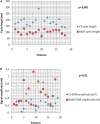
Right Atrial Appendage Firing (RAAF) EGM analysis. The cycle length analysis shows fast drivers with a mean cycle 1.94 shorter than the CS CL (p < 0.001) (A). The RAAF cycle lengths were found between 76 and 130 ms. High voltage signals observed in the thick areas of the RAA and crista terminalis (B). All the data were calculated as the average of ten consecutive measures.
FIGURE 5
FIGURE 5
Right atrial appendage firing predictive factors. Clinical factors tested for RAAF. None of the studied factors were significantly associated with RAAF. LA, left atrium; RA, right atrium, RAAF, right atrial appendage firing; AF, atrial fibrillation, CAD, coronary artery disease; SHD, structural heart disease; LVEF, left ventricular ejection fraction.

References

    1. Haissaguerre M, Hocini M, Denis A, Shah AJ, Komatsu Y, Yamashita S, et al. Driver domains in persistent atrial fibrillation. Circulation. (2014) 130:530–8. - PubMed
    1. Haissaguerre M, Jais P, Shah DC, Takahashi A, Hocini M, Quiniou G, et al. Spontaneous initiation of atrial fibrillation by ectopic beats originating in the pulmonary veins. N Engl J Med. (1998) 339:659–66. - PubMed
    1. Mandapati R, Skanes A, Chen J, Berenfeld O, Jalife J. Stable microreentrant sources as a mechanism of atrial fibrillation in the isolated sheep heart. Circulation. (2000) 101:194–9. 10.1161/01.cir.101.2.194 - DOI - PubMed
    1. Patterson E, Jackman WM, Beckman KJ, Lazzara R, Lockwood D, Scherlag BJ, et al. Spontaneous pulmonary vein firing in man: relationship to tachycardia-pause early afterdepolarizations and triggered arrhythmia in canine pulmonary veins in vitro. J Cardiovasc Electrophysiol. (2007) 18:1067–75. 10.1111/j.1540-8167.2007.00909.x - DOI - PubMed
    1. Leone O, Boriani G, Chiappini B, Pacini D, Cenacchi G, Martin Suarez S, et al. Amyloid deposition as a cause of atrial remodelling in persistent valvular atrial fibrillation. Eur Heart J. (2004) 25:1237–41. 10.1016/j.ehj.2004.04.007 - DOI - PubMed

LinkOut - more resources