Case Reports
. 2022 Oct 27;18(1):64-69.
doi: 10.1016/j.radcr.2022.09.094. eCollection 2023 Jan.

Juvenile psammomatoid ossifying fibroma in paranasal sinuses: A case report and literature review

Affiliations
Case Reports

Juvenile psammomatoid ossifying fibroma in paranasal sinuses: A case report and literature review

Nguyen Ngoc Trung et al. Radiol Case Rep. .

Abstract

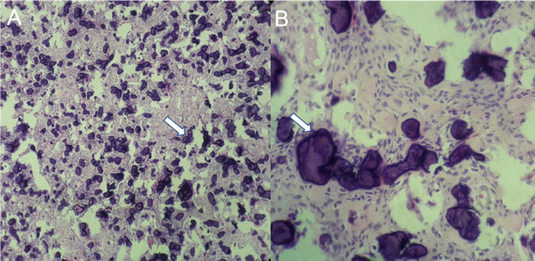
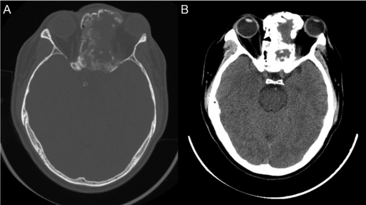
Juvenile psammomatoid ossifying fibroma (JPOF) is a rare, benign type of ossifying fibroma. JPOFs predominantly present as rapidly growing masses with a high recurrence rate. We report a 40-year-old male patient who suffered from a large tumor with multiple invasions into the paranasal sinuses. Total excision was performed, and significant relief of clinical symptoms was recorded after 4 months of follow-up. Multi-departmental management involving radiologists, neurology surgeons, craniofacial surgeons, pathologists, and otolaryngologists is vital for JPOF treatment. First-line treatment options include total or partial resection, depending on the patient's condition.

Keywords: Computed tomography; Juvenile psammomatoid ossifying fibroma; Magnetic resonance imaging; Paranasal sinuses.


Figures

Fig 1
Fig. 1
Axial pre- and post-contrast computed tomography (CT) images. Pre-contrast CT scans on the bone window (A) and the soft-tissue window (C) demonstrated a paranasal lesion with a thick capsule and multiple internal “ground-glass” nodules (arrows). Central hypodensity (*) and a well-defined margin were noticed (open arrows). Post-contrast CT scans on the bone window (B) and the soft tissue window (D) showed no enhancement.
Fig 2
Fig. 2
Pre- and post-gadolinium magnetic resonance imaging (MRI). (A) Coronal T2-weighted (T2W), (C) sagittal T1-weighted (T1W), and (E) axial fluid-attenuated inversion recovery sequences demonstrated a paranasal mass with a hypointense cortical area (arrows) and a fluidized, hyperintense center area (*). A well-defined margin with an adjacent bone structure and soft tissue were also noted. (B) Post-gadolinium coronal T1W, (D) post-gadolinium sagittal T1W, and (F) post-gadolinium axial T1W sequences demonstrated ring-like and septal enhancement. G) Axial diffusion-weighted imaging and (H) axial apparent diffusion coefficient showed no restricted diffusion.
Fig 3
Fig. 3
Microscopic histopathologic images. (A) A 10× magnification image showed large cytoplasmic spindle cells arranged in rafts with interspersed psammomatoid bodies interspersed (arrows). (B) A 40× magnification image showing psammomatoid bodies (arrows).
Fig 4
Fig. 4
Post-surgical computed tomography (CT) images. (A) Bone window and (B) soft tissue window showed significant size reductions, correlated with clinical symptom relief.

References

    1. Mohsenifar Z, Nouhi S, Abbas FM, Farhadi S, Abedin B. Ossifying fibroma of the ethmoid sinus: report of a rare case and review of literature. J Res Med Sci. 2011;16(6):841–847. - PMC - PubMed
    1. El-Naggar A.K., Chan J.K.C., Grandis J.R., Takata T., Slootweg P.J. 4th ed. 2017. WHO classification of head and neck tumours; pp. 251–253.
    1. Diniz JA, Siqueira ADS, Araújo GM, Faro TF, Torres LHS, Oliveira E Silva ED, et al. Intraoral Approach for surgical treatment of psammomatoid juvenile ossifying fibroma. J Craniofac Surg. 2020;31(3):e306–e309. doi: 10.1097/SCS.0000000000006171. - DOI - PubMed
    1. Gantala R, Vemula AY, Kubbi JR, Sekhar MM, Jhawar D. Psammomatoid juvenile ossifying fibroma involving upper jaw: a rare case report. J Clin Diagn Res JCDR. 2015;9(7):ZD17–ZD19. doi: 10.7860/JCDR/2015/14603.6199. - DOI - PMC - PubMed
    1. Keles B, Duran M, Uyar Y, Azimov A, Demirkan A, Esen HH. Juvenile ossifying fibroma of the mandible: a case report. J Oral Maxillofac Res. 2010;1(2) doi: 10.5037/jomr.2010.1205. - DOI - PMC - PubMed

Publication types

LinkOut - more resources