. 2022 Oct 12:10:1008271.
doi: 10.3389/fbioe.2022.1008271. eCollection 2022.

Genetic profiling of human bone marrow mesenchymal stromal cells after in vitro expansion in clinical grade human platelet lysate

Affiliations

Genetic profiling of human bone marrow mesenchymal stromal cells after in vitro expansion in clinical grade human platelet lysate

Ann De Becker et al. Front Bioeng Biotechnol. .

Abstract

Mesenchymal stromal cells (MSCs) are non-hematopoietic cells that have a broad therapeutic potential. To obtain sufficient cells for clinical application, they must be expanded ex vivo. In the initial expansion protocols described, fetal calf serum (FCS) was used as the reference growth supplement, but more recently different groups started to replace FCS with platelet lysate (PL). We investigated in this study the impact of the culture supplement on gene expression of MSCs. Human bone marrow derived MSCs were expanded in vitro in FCS and PL supplemented medium. We found that MSCs expanded in PL-containing medium (PL-MSCs) express typical MSC immunomorphological features and can migrate, as their counterparts expanded in FCS-containing medium, through a layer of endothelial cells in vitro. Additionally, they show an increased proliferation rate compared to MSCs expanded in FCS medium (FCS-MSCs). RNA sequencing performed for MSCs cultured in both types of expansion medium revealed a large impact of the choice of growth supplement on gene expression: 1974 genes were at least twofold up- or downregulated. We focused on impact of genes involved in apoptosis and senescence. Our data showed that PL-MSCs express more anti-apoptotic genes and FCS-MSCs more pro-apoptotic genes. FCS-MSCs showed upregulation of senescence-related genes after four passages whereas this was rarer in PL-MSCs at the same timepoint. Since PL-MSCs show higher proliferation rates and anti-apoptotic gene expression, they might acquire features that predispose them to malignant transformation. We screened 10 MSC samples expanded in PL-based medium for the presence of tumor-associated genetic variants using a 165 gene panel and detected only 21 different genetic variants. According to our analysis, none of these were established pathogenic mutations. Our data show that differences in culture conditions such as growth supplement have a significant impact on the gene expression profile of MSCs and favor the use of PL over FCS for expansion of MSCs.

Keywords: expansion; gene expression; mesenchymal stromal cell; platelet lysate; transformation.


Conflict of interest statement

The authors declare that the research was conducted in the absence of any commercial or financial relationships that could be construed as a potential conflict of interest.

Figures

FIGURE 1
FIGURE 1
Characterisation of in vitro expanded MSCs. (A,B) Representative photographs of MSC’s cultured in PL-based (A) and FCS-based (B) expansion medium (30×); (C): Phenotype MSCs expanded in PL-based medium: the first peak represents the negative control antibody whereas the second peak represents the antigen-specific antibody; (D): Proliferation capacity of MSCs expanded in PL-based medium versus FCS-based medium (n = 3), the number of cells is presented according to the number of passages; (E): In vitro migration of MSCs cultured in FCS-based medium and PL-based medium (n = 5). Migration is expressed as mean relative fluorescence intensity (RFI). (p= 0.69, 95% CI, Mann Whitney).
FIGURE 2
FIGURE 2
RNA sequencing of FCS-MSCs and PL-MSCs. (A): Overview of the number of transcripts detected per sample for 4 different donors. The green bars represent coding genes, dark blue bars long non coding RNA and light blue bars other RNA species. (B): Volcano plot showing -log10 adjusted p-values in function of the log2 fold change for FCS-MSCs versus PL-MSCs. Pink points indicate significantly differential expressed genes at FDR <0.05. FDR: false discovery rate. (C): Heatmap showing the top 500 differentially expressed genes according to adjusted p-values at FDR <0.05 for FCS-MSCs versus PL-MSCs.
FIGURE 3
FIGURE 3
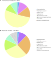
Gene Ontology analysis: processes enriched in (A) FCS-MSCs and (B) PL-MSCs (q ≤ 0,05, false discovery rate adjusted p value ≤ 0,05). Enriched processes are grouped in 8 major categories to get a better overview of processes impacted by differential expression due to use of a different growth supplement.
FIGURE 4
FIGURE 4
Heatmap generated based on FPKM of the 221 genes included in the Gene Ontology term ‘regulation of cell population proliferation’ (GO:004212). Rows are centered and unit variance scaling is applied to the rows. Both rows and columns are clustered using correlation distance and average linkage.
FIGURE 5
FIGURE 5
β-galactosidase staining of cultured MSCs. Cells from a representative donor were cultured in both media and stained after 4 passages. More β-galactosidase positive (blue colorored) cells were found in MSC cultures with FCS based expansion medium versus PL-based medium (4 × magnification) (P4 = passage 4).

Similar articles

Cited by

References

    1. Azuma K., Umezu T., Imanishi S., Asano M., Yoshizawa S., Katagiri S., et al. (2017). Genetic variations of bone marrow mesenchymal stromal cells derived from acute leukemia and myelodysplastic syndrome by targeted deep sequencing. Leuk. Res. 62, 23–28. 10.1016/j.leukres.2017.09.008 - DOI - PubMed
    1. Barkholt L., Flory E., Jekerle V., Lucas-Samuel S., Ahnert P., Bisset L., et al. (2013). Risk of tumorigenicity in mesenchymal stromal cell-based therapies-bridging scientific observations and regulatory viewpoints. Cytotherapy 15, 753–759. 10.1016/j.jcyt.2013.03.005 - DOI - PubMed
    1. Bashir J., Sherman A., Lee H., Kaplan L., Hare J. M. (2014). Mesenchymal stem cell therapies in the treatment of musculoskeletal diseases. PM&R 6, 61–69. 10.1016/j.pmrj.2013.05.007 - DOI - PubMed
    1. Bieback K., Hecker A., Kocaomer A., Lannert H., Schallmoser K., Strunk D., et al. (2009). Human alternatives to fetal bovine serum for the expansion of mesenchymal stromal cells from bone marrow. Stem Cells 27, 2331–2341. 10.1002/stem.139 - DOI - PubMed
    1. Bruckheimer E. M., Cho S. H., SarkissM., Herrmann J., McDonnell T. J. (1998). The Bcl-2 gene family and apoptosis. Adv. Biochem. Eng. Biotechnol. 62, 75–105. 10.1007/BFb0102306 - DOI - PubMed

LinkOut - more resources