

The development of a new, ultra-fine, and flexible neuroendoscope for intracranial observation
- PMID: 36324912
- PMCID: PMC9609956
- DOI: 10.25259/SNI_748_2022
The development of a new, ultra-fine, and flexible neuroendoscope for intracranial observation
Abstract
Background: A neuroendoscope is a technical advance that allows surgeons to visualize certain regions of the brain that was previously inaccessible through the use of a surgical microscope. Several neuroendoscope designs have been implemented by other neurosurgeons over the past 5 years. The advantage of a neuroendoscope is the addition of a flexible and narrow tip that allows for safe entry into intracranial structures for clinical observation. However, there are some limitations to this approach. Here, we report the use of a modified angioscope as a newly developed neuroendoscope to be employed in observing intracranial structures.
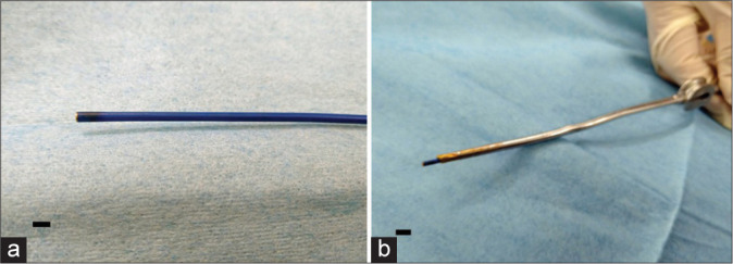
Methods: We report the use of an angioscope that is 1.8 mm in diameter and has both a thin and flexible tip. In this study, the angioscope was inserted into the lumen of an aspirator tube, and the tip of the device was placed at the intracranial area of intended observation area. Image findings were evaluated using an established in vivo goat brain model.
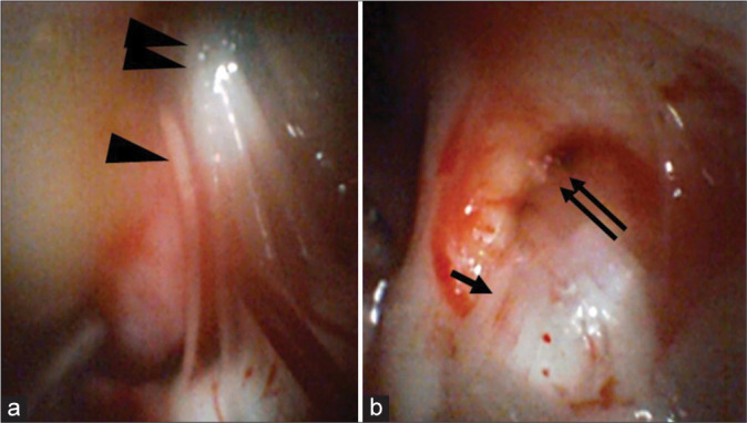
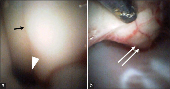
Results: The angioscope was light in weight and maneuverable and could be reached and observed in the blind spot using a surgical microscope. From the cerebellopontine angle, the lower cranial nerves and trigeminal nerve could be observed, and from the cisterna magna, the floor of the fourth ventricle and the aqueduct could be seen.
Conclusion: The angioscope is a useful instrument to observe intracranial locations safely and effectively even within a limited surgical field. Further modifications will be required to use the angioscope in various craniotomy procedures.
Keywords: Angioscope; Intracranial; Neuroendoscope; Neurosurgery.
Copyright: © 2022 Surgical Neurology International.
Conflict of interest statement
There are no conflicts of interest.
Figures
References
-
- Almeida JP, de Albuquerque LA, Fabbro MD, Sampaio M, Medina R, Chacon M, et al. Endoscopic skull base surgery: Evaluation of current clinical outcomes. J Neurosurg Sci. 2019;63:88–95. - PubMed
-
- Azab WA, Abdelrahman AY, Alsheikh TM, Najibullah M. Neuroendoscopy in Kuwait: Evolution, current status and future directions. World Neurosurg. 2016;92:298–302. - PubMed
-
- Fischer G, Oertel J, Perneczky A. Endoscopy in aneurysm surgery. Neurosurgery. 2012;70(Suppl 2):184–91. - PubMed
-
- Hatada A, Takagaki Y, Yamamoto S, Okamura Y, Nishimura Y. High-resolution angioscopic observation of deep vein thrombosis after catheter-directed venous thrombolysis. Circ J. 2020;84:1885. - PubMed
-
- Ho CL, Peter Y Hwang PY. Endoscope-assisted transorbital keyhole surgical approach to ruptured supratentorial aneurysms. J Neurol Surg A Cent Eur Neurosurg. 2015;76:376–83. - PubMed
LinkOut - more resources
Full Text Sources
Miscellaneous