Review
. 2022 Oct 6:7:285-316.
doi: 10.1016/j.cnp.2022.09.003. eCollection 2022.

Myoclonus and other jerky movement disorders

Affiliations
Review

Myoclonus and other jerky movement disorders

Sterre van der Veen et al. Clin Neurophysiol Pract. .

Abstract

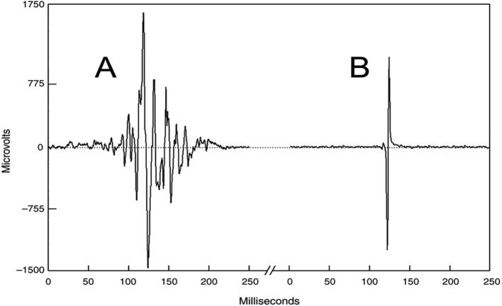
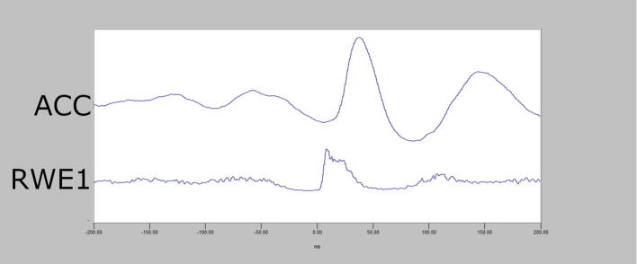
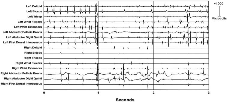
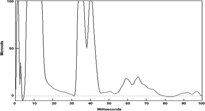
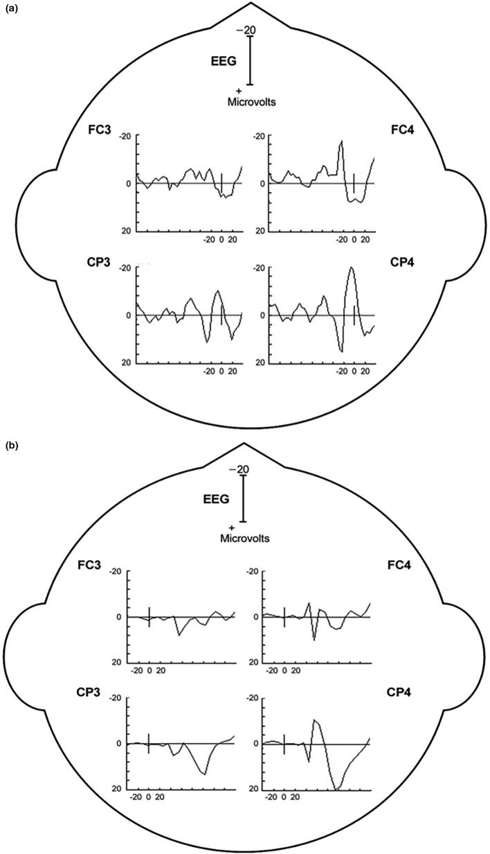
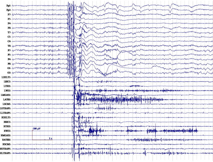
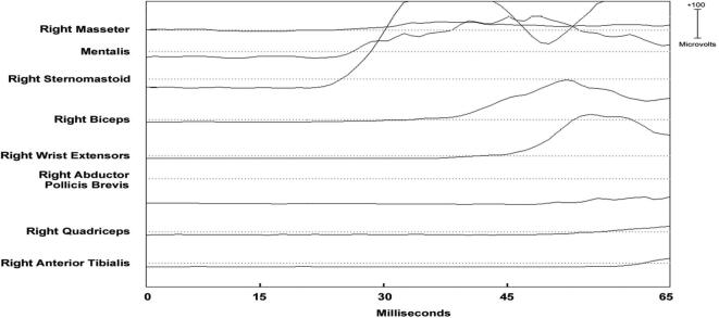
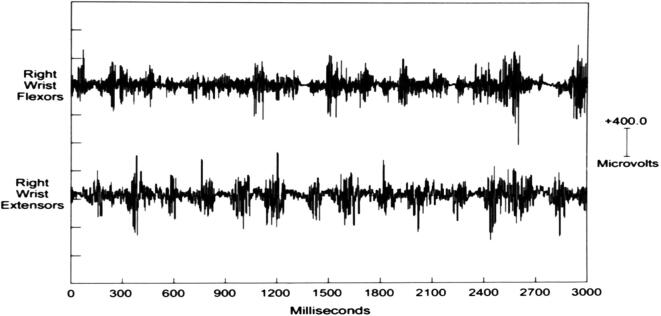
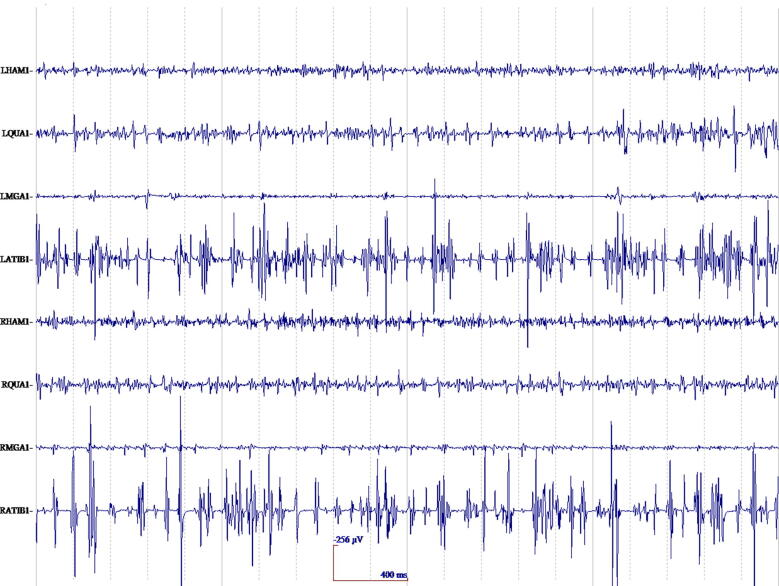
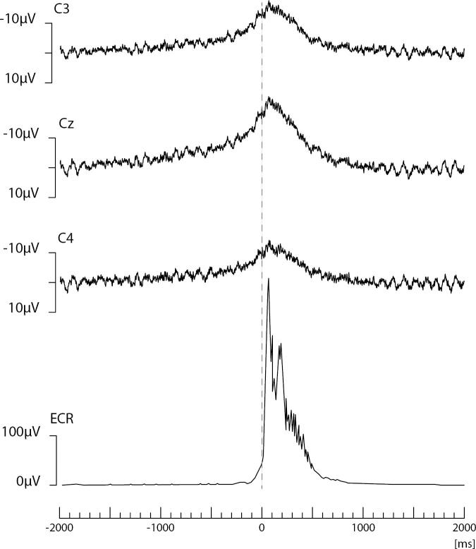
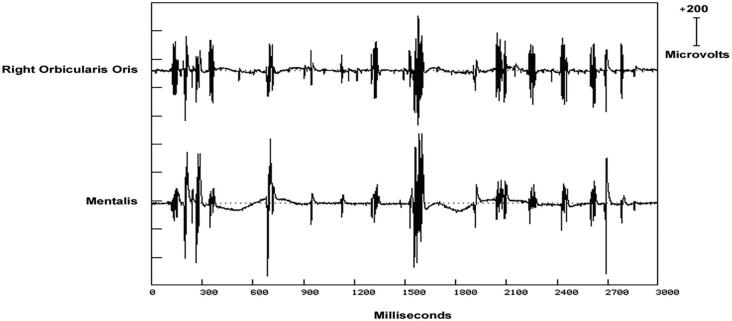
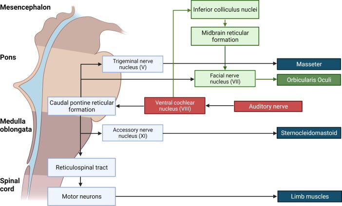
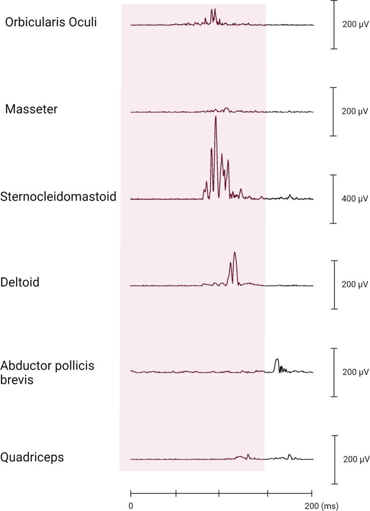
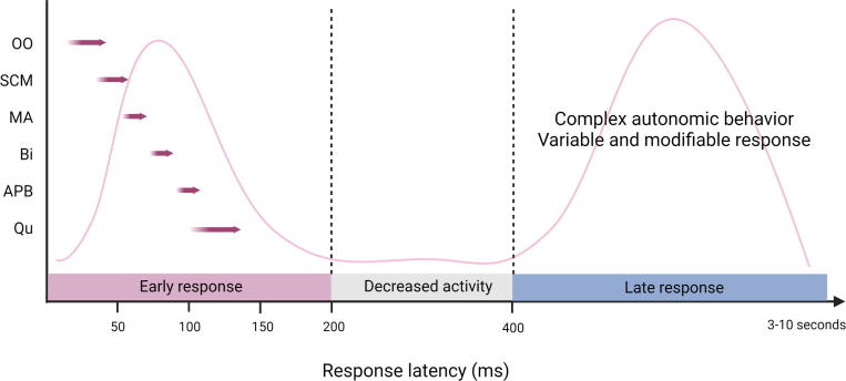
Myoclonus and other jerky movements form a large heterogeneous group of disorders. Clinical neurophysiology studies can have an important contribution to support diagnosis but also to gain insight in the pathophysiology of different kind of jerks. This review focuses on myoclonus, tics, startle disorders, restless legs syndrome, and periodic leg movements during sleep. Myoclonus is defined as brief, shock-like movements, and subtypes can be classified based the anatomical origin. Both the clinical phenotype and the neurophysiological tests support this classification: cortical, cortical-subcortical, subcortical/non-segmental, segmental, peripheral, and functional jerks. The most important techniques used are polymyography and the combination of electromyography-electroencephalography focused on jerk-locked back-averaging, cortico-muscular coherence, and the Bereitschaftspotential. Clinically, the differential diagnosis of myoclonus includes tics, and this diagnosis is mainly based on the history with premonitory urges and the ability to suppress the tic. Electrophysiological tests are mainly applied in a research setting and include the Bereitschaftspotential, local field potentials, transcranial magnetic stimulation, and pre-pulse inhibition. Jerks due to a startling stimulus form the group of startle syndromes. This group includes disorders with an exaggerated startle reflex, such as hyperekplexia and stiff person syndrome, but also neuropsychiatric and stimulus-induced disorders. For these disorders polymyography combined with a startling stimulus can be useful to determine the pattern of muscle activation and thus the diagnosis. Assessment of symptoms in restless legs syndrome and periodic leg movements during sleep can be performed with different validated scoring criteria with the help of electromyography.

Keywords: Deep Brain Stimulation; EEG; EMG; Local field potentials; Myoclonus; Neurophysiology; PLMS; RLS; Startle; Tics; Tourette disorder; Transcranial Magnetic Stimulation.


Conflict of interest statement

The authors declare that they have no known competing financial interests or personal relationships that could have appeared to influence the work reported in this paper.

Figures

Fig. 1
Fig. 1
A shows the surface electromyographic (EMG) pattern from a normal voluntary ballistic movement of wrist flexion. The subject was instructed to perform the wrist extension as quick and as brief as possible. In B, there is a myoclonus surface EMG wrist extension discharge from a patient with multifocal action myoclonus. Despite the fact that the normal ballistic movement was performed as brief as possible, note that there is still a more gradual build-up of activity when compared to the involuntary myoclonus EMG discharge. Modified from (Caviness, 1996).
Fig. 2
Fig. 2
Top: Averaged accelerometer (ACC) deflection. Bottom: Averaged rectified right wrist extensor electromyographic (RWE1) discharge of less than 50 ms. A time-locked relationship between myoclonus electromyographic discharge and the sudden, brief movement, through signal averaging is shown.
Fig. 3
Fig. 3
Multichannel surface electromyographic (EMG) recording in upper extremities during postural activation from a patient with cortical myoclonus. There are myoclonus EMG discharges that occur with almost synchronous timing bilaterally.
Fig. 4
Fig. 4
Cortical myoclonus. A: Multiple brief myoclonus electromyographic (EMG) discharges seen in the lower trace during postural activation of the right wrist extensor musculature (RWE1). The contralateral EEG motor area (C3) shows no gross encephalographic (EEG) transient correlate in the upper trace. B: Cortical myoclonus. Using 100 surface myoclonus EMG discharges to perform EEG-EMG back-averaging to increase signal to noise ratio, a sharp EEG transient is elicited at C3. The averaged EEG transient at C4 is smaller, has different configuration, and not significantly different from the C4 EEG waves before and after the trigger (time 0). These findings together demonstrate a cortical myoclonus physiology.
Fig. 5
Fig. 5
Enhanced Abductor Pollicis Brevis long latency electromyographic (EMG) reflexes at rest to median nerve stimulation at 53 msec and a smaller wave at 84 msec. This demonstrates an enhance transcortical reflex response.
Fig. 6
Fig. 6
A: Back-averaging of a focal cortical transient preceding averaged left arm myoclonus electromyographic (EMG) discharges in a patient with cortical myoclonus. B: Enlarged cortical somatosensory-evoked potentials (SEP) from the same patient. Note the similarity of the P25-N33 wave dipole in both A and B. In both A and B, there is a positive wave in the CP4 electrode with a simultaneous FC4 negative wave. Averaged ear reference was used.
Fig. 7
Fig. 7
Generalized myoclonus electromyographic (EMG) discharges with a typical cortical-subcortical physiology is shown with polyspike encephalographic (EEG) discharges occurring with the myoclonus. Also in this patient, there were common interictal spike and wave EEG discharges without myoclonus. These cortical-subcortical electrophysiology findings should be noted as a strike contrast to cortical myoclonus, despite the fact that EEG discharges are present in both.
Fig. 8
Fig. 8
Expanded time scale that shows a simultaneous ascending and descending order of recruitment from a presumed lower brainstem source. Note that the sternomastoid muscle is recruited first. The electromyographic (EMG) discharges are rectified and averaged from 20 trials. Time zero marks the time that the examiner touched the shoulder to elicit the generalized jerks.
Fig. 9
Fig. 9
Right wrist multichannel surface electromyographic (EMG) recording from a patient with myoclonus –dystonia syndrome during postural activation eliciting the right arm myoclonus. Note the long duration of the shortest EMG discharges (100–200 ms) in addition to longer duration discharges. The myoclonus EMG discharges are irregular with respect to amplitude, duration, and timing between agonist and antagonist muscles.
Fig. 10
Fig. 10
Multichannel surface electromyographic (EMG) recording from the face of a patient with hemifacial spasm during a train of right facial myoclonic jerks and their corresponding EMG discharges. Similar discharges are seen across all facial (CN VII) nerve innervated muscles. EMG discharge duration variability with the facial myoclonus is demonstrated. This patient also had much longer EMG discharges associated with sustained spasms (not shown).
Fig. 11
Fig. 11
Representative recording of an individual case with jerky movements in the arm of functional origin. Four seconds of raw electroencephalographic (EEG) and electromyographic (EMG) data of the extensor carpi radialis (ECR). Note the long duration EMG bursts (+/- 500 ms). After back-averaging of 70 epochs of jerks, a Bereitschaftspotential can be seen be on C3, Cz and C4, which starts approximately 1 s before jerk onset.
Fig. 12
Fig. 12
The electromyographic (EMG) discharges have a highly variable duration from 25 to 100 ms while showing both synchronous and asynchronous discharge relationships between muscles, giving a disorganized appearance to the EMG polygraphy recording. This irregular pattern correlates with lower extremity myoclonus and thus “orthostatic myoclonus”.
Fig. 13
Fig. 13
The circuit of the startle reflex and blink reflex elicit by the auditory stimulus. In red, the afferent pathway of the auditory stimulus is shown. After the signal has reached the ventral cochlear nucleus, the pathway of the blink reflex is shown in green and the startle reflex in blue. The orbicularis oculi is thought to be elicited twice: first as part of the blink reflex, second as part of the startle reflex. Created with BioRender.com. Based on (Valls-Solé et al., 2008). (For interpretation of the references to colour in this figure legend, the reader is referred to the web version of this article.)
Fig. 14
Fig. 14
Rectified electromyographic (EMG) (single trial) muscle responses of a 13-year-old boy following 104 dB tone. The latency of the abductor pollicis brevis (APB) muscle is disproportionately long as the onset is seen after the quadriceps response Although not explained in the original article, the auditory blink reflex does not seem to be present in this EMG (Bakker et al., 2009; Dreissen et al., 2012).
Fig. 15
Fig. 15
A schematic diagram to depict the early and late components of the typical normal human startle response. The sequence of muscle recruitment consisting of the orbicularis oculi (OO), the sternocleidomastoid (SCM), masseter (MA), biceps (Bi), abductor pollicis brevis (APB) and quadriceps (Qu) can be seen with a latency to onset between 20 and 150 ms. No electromyographic (EMG) activity is visible in the period between the early and late response (200–400 ms). No typical muscle recruitment exists for the late response lasting up to 3–10 s, as this is thought to be complex autonomic behavior. Created with BioRender.com. Based on (Saini and Pandey, 2020).

References

    1. Allen R.P. Restless leg syndrome/Willis-Ekbom disease pathophysiology. Sleep Med. Clin. 2015;10:207–214. doi: 10.1016/j.jsmc.2015.05.022. - DOI - PMC - PubMed
    1. Allen R.P., Earley C.J. Validation of the Johns Hopkins restless legs severity scale. Sleep Med. 2001;2:239–242. doi: 10.1016/S1389-9457(00)00080-0. - DOI - PubMed
    1. Allen R.P., Picchietti D., Hening W.A., Trenkwalder C., Walters A.S., Montplaisi J., et al. Restless legs syndrome: Diagnostic criteria, special considerations, and epidemiology. A report from the restless legs syndrome diagnosis and epidemiology workshop at the National Institutes of Health. Sleep Med. 2003;4:101–119. doi: 10.1016/S1389-9457(03)00010-8. - DOI - PubMed
    1. Allen R.P., Walters A.S., Montplaisir J., Hening W., Myers A., Bell T.J., Ferini-Strambi L. Restless legs syndrome prevalence and impact: REST general population study. Arch. Intern. Med. 2005;165(11):1286. doi: 10.1001/archinte.165.11.1286. - DOI - PubMed
    1. Allen R.P., Stillman P., Myers A.J. Physician-diagnosed restless legs syndrome in a large sample of primary medical care patients in western Europe: Prevalence and characteristics. Sleep Med. 2010;11:31–37. doi: 10.1016/j.sleep.2009.03.007. - DOI - PubMed

LinkOut - more resources