. 2022 Oct 17:13:930511.
doi: 10.3389/fimmu.2022.930511. eCollection 2022.

Lactobacillus (LA-1) and butyrate inhibit osteoarthritis by controlling autophagy and inflammatory cell death of chondrocytes

Affiliations

Lactobacillus (LA-1) and butyrate inhibit osteoarthritis by controlling autophagy and inflammatory cell death of chondrocytes

Keun-Hyung Cho et al. Front Immunol. .

Abstract

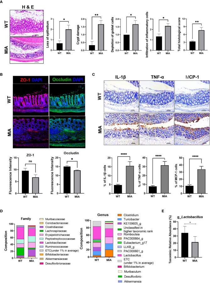
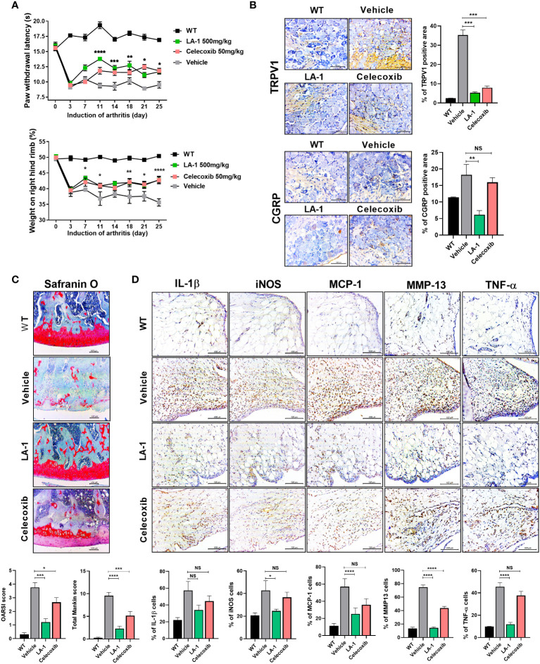
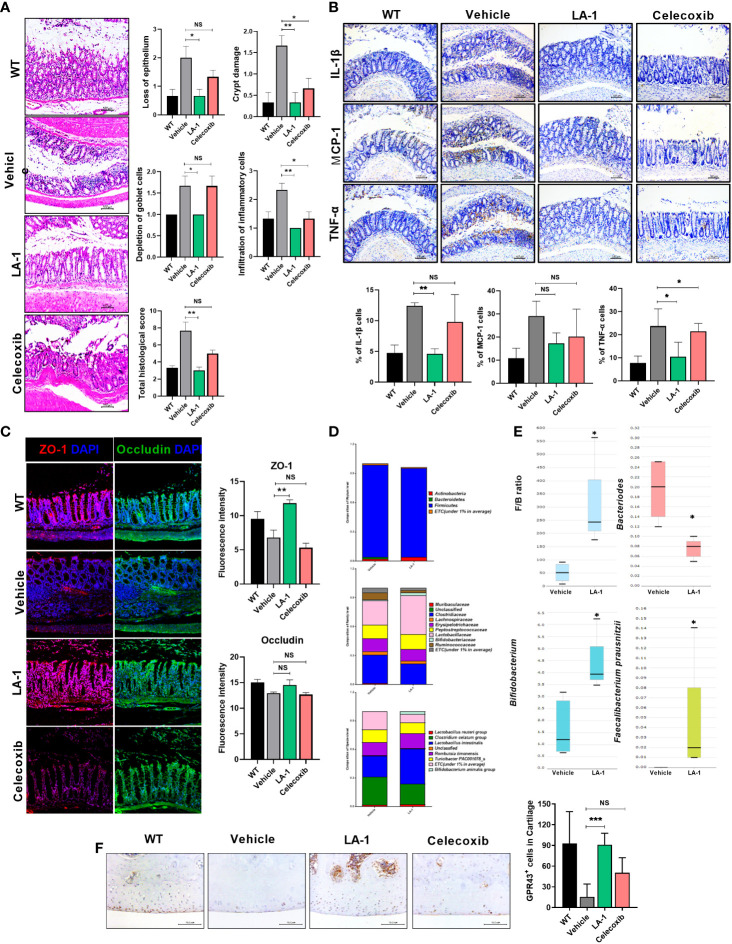
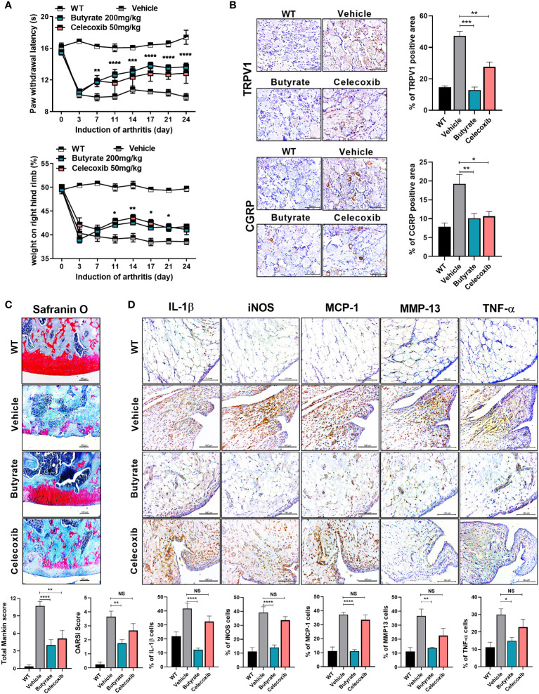
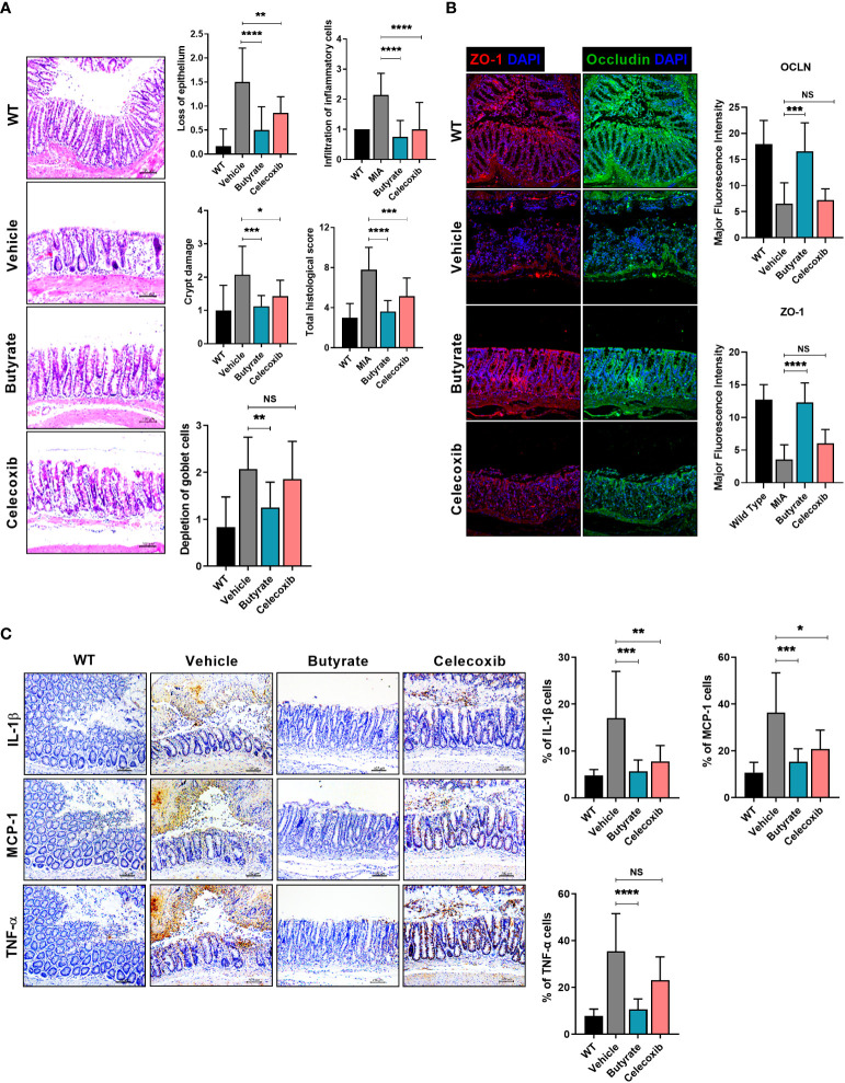
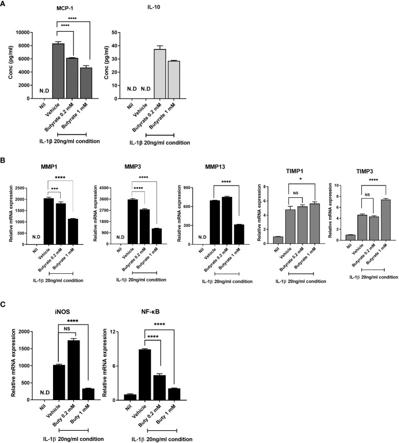
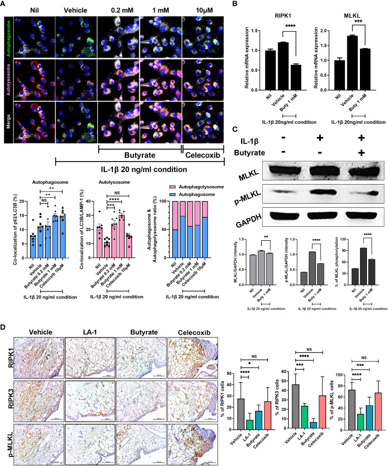
Osteoarthritis (OA) reduces the quality of life as a result of the pain caused by continuous joint destruction. Inactivated Lactobacillus (LA-1) ameliorated osteoarthritis and protected cartilage by modulating inflammation. In this study, we evaluated the mechanism by which live LA-1 ameliorated OA. To investigate the effect of live LA-1 on OA progression, we administered LA-1 into monosodium iodoacetate (MIA)-induced OA animals. The pain threshold, cartilage damage, and inflammation of the joint synovial membrane were improved by live LA-1. Furthermore, the analysis of intestinal tissues and feces in the disease model has been shown to affect the systems of the intestinal system and improve the microbiome environment. Interestingly, inflammation of the intestinal tissue was reduced, and the intestinal microbiome was altered by live LA-1. Live LA-1 administration led to an increase in the level of Faecalibacterium which is a short-chain fatty acid (SCFA) butyrate-producing bacteria. The daily supply of butyrate, a bacterial SCFA, showed a tendency to decrease necroptosis, a type of abnormal cell death, by inducing autophagy and reversing impaired autophagy by the inflammatory environment. These results suggest that OA is modulated by changes in the gut microbiome, suggesting that activation of autophagy can reduce aberrant cell death. In summary, live LA-1 or butyrate ameliorates OA progression by modulating the gut environment and autophagic flux. Our findings suggest the regulation of the gut microenvironment as a therapeutic target for OA.

Keywords: LA-1; butyrate; inflammation; microbiota; necroptosis; osteoarthritis.


Conflict of interest statement

The authors declare that the research was conducted in the absence of any commercial or financial relationships that could be construed as a potential conflict of interest.

Figures

Figure 1
Figure 1
Osteoarthritis (OA) leads to an imbalanced intestinal environment and an altered microbiome. (A) Intestinal epithelial tissue images of the healthy (WT) and diseased (monosodium iodoacetate, MIA) groups at 3 weeks after OA induction (magnification ×200). (B) Intestinal tight junction protein expression (magnification ×200). (C) Expression of inflammatory factors by immunohistochemistry (IHC) (magnification ×200). (D) The microbiome of fecal samples in WT and MIA. (E) The abundance of Lactobacillus in WT and MIA. Data are means ± SD (*p < 0.05, **p < 0.01, ****p < 0.001), ns, not significant.
Figure 2
Figure 2
Supplementation of live Lactobacillus acidophilus (LA-1) reduces OA-mediated pain and inflammation. (A) Pain withdrawal latency and weight on the right hind limb were measured for 25 days in the WT, vehicle, LA-1, and celecoxib groups. (B) IHC of TRPV1 and CGRP in the dorsal root ganglion (DRG) of rats treated with LA-1 for 3 weeks. (C) After 3 weeks of MIA injection, OA-induced knee joints were sectioned and subjected to Safranin O staining. The OARSI score and total Mankin score were calculated (magnification ×200). (D) Analysis of positive cell expression in the synovial membrane of the same tissue for inflammatory factors (IL-1β, iNOS, MCP-1, TNF-α) and catabolic factors (MMP13), accompanying the occurrence of arthritis, was performed by IHC (magnification ×400). Data are means ± SD (*p < 0.05, **p < 0.01, ***p < 0.005, ****p < 0.001), ns, not significant.
Figure 3
Figure 3
LA-1 improves the imbalanced intestinal environment and microbiome. (A) Representative images show H&E staining of the intestine in the WT, vehicle, LA-1, and celecoxib groups (magnification ×200). (B) Inflammatory factor expression in intestinal epithelial cells was determined by IHC in each group (magnification ×200). (C) Tight junction protein (TJP) expression in intestinal epithelial cells was increased by LA-1 (magnification ×200). (D) The feces of OA rats had different phyla, family, and species phenotypes from those treated with LA-1, and a relatively high proportion of beneficial bacteria was found in the feces of LA-1-treated rats. (E) LA-1 altered the F/B ratio and the population of butyrate-producing bacteria. (F) LA-1 increased the level of the short-chain fatty acid receptor G-protein-coupled receptor 43 (magnification ×400). Data are means ± SD (*p < 0.05, **p < 0.01, ***p < 0.005), ns, not significant.
Figure 4
Figure 4
Butyrate reduces OA pain and exerts a chondroprotective effect. (A) Butyrate improved rat paw withdrawal latency and weight bearing at 3 weeks after induction of MIA, indicating pain suppression. (B) Butyrate reduced the levels of neurotransmitters in rat dorsal root ganglion (magnification ×400). (C) Three weeks after OA induction, butyrate protected the rat knee joint from OA-associated degradation (magnification ×200). (D) IHC analysis confirmed the regulation of butyrate-mediated inflammatory factors (IL-1β, iNOS, MCP-1, TNF-α) and catabolic factors (MMP13) in the same synovial tissue site as determined by IHC (magnification ×400). Data are means ± SD (*p < 0.05, **p < 0.01, ***p < 0.005, ****p < 0.001), ns, not significant.
Figure 5
Figure 5
Butyrate restores tight junction proteins, inhibits intestinal damage, and reduces inflammatory factor levels. (A) Three weeks after MIA injection, the degree of intestinal damage in rats was evaluated by H&E staining (magnification ×200). (B) Effect of butyrate on tight junction protein (TJP) expression by immunofluorescence staining (magnification ×200). (C) Effect of butyrate on the expression levels of inflammatory factors in rat intestine by IHC (magnification ×200). Data are means ± SD (*p < 0.05, **p < 0.01, ***p < 0.005, ****p < 0.001), ns, not significant.
Figure 6
Figure 6
Butyrate rebalances anabolic/catabolism and suppresses the expression of inflammatory factors to prevent exacerbation of OA. (A) Protein levels of monocyte chemoattractant protein 1 (MCP-1) and interleukin 10 (IL-10) in chondrocytes cultured in serum-free medium with 20 ng/ml of IL-1β for 24 h. (B) To determine the level of anabolic/catabolism expressed from chondrocytes upon butyrate treatment within an inflammatory environment, quantitative PCR was performed to determine mRNA levels. (C) Inducible nitric oxide synthase (iNOS) and nuclear factor kappa-light-chain-enhancer of activated B-cell (NF-κB) expression levels of chondrocytes under inflammatory conditions were analyzed by quantitative PCR. Data are means ± SD (*p < 0.05, ***p < 0.005, ****p < 0.001), nd, not detected; ns, not significant.
Figure 7
Figure 7
Butyrate reduced inflammatory cell death by restoring abnormal autophagy of chondrocytes in an inflammatory environment. (A) Effect on autophagy of butyrate in chondrocytes cultured in medium with IL-1β at 20 ng/ml (magnification ×200). (B) Effect of butyrate on the expression levels of necrosis factors of chondrocytes under inflammatory conditions. (C) In chondrocytes, the effect of butyrate on MLKL phosphorylation in chondrocytes under inflammatory conditions. (D) IHC showed that live LA-1 and butyrate modulated the levels of necroptosis factors in OA synovial tissue (magnification ×400). Data are means ± SD (*p < 0.05, **p < 0.01, ***p < 0.005, ****p < 0.001), ns, not significant.

References

    1. Pereira D, Peleteiro B, Araujo J, Branco J, Santos RA, Ramos E. The effect of osteoarthritis definition on prevalence and incidence estimates: a systematic review. Osteoarthritis Cartilage (2011) 19(11):1270–85. doi: 10.1016/j.joca.2011.08.009 - DOI - PubMed
    1. Briggs AM, Cross MJ, Hoy DG, Sanchez-Riera L, Blyth FM, Woolf AD, et al. . Musculoskeletal health conditions represent a global threat to healthy aging: A report for the 2015 world health organization world report on ageing and health. Gerontologist (2016) 56 Suppl 2:S243–55. doi: 10.1093/geront/gnw002 - DOI - PubMed
    1. Hunter DJ, Bierma-Zeinstra S. Osteoarthritis. Lancet (2019) 393(10182):1745–59. doi: 10.1016/S0140-6736(19)30417-9 - DOI - PubMed
    1. Martel-Pelletier J, Barr AJ, Cicuttini FM, Conaghan PG, Cooper C, Goldring MB, et al. . Osteoarthritis. Nat Rev Dis Primers (2016) 2:16072. doi: 10.1038/nrdp.2016.72 - DOI - PubMed
    1. Tonge DP, Pearson MJ, Jones SW. The hallmarks of osteoarthritis and the potential to develop personalised disease-modifying pharmacological therapeutics. Osteoarthritis Cartilage (2014) 22(5):609–21. doi: 10.1016/j.joca.2014.03.004 - DOI - PubMed

Publication types