

Characterization of clinicopathologic and abdominal ultrasound findings in dogs with glucocorticoid deficient hypoadrenocorticism
- PMID: 36326216
- PMCID: PMC9708419
- DOI: 10.1111/jvim.16564
Characterization of clinicopathologic and abdominal ultrasound findings in dogs with glucocorticoid deficient hypoadrenocorticism
Abstract
Background: Clinical findings of glucocorticoid-deficient hypoadrenocorticism (GDH) can overlap with other diseases, presenting a diagnostic challenge.
Objectives: Describe clinicopathologic and ultrasonographic features of dogs with GDH and those suspected of having GDH that had the disease ruled out.
Animals: Six hundred twenty-three dogs.
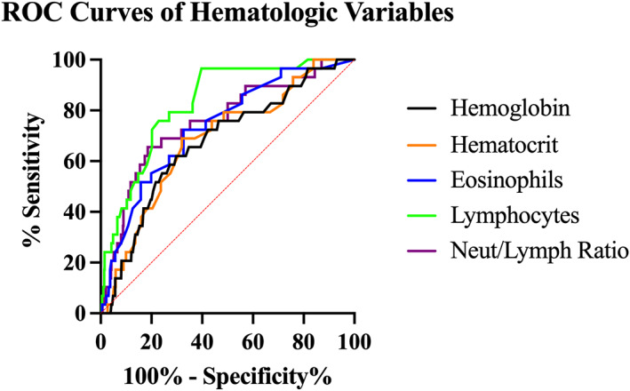
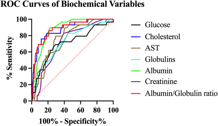
Methods: Records from dogs with suspected GDH between 2003 and 2018 were reviewed. Dogs with hyperkalemia or hyponatremia were excluded. Dogs were categorized as controls when the resting serum cortisol or post-ACTH cortisol concentration were > 2 μg/dL. Clinicopathologic and ultrasonographic features were compared between groups. The optimal cut-point, area under the receiver operating characteristic curve (AUC), sensitivity, and specificity were calculated for individual features used to detect GDH.
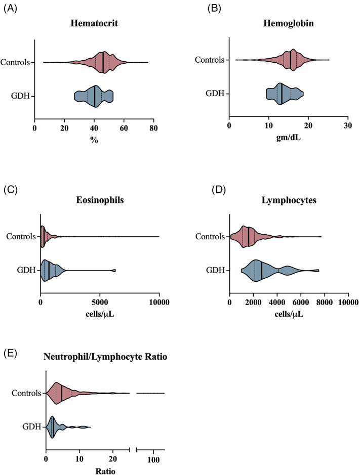
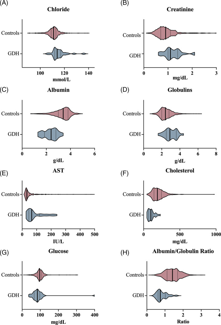
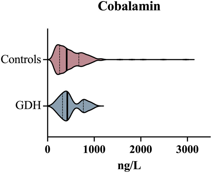
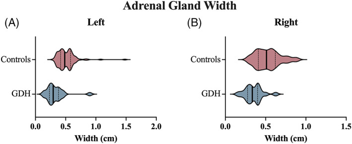
Results: Dogs were categorized as GDH (n = 29) or controls (n = 594). Lymphocyte count (>1750 cells/L; sensitivity, 96.6%; 95% confidence interval [CI], 82.8%-99.8%; specificity, 60.3%; 95% CI, 56.3%-64.1%; AUC, 0.828; 95% CI, 0.762-0.894) and albumin/globulin ratio (<1.081; sensitivity, 86.2%; 95% CI, 69.4%-94.5%; specificity, 78.8%; 95% CI, 75.3%-81.9%; AUC, 0.886; 95% CI, 0.827-0.944) had the highest discriminatory power between groups. Left adrenal gland width <0.39 cm was 80% (95% CI, 58.4%-91.9%) sensitive and 82.4% (95% CI, 74.2-88.4) specific for GDH. Serum cobalamin concentrations and ultrasonographic abnormalities of the GI tract were not different between groups.
Conclusion and clinical importance: No single variable could be used to confidently rule out GDH and obviate the need for cortisol testing in dogs with a clinical presentation consistent with GDH.
Keywords: Addison's disease; adrenal gland; atypical Addison's; cortisol.
© 2022 The Authors. Journal of Veterinary Internal Medicine published by Wiley Periodicals LLC on behalf of American College of Veterinary Internal Medicine.
Conflict of interest statement
Authors declare no conflicts of interest.
Figures
References
-
- Kelch WJ. Canine Hypoadrenocorticism (Canine Addison's Disease): History, Contemporary Diagnosis by Practicing Veterinarians, and Epidemiology. Knoxville, TN: The University of Tennessee; 1996.
-
- Peterson ME, Kintzer PP, Kass PH. Pretreatment clinical and laboratory findings in dogs with hypoadrenocorticism: 225 cases (1979‐1993). J Am Vet Med Assoc. 1996;208:85‐91. - PubMed
-
- Borin‐Crivellenti S, Garabed RB, Moreno‐Torres KI, Wellman ML, Gilor C. Use of a combination of routine hematologic and biochemical test results in a logistic regression model as a diagnostic aid for the diagnosis of hypoadrenocorticism in dogs. Am J Vet Res. 2017;78:1171‐1181. - PubMed
