Review
. 2023 May;36(5):e4867.
doi: 10.1002/nbm.4867. Epub 2022 Dec 14.

Off-resonance artifact correction for MRI: A review

Affiliations
Review

Off-resonance artifact correction for MRI: A review

Melissa W Haskell et al. NMR Biomed. 2023 May.

Abstract

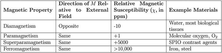
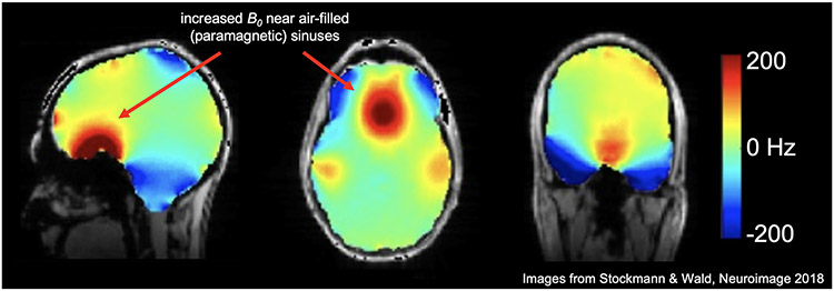
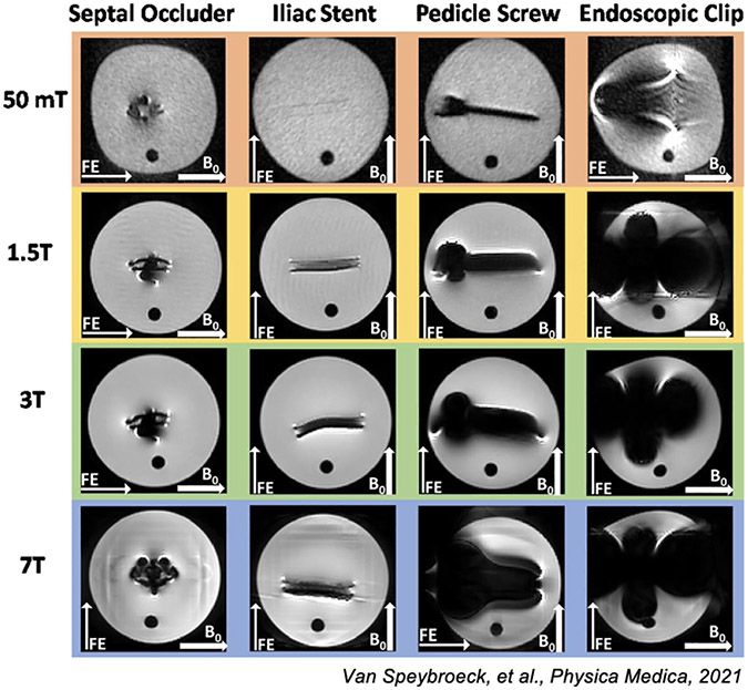
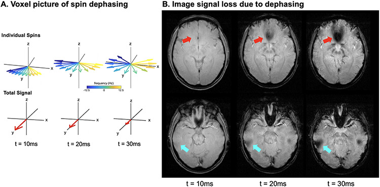
In magnetic resonance imaging (MRI), inhomogeneity in the main magnetic field used for imaging, referred to as off-resonance, can lead to image artifacts ranging from mild to severe depending on the application. Off-resonance artifacts, such as signal loss, geometric distortions, and blurring, can compromise the clinical and scientific utility of MR images. In this review, we describe sources of off-resonance in MRI, how off-resonance affects images, and strategies to prevent and correct for off-resonance. Given recent advances and the great potential of low-field and/or portable MRI, we also highlight the advantages and challenges of imaging at low field with respect to off-resonance.


Figures

Figure 1:
Figure 1:. Magnetic Susceptibility.
Adapted from https://mriquestions.com/what-is-susceptibility.html [36]
Figure 2:
Figure 2:. Bo magnetic field in the brain at 7T with 2nd order shimming.
This fieldmap shows the spatially varying patterns of the B0 field in the human brain (superimposed over an anatomical image). Even after 2nd order shimming has been applied to flatten the field, higher order field inhomogeneities persist. Arrows show that near the air-filled, and therefore paramagnetic, sinuses, the B0 field is higher than areas in the center of the mostly diamagnetic brain tissue. (Images reused from Stockmann & Wald, 2018 [35] with permission.)
Figure 3:
Figure 3:. Metal artifact from 50mT to 7T.
Here we show Figure 7 from Van Speybroek, et al., 2021 [37] where the “worst-case” scenario images for four types of metal implants are demonstrated. (Images reused via open access license.)
Figure 4:
Figure 4:. Signal Loss from Spin Dephasing.
A. Voxel view of spin dephasing. Spins with different off-resonance will gain different amounts of phase during signal readout leading them to “fan out”, and eventually completely cancel each other. B. A gradient echo image is shown for two different slices at three different echo times (TE). At the later echo times, signal loss due to dephasing is present as indicated by the arrows.
Figure 5:
Figure 5:. Ideal vs. actual encoding phase at a single k-space location.
A. Simulated off-resonance fieldmap in Hz. B. 1D plots of the encoding fields as a function of position, where the red dotted line correspond to the dotted red line in A. C. The ideal k-space encoding phase for (kx,ky)=(0,0.426cm1). D. The actual k-space encoding phase for (kx,ky)=(0,0.426cm1). Combined with k-space encoding errors at other k-space sampling locations, this phase error will lead to off-resonance image artifacts.
Figure 6:
Figure 6:. Image simulations: Cartesian vs. non-Cartesian.
Here we show the different off-resonance effects from EPI vs. spiral acquisition. In the EPI image, geometric distortion of the brain is visible with a distortion sensitivity of 75 Hz/cm (see Eq. (9)), a typical value, resulting in the brain being stretched compared to the original Cartesian spin-warp image (blue arrows). In the spiral image, there is a large amount of blurring, noted by the red arrow, but there is less geometric distortion than in EPI.
Figure 7:
Figure 7:. Common B0 artifact mitigation strategies.
Figure 8:
Figure 8:. Illustration of EPI distortion correction using Eq. (14).
(A) Ground-truth (undistorted) object and (B) fieldmap. (C,D) K+,i and K,i for the dashed lines in (E,F). (E,F) Distorted images f+ and f. (G) Distortion correction attempt using only f+. (H) Successful distortion correction using Eq. (14). (Images reused from Andersson et al., 2003 [75] with permission.)
Figure 9:
Figure 9:. CP and MB reconstruction at 50mT.
Compared to a standard iFFT reconstruction, CP and MB reconstructions both correct for geometric distortions due to off-resonance, especially in the right side of the brain where the magnetic field off-resonance is stronger. MB also shows more uniform signal intensity. (Images reused from Koolstra, et al., 2021 [82] via open access license.)
Figure 10:
Figure 10:
Comparison of standard spin echo imaging (top row), VAT (middle), and SEMAC (bottom row) methods for a patient with a spinal implant. The right column is reformatted so slice direction is along the horizontal axis. The SE image is badly distorted in both frequency and slice directions, the VAT images are partially corrected in the frequency direction with substantial slice distortion, while the SEMAC image is corrected in both directions. (Images reused from [120] with permission.)

References

    1. Lüdeke KM, Röschmann P, and Tischler R. Susceptibility artefacts in NMR imaging. Magnetic Resonance Imaging, 3(4):329–343, January 1985. - PubMed
    1. O’Donnell M and Edelstein WA. NMR imaging in the presence of magnetic field inhomogeneities and gradient field nonlinearities. Medical Physics, 12(1):20–26, January 1985. - PubMed
    1. Czervionke LF, Daniels DL, Wehrli FW, Mark LP, Hendrix LE, Strandt JA, Williams AL, and Haughton VM. Magnetic susceptibility artifacts in gradient-recalled echo MR imaging. AJNR. American journal of neuroradiology, 9(6):1149–55, 1988. - PMC - PubMed
    1. Michiels J, Bosmans H, Pelgrims P, Vandermeulen D, Gybels J, Marchal G, and Suetens P. On the problem of geometric distortion in magnetic resonance images for stereotactic neurosurgery. Magnetic Resonance Imaging, 12(5):749–765, 1994. - PubMed
    1. Ladd Mark E., Erhart Peter, Debatin Jörg F., Romanowski Benjamin J., Boesiger Peter, and Graeme C McKinnon. Biopsy needle susceptibility artifacts. Magnetic Resonance in Medicine, 36(4):646–651, October 1996. - PubMed

Publication types