. 2022 Nov 1;63(12):5.
doi: 10.1167/iovs.63.12.5.

Zfp503/Nlz2 Is Required for RPE Differentiation and Optic Fissure Closure

Affiliations

Zfp503/Nlz2 Is Required for RPE Differentiation and Optic Fissure Closure

Elangovan Boobalan et al. Invest Ophthalmol Vis Sci. .

Abstract

Purpose: Uveal coloboma is a congenital eye malformation caused by failure of the optic fissure to close in early human development. Despite significant progress in identifying genes whose regulation is important for executing this closure, mutations are detected in a minority of cases using known gene panels, implying additional genetic complexity. We have previously shown knockdown of znf503 (the ortholog of mouse Zfp503) in zebrafish causes coloboma. Here we characterize Zfp503 knockout (KO) mice and evaluate transcriptomic profiling of mutant versus wild-type (WT) retinal pigment epithelium (RPE)/choroid.

Methods: Zfp503 KO mice were generated by gene targeting using homologous recombination. Embryos were characterized grossly and histologically. Patterns and level of developmentally relevant proteins/genes were examined with immunostaining/in situ hybridization. The transcriptomic profile of E11.5 KO RPE/choroid was compared to that of WT.

Results: Zfp503 is dynamically expressed in developing mouse eyes, and loss of its expression results in uveal coloboma. KO embryos exhibit altered mRNA levels and expression patterns of several key transcription factors involved in eye development, including Otx2, Mitf, Pax6, Pax2, Vax1, and Vax2, resulting in a failure to maintain the presumptive RPE, as evidenced by reduced melanin pigmentation and its differentiation into a neural retina-like lineage. Comparison of RNA sequencing data from WT and KO E11.5 embryos demonstrated reduced expression of melanin-related genes and significant overlap with genes known to be dynamically regulated at the optic fissure.

Conclusions: These results demonstrate a critical role of Zfp503 in maintaining RPE fate and optic fissure closure.


Conflict of interest statement

Disclosure: E. Boobalan, None; A.H. Thompson, None; R.P. Alur, None; D.M. McGaughey, None; L. Dong, None; G. Shih, None; E.R. Vieta-Ferrer, None; I.F. Onojafe, None; V.K. Kalaskar, None; G. Arno, None; A.J. Lotery, None; B. Guan, None; C. Bender, None; O. Memon, None; L. Brinster, None; C. Soleilhavoup, None; L. Panman, None; T.C. Badea, None; A. Minella, None; A.J. Lopez, None; S.M. Thomasy, None; A. Moshiri, None; D. Blain, None; R.B. Hufnagel, None; T. Cogliati, None; K. Bharti, None; B.P. Brooks, None

Figures

Figure 1.
Figure 1.
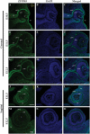
ZFP503 protein is expressed in the developing mouse eye. ZFP503 expression (green) by immunofluorescence in representative coronal (A–I) and sagittal (J–O) sections of WT mouse embryos. The three developmental time points correspond to “before closure” at E10.5, “during closure” (E11.5), and “after closure” (E12.5) of the optic fissure (n = 3 embryos each). DAPI nuclear staining in blue. Confocal microscope exposure and parameters were maintained equal for comparison purposes. Compasses in (A) and (J) apply to panels A–I and J–O, respectively: D, dorsal; Dis, distal; P, proximal; V, ventral. L, lens; LV, lens vesicle; OS, optic stalk; SE, surface ectoderm. Scale bar: 100 µm.
Figure 2.
Figure 2.
Homozygous deletion of Zfp503 results in relatively normal-appearing E18.5 embryos with characteristic organ pathologies. β-Galactosidase staining (indicative of Zfp503 localization) in representative Zfp503/ embryos was present in the developing brain, spinal cord, eye, and branchial arches (A, B, n = 4/genotype). Loss of ZFP503 resulted in smaller lungs (C, D, n = 4/4 from two litters) and faulty alveolar formation in the lung (E, F, n = 3 sections through each of four embryos) visualized by H&E staining in representative histology sections. The sternum of Zfp503/ embryos visualized with Alcian blue was shorter than in WT embryos, accompanied by abnormal rib morphology (G, H, frontal view, arrow, n = 3/genotype) and (I, J, lateral view). Scale bars: 1 mm (C, D), 50 µm (E, F), and 0.9 mm (GJ).
Figure 3.
Figure 3.
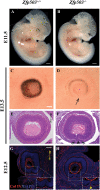
Zfp503 deletion results in hypopigmentation of the RPE and uveal coloboma. Photomicrographs of representative WT embryos at E11.5 (A) and E13.5 (C) showed normal, circumferential, and continuous pigmentation of the RPE that was almost completely absent in age-matched Zfp503−/− embryos (B, D, arrows, n = 5/genotype). Sagittal sections of representative E13.5 embryos stained with H&E revealed that the edges of the optic fissure were fused and no longer discernible in WT embryos (E), whereas representative Zfp503/ embryos showed distinct nonfusion of fissure edges (F, arrowhead). Note the hyperplasia, resembling pNR, flanking the fissure (arrows). Whereas the basement membrane of the optic fissure edges was no longer detectable by collagen IV immunostaining (red) in E13.5 WT embryos (G), a clear separation of the optic fissure edges could be seen in Zfp503/ littermates (H, arrowhead, n = 2/genotype). DAPI nuclear staining in blue. Confocal microscope exposure and parameters were maintained equal for comparison purposes. Compass in (E, G) applies to E–H: D, dorsal; V, ventral. Scale bars: 500 µm (A, B); 100 µm (C–H).
Figure 4.
Figure 4.
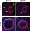
VSX2-positive cellular hyperplasia is observed in the pRPE of Zfp503/ developing eyes. VSX2/CHX10 immunostaining in representative coronal sections of E12.5 WT (A) and Zfp503/ embryos (B) demonstrates fluorescent signal (red) in a hyperplastic ventro-proximal population of KO pRPE (arrow), as well as in the pNR (as expected) in both genotypes (n = 2/genotype). By E14.5, this VSX2-positive hyperplasia extends further distally and temporally (arrow, D) in KO embryos and is absent in WT embryos (C) (n = 2/genotype, coronal sections). DAPI nuclear staining in blue. Confocal microscope exposure and parameters were maintained equal for comparison purposes. Compass in (A) applies to A and B, in (C) applies to C and D: D, dorsal; Dis, distal; P, proximal; V, ventral. L, lens; OS, optic stalk. Scale bars: 100 µm (A–D); 200 µm (E, F).
Figure 5.
Figure 5.
Loss of Zfp503 is associated with reduced expression of MITF and OTX2. Sagittal sections from representative E11.5 WT (A, B, D, E) and Zfp503/ (G, H, J, K) embryos immunostained with anti-MITF antibody (green) and anti-OTX2 are shown in distal to proximal series. Note the open optic fissure (arrow) in Zfp503/ section (I). Qualitative reduction of MITF expression in Zfp503/ was particularly evident in proximal sections, near the optic stalk (L, arrow). All panels were imaged with the same exposure (n = 3/genotype). Compass in (A) refers to all panels: D, dorsal; V, ventral. L, lens. Scale bar: 50 µm.
Figure 6.
Figure 6.
Loss of Zfp503 expression is associated with an abnormal expression pattern of transcription factors important for RPE differentiation. Immunofluorescence staining for PAX2 (red) and PAX6 (green) of representative E11.5 sagittal sections of WT (A, D, G and B, E, H) and Zfp503/ (J, M, P and K, N, Q) eyes are shown. PAX6 expression was increased and expanded into the proximal/dorsal pRPE of Zfp503/ eyes (compare K, N, Q with B, E, H, arrowhead). PAX2 expression in Zfp503/ eyes extended dorsally and distally into the ventral pRPE (compare J, M, P with A, D, G, arrow). DAPI nuclear staining in blue. Confocal microscope exposure and parameters were maintained equal for comparison purposes. Compass in (A) applies to all the panels. D, dorsal; V, ventral. L, lens; OS, optic stalk. Scale bars: 50 µm (n = 3/genotype).
Figure 7.
Figure 7.
Loss of Zfp503 is associated with abnormal Vax1 and Vax2 mRNA expression at E11.5. Microphotographs of representative sagittal section series, distal to proximal, of developing mouse eyes at E11.5 showing Vax1 (red, A, D, G, J, M, P, S, V) and Vax2 (green, B, E, H, K, N, Q, T, W) expression using in situ hybridization. Overlays are shown in C, F, I, L, O, R, U, and X with DAPI nuclear staining in blue. In WT eyes, Vax1 expression was limited to the very proximal ventral optic cup and the ventral optic stalk (A, D, G, J), while Vax2 signal was mostly in the ventral retinal neuroblasts of the optic cup (B, E, H, K). In Zfp503/ embryos, Vax1 in situ hybridization extended more dorsally and further into the proximal optic cup (M, P, S, V). Vax2 expression was more prominent in ventral pRPE in addition to retinal neuroblasts and extended further into the optic stalk in KO embryos (N, Q, T, W). The images are from a single plane (n = 2/genotype). Confocal microscope exposure and parameters were maintained equal for comparison purposes. Compass in (A) is applies to all panels. D, dorsal; V, ventral. Scale bar: 50 µm.
Figure 8.
Figure 8.
RNA-seq from RPE at E11.5. Quantitation of several key RPE and retina mRNA transcripts in Zfp503/ versus Zfp50+/+ embryos demonstrated changes consistent with the qualitative differences noted in immunofluorescence experiments (A). Volcano plot of differentially regulated genes showing changes in several key transcription factor, melanin pigmentation, and genes previously identified as changing during the time of optic fissure closure (B).
Figure 9.
Figure 9.
Overview of key transcription factor changes in Zfp503/ embryos. Schematics of coronal (upper panels) and sagittal (lower panels) sections of mouse embryo eyes around the time of optic fissure closure. Three sagittal sections are shown, proximal to distal from the optic stalk (panels i, ii, and iii, respectively). Reduction in OTX2 and MITF expression is accompanied by abnormal expansions of PAX2, PAX6, Vax1, and Vax2 and the adoption of a neural retina–like phenotype in the pRPE, as evidenced by VSX2 expression. Note that OTX2 and MITF changes in the presumptive RPE are very similar (and shown in one color), with the exception of possible changes in the level of OTX2 in the presumptive neural retina (denoted with an asterisk).

References

    1. Fuhrmann S. Eye morphogenesis and patterning of the optic vesicle. Curr Top Dev Biol. 2010; 93: 61–84. - PMC - PubMed
    1. Patel A, Sowden JC.. Genes and pathways in optic fissure closure. Semin Cell Dev Biol. 2019; 91: 55–65. - PubMed
    1. Hero I. The optic fissure in the normal and microphthalmic mouse. Exp Eye Res. 1989; 49: 229–239. - PubMed
    1. Williamson KA, FitzPatrick DR.. The genetic architecture of microphthalmia, anophthalmia and coloboma. Eur J Med Genet. 2014; 57: 369–380. - PubMed
    1. Hsu P, Ma A, Wilson M, et al. .. CHARGE syndrome: a review. J Paediatr Child Health. 2014; 50: 504–511. - PubMed

Publication types