. 2023 Jan 18;15(679):eabq4064.
doi: 10.1126/scitranslmed.abq4064. Epub 2023 Jan 18.

S-217622, a SARS-CoV-2 main protease inhibitor, decreases viral load and ameliorates COVID-19 severity in hamsters

Affiliations

S-217622, a SARS-CoV-2 main protease inhibitor, decreases viral load and ameliorates COVID-19 severity in hamsters

Michihito Sasaki et al. Sci Transl Med. .

Abstract

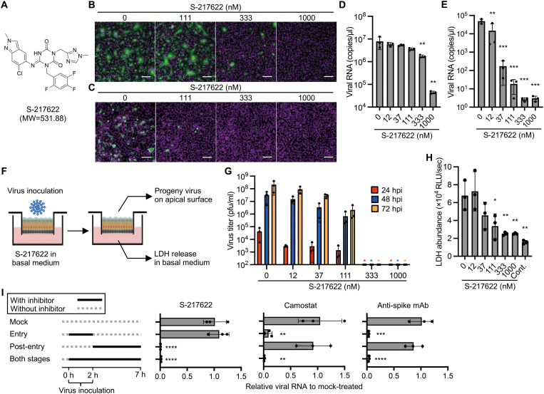
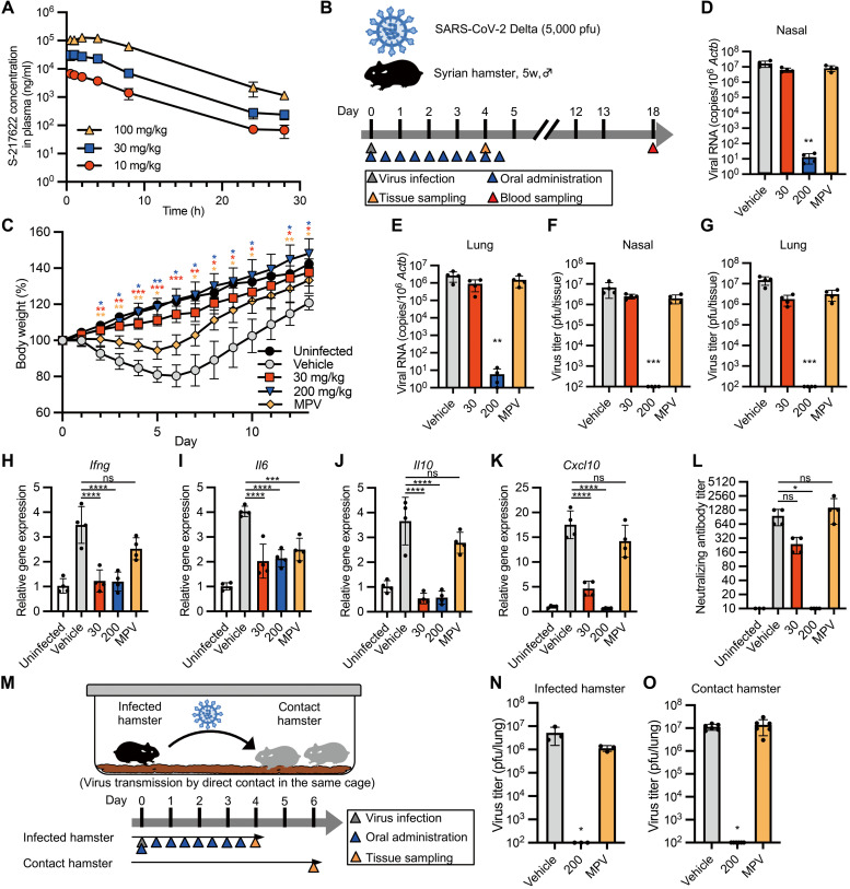
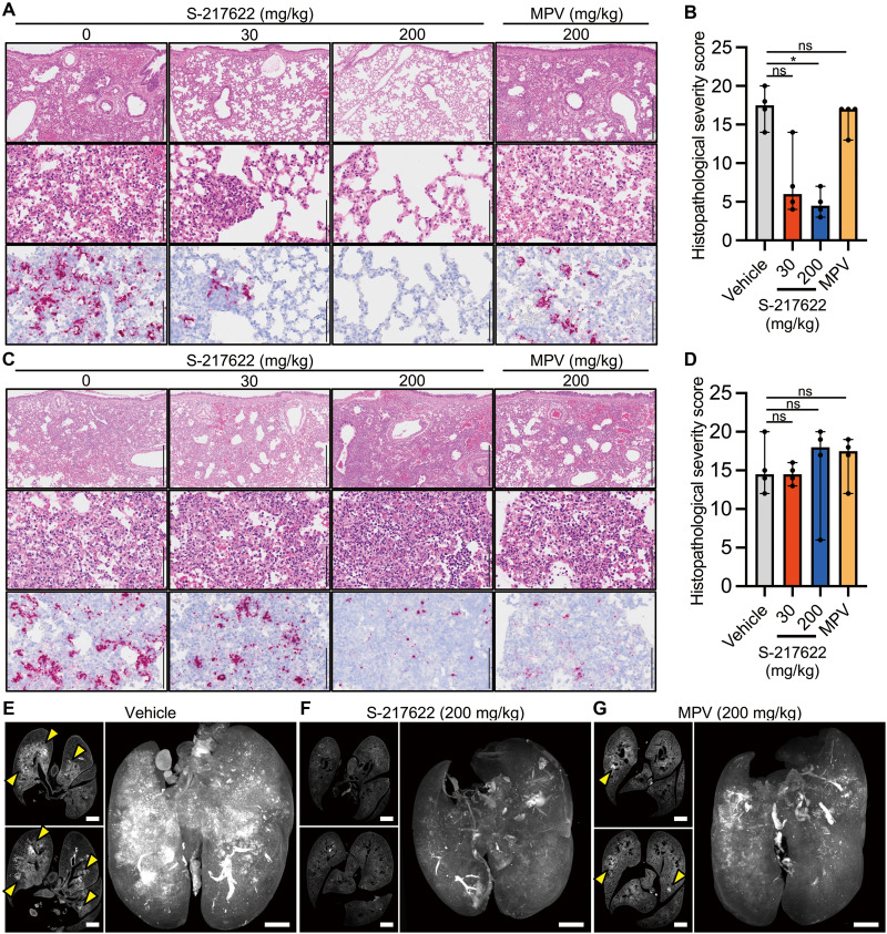
In parallel with vaccination, oral antiviral agents are highly anticipated to act as countermeasures for the treatment of the coronavirus disease 2019 (COVID-19) pandemic caused by severe acute respiratory syndrome coronavirus 2 (SARS-CoV-2). Oral antiviral medication demands not only high antiviral activity but also target specificity, favorable oral bioavailability, and high metabolic stability. Although a large number of compounds have been identified as potential inhibitors of SARS-CoV-2 infection in vitro, few have proven to be effective in vivo. Here, we show that oral administration of S-217622 (ensitrelvir), an inhibitor of SARS-CoV-2 main protease (Mpro; also known as 3C-like protease), decreases viral load and ameliorates disease severity in SARS-CoV-2-infected hamsters. S-217622 inhibited viral proliferation at low nanomolar to submicromolar concentrations in cells. Oral administration of S-217622 demonstrated favorable pharmacokinetic properties and accelerated recovery from acute SARS-CoV-2 infection in hamster recipients. Moreover, S-217622 exerted antiviral activity against SARS-CoV-2 variants of concern, including the highly pathogenic Delta variant and the recently emerged Omicron BA.5 and BA.2.75 variants. Overall, our study provides evidence that S-217622, an antiviral agent that is under evaluation in a phase 3 clinical trial (clinical trial registration no. jRCT2031210350), has remarkable antiviral potency and efficacy against SARS-CoV-2 and is a prospective oral therapeutic option for COVID-19.


Figures

Fig. 1.
Fig. 1.. S-217622 inhibits SARS-CoV-2 infection in vitro at the post-entry stage.
(A)Chemical structure formula of S-217622. (B and C) Immunofluorescence staining of SARS-CoV-2-infected cells. Vero-TMPRSS2 (B) and Calu-3 (C) cells were inoculated with a SARS-CoV-2 Delta variant at MOIs of 0.1 or 10, respectively, and then cultured in the presence of S-217622 for 24 hours and 72 hours, respectively. Cells were stained with anti-SARS-CoV-2 nucleocapsid antibody (green) and Hoechst 33342 (magenta). Scale bars, 100 μm. (D) Viral RNA concentrations were measured in the culture supernatant of Vero-TMPRSS2 cells at 24 hours post-infection (hpi) with a SARS-CoV-2 Delta variant at an MOI of 0.01. (E) Viral RNA concentrations were measured in the culture supernatant of Calu-3 cells at 72 hpi with SARS-CoV-2 Delta variant at an MOI of 0.1. (F) Schematic representation of the SARS-CoV-2 infection experiment in a human airway tissue model. Cells were inoculated at the apical surface with 5,000 pfu of SARS-CoV-2. Viral growth was monitored by titration of progeny virus in the mucus layer on apical surface. The tissue damage by viral infection was estimated by measurement of lactate dehydrogenase (LDH) released from cells into the basal culture medium. (G) Growth of the SARS-CoV-2 Delta variant in a human airway tissue model. Basal culture medium was supplemented with the indicated concentration of S-217622. (H) The abundance of LDH in the basal culture medium was measured by LDH-Glo luminescent assay. (I) A time of the addition assay was conducted using S-217622. Calu-3 cells were treated with S-217622 during inoculation (entry stage), after inoculation (post-entry stage), or both entry and post-entry stages of SARS-CoV-2 at an MOI of 3. The inhibitory effect of each treatment was evaluated by measurement of intracellular viral RNA concentrations at 7 hpi. Camostat and anti-SARS-CoV-2 spike protein antibody were used as entry inhibitors for assay controls. The values shown are mean ± standard deviation (SD) of triplicate samples. *p < 0.05, **p < 0.01, ***p < 0.001, ****p < 0.0001 by one-way ANOVA with Dunnett’s test (D, E, H, I) or Kruskal-Wallis test with Dunn’s test (G).
Fig. 2.
Fig. 2.. Prophylactic treatment of S-217622 controls viral burden and disease in hamsters inoculated with SARS-CoV-2.
(A)The plasma concentration profile of S-217622 after a single oral administration in hamsters is shown (n = 3 for each treatment). Plasma samples were harvested at the indicated time points and analyzed by LC-MS/MS. (B) Schematic of the experimental design for prophylactic treatment in a hamster model. Hamsters were intranasally inoculated with 5,000 pfu of SARS-CoV-2 Delta variant. For prophylactic treatment, the hamsters were treated with oral administration of S-217622 or vehicle (0 mg/kg) twice daily (b.i.d.) from the time of infection (0 hpi) to 4 dpi. Molnupiravir (MPV) was used as a comparator drug. A group of hamsters (n = 4 for each treatment) was euthanized at 4 dpi for tissue collection. Another subset of hamsters (n = 4 for each treatment; n = 3 for uninfected) was monitored for 13 days for body weight change and then euthanized at 18 dpi for serum collection. (C) Body weight changes in uninfected hamsters (n = 3) and SARS-CoV-2-infected hamsters treated with S-217622 (30 mg/kg and 200 mg/kg), vehicle, or MPV (200 mg/kg) is shown (n = 4 for each group). (D and E) Viral RNA concentrations were measured in nasal turbinates (D) and lungs (E) isolated from hamsters at 4 dpi. Each group of hamsters was treated with vehicle (red), 30 mg/kg (blue), and 200 mg/kg (orange) of S-217622 or MPV (green) (n = 4 for each group). Relative viral RNA abundance in lungs as compared with lungs from vehicle-treated hamsters were examined. Data were normalized to β-actin. (F and G) Virus titers were measured in nasal turbinates (F) and lungs (G) isolated from hamsters at 4 dpi as determined by plaque assay. Each group of hamsters was treated with vehicle (red), 30 mg/kg (blue) and 200 mg/kg (orange) of S-217622, or MPV (green) (n = 4 for each group). (H to K) Cytokine gene expression was measured in lungs isolated from hamsters at 4 dpi (n = 4 for each group). Relative gene expression of Ifng (H), Il6 (I), Il10 (J), and Cxcl10 (K) in the lungs was compared to lungs from uninfected hamsters. Data were normalized to Actb. (L) Neutralizing antibody titers were measured in hamster serum at 18 dpi. (M) A schematic of the experimental design for the virus transmission experiment is shown. One hamster per cage was inoculated with 5,000 pfu of SARS-CoV-2 Delta variant (infected hamster). Two naïve hamsters (contact hamsters) were co-housed with the infected hamster. Only the infected hamsters were prophylactically treated with S-217622 or MPV from the time of infection (0 hpi) (b.i.d.) Each treatment group consists of three infected hamsters and six contact hamsters in three cages. (N and O) Virus titers were measured in lungs isolated from the infected hamsters at 4 dpi (N) and isolated from the contact hamsters at 6 days after co-housing (O). Each group of the infected hamsters was treated with vehicle (red), 200 mg/kg (blue), or MPV (green). The values shown are mean ± SD. ns, not significant; *p < 0.05, **p < 0.01, ***p < 0.001, ****p < 0.0001 by two-way ANOVA with Dunnett’s test (C), Kruskal-Wallis test with Dunn’s test (D to G, L, N, O) or one-way ANOVA with Tukey’s test (H to K).
Fig. 3.
Fig. 3.. S-217622 retains antiviral activity against SARS-CoV-2 VOCs in vivo.
Hamsters were intranasally inoculated with 5,000 pfu of SARS-CoV-2 Alpha, Gamma, and Omicron variants. The hamsters were treated with oral administration of S-217622 (200 mg/kg) or vehicle (0 mg/kg) twice a day from the time of inoculation (0 hpi) to 3 dpi (n = 4 for each group). Lung tissues were harvested at 4 dpi. (A) Relative viral RNA abundance in the lungs as compared with lungs from vehicle-treated hamsters were examined. Data were normalized to Actb. (B) Virus titers in the lungs were measured by plaque assay. The values shown are mean ± SD. *p < 0.05 by two-tailed Mann-Whitney test.
Fig. 4.
Fig. 4.. Therapeutic treatment with S-217622 decreased the viral load of SARS-CoV-2 and reduces disease severity in hamsters.
(A) The experimental design for therapeutic treatment in a hamster model is shown. Hamsters were intranasally inoculated with 5,000 pfu of SARS-CoV-2 Delta variant. For therapeutic treatment, the hamsters were treated with oral administration of S-217622 or vehicle twice daily from 24 hpi to 5 dpi. MPV was used as a comparator drug. A group of hamsters (n = 4 for each treatment) was euthanized at 4 dpi for tissue collection. Another subset of hamsters (n = 4 for each treatment; n = 3 for uninfected) was monitored for 13 dpi for body weight change and then euthanized at 18 dpi for serum collection. (B) Body weight change is shown for uninfected hamsters (n = 3) and SARS-CoV-2-infected hamsters treated with S-217622 (30 mg/kg and 200 mg/kg), vehicle, or MPV (200 mg/kg) (n = 4 for each group). (C and D) Viral RNA concentrations were measured in nasal turbinates (C) and lungs (D) isolated from hamsters at 4 dpi. Each group of hamsters was treated with vehicle (red), 30 mg/kg (blue) and 200 mg/kg (orange) of S-217622, or MPV (green) from 24 hpi (n = 4 for each group). Relative viral RNA concentrations in lungs were compared with concentrations in lungs from vehicle-treated hamsters. Data were normalized to Actb. (E and F) Virus titers were measured in nasal turbinates (E) and lungs (F) isolated from hamsters after 4 dpi with SARS-CoV-2 by plaque assay. Each group of hamsters was treated with vehicle (red), 30 mg/kg (blue) and 200 mg/kg (orange) of S-217622, or MPV (green) from 24 hpi (n = 4 for each group). (G to J) Cytokine gene expression profiles in lungs from hamsters at 4 dpi with SARS-CoV-2 (n = 4 for each group). Relative gene expression of Ifng (G), Il6 (H), Il10 (I), and Cxcl10 (J) in the lungs was compared with lungs from uninfected hamsters. Data were normalized to Actb. (K) Neutralizing antibody titers were measured in hamster serum at 18 dpi. The values shown are mean ± SD. ns, not significant; *p < 0.05, **p < 0.01, ***p < 0.001 by two-way ANOVA with Dunnett’s test (B), Kruskal-Wallis test with Dunn’s test (C to F, K) or one-way ANOVA with Tukey’s test (G to J).
Fig. 5.
Fig. 5.. Histopathological analysis of the lungs isolated from SARS-CoV-2-infected, antiviral-treated hamsters.
Hamsters were infected with 5,000 pfu of SARS-CoV-2 Delta variant and euthanized at 4 dpi for histopathological examination. S-217622 (30 mg/kg or 200 mg/kg), molnupiravir (200 mg/kg), or vehicle control was administered from 0 to 3 dpi in the prophylactic setting (A and B) or 1 to 3 dpi in the therapeutic setting (C to G) twice a day following the schedules shown in Fig. 2B and Fig. 3A, respectively. (A and C) Representative histopathological images are shown for lung sections obtained from the animals treated with the indicated antivirals (n = 4 for each group). Upper and middle panels, hematoxylin and eosin (H&E) staining. Lower panels, ISH targeting the n gene of SARS-CoV-2. Scale bars in upper panels, 500 μm. Scale bars in middle and lower panels, 100 μm. (B and D) Histopathological severity score of pneumonia was calculated based on the percentage of alveolitis area in a given section. Data are shown as the median score ± 95% confidential interval with each dot representing the score of each animal (n = 4). ns, not significant; *p < 0.05 by Kruskal-Wallis test with Dunn’s test. (E to G) Shown are cross-sectional images of lungs and 3D image reconstruction of whole lungs isolated from hamsters at 4 dpi. Infected hamsters were treated with vehicle (E), S-217622 (F), or MPV (G). The whole lung tissues were stained with anti-SARS-CoV-2 spike protein antibody and scanned by light sheet microscopy. Arrowheads indicate foci of SARS-CoV-2-positive alveoli. Scale bars, 2 mm.

References

    1. Tao K., Tzou P. L., Nouhin J., Bonilla H., Jagannathan P., Shafer R. W., SARS-CoV-2 Antiviral Therapy. Clin. Microbiol. Rev. 34, e00109–e00121 (2021). - PMC - PubMed
    1. Fan H., Lou F., Fan J., Li M., Tong Y., The emergence of powerful oral anti-COVID-19 drugs in the post-vaccine era. Lancet Microbe 3, e91 (2022). - PMC - PubMed
    1. Owen D. R., Allerton C. M. N., Anderson A. S., Aschenbrenner L., Avery M., Berritt S., Boras B., Cardin R. D., Carlo A., Coffman K. J., Dantonio A., Di L., Eng H., Ferre R., Gajiwala K. S., Gibson S. A., Greasley S. E., Hurst B. L., Kadar E. P., Kalgutkar A. S., Lee J. C., Lee J., Liu W., Mason S. W., Noell S., Novak J. J., Obach R. S., Ogilvie K., Patel N. C., Pettersson M., Rai D. K., Reese M. R., Sammons M. F., Sathish J. G., Singh R. S. P., Steppan C. M., Stewart A. E., Tuttle J. B., Updyke L., Verhoest P. R., Wei L., Yang Q., Zhu Y., An oral SARS-CoV-2 Mpro inhibitor clinical candidate for the treatment of COVID-19. Science 374, 1586–1593 (2021). - PubMed
    1. Wahl A., Gralinski L. E., Johnson C. E., Yao W., Kovarova M., Dinnon K. H., Liu H., Madden V. J., Krzystek H. M., De C., White K. K., Gully K., Schäfer A., Zaman T., Leist S. R., Grant P. O., Bluemling G. R., Kolykhalov A. A., Natchus M. G., Askin F. B., Painter G., Browne E. P., Jones C. D., Pickles R. J., Baric R. S., Garcia J. V., SARS-CoV-2 infection is effectively treated and prevented by EIDD-2801. Nature 591, 451–457 (2021). - PMC - PubMed
    1. Rosenke K., Hansen F., Schwarz B., Feldmann F., Haddock E., Rosenke R., Barbian K., Meade-White K., Okumura A., Leventhal S., Hawman D. W., Ricotta E., Bosio C. M., Martens C., Saturday G., Feldmann H., Jarvis M. A., Orally delivered MK-4482 inhibits SARS-CoV-2 replication in the Syrian hamster model. Nat. Commun. 12, 2295 (2021). - PMC - PubMed

Publication types

Supplementary concepts