. 2022 Dec:327:111560.
doi: 10.1016/j.pscychresns.2022.111560. Epub 2022 Oct 26.

Neural correlates linking trauma and physical symptoms

Affiliations

Neural correlates linking trauma and physical symptoms

Justin J Choi et al. Psychiatry Res Neuroimaging. 2022 Dec.

Abstract

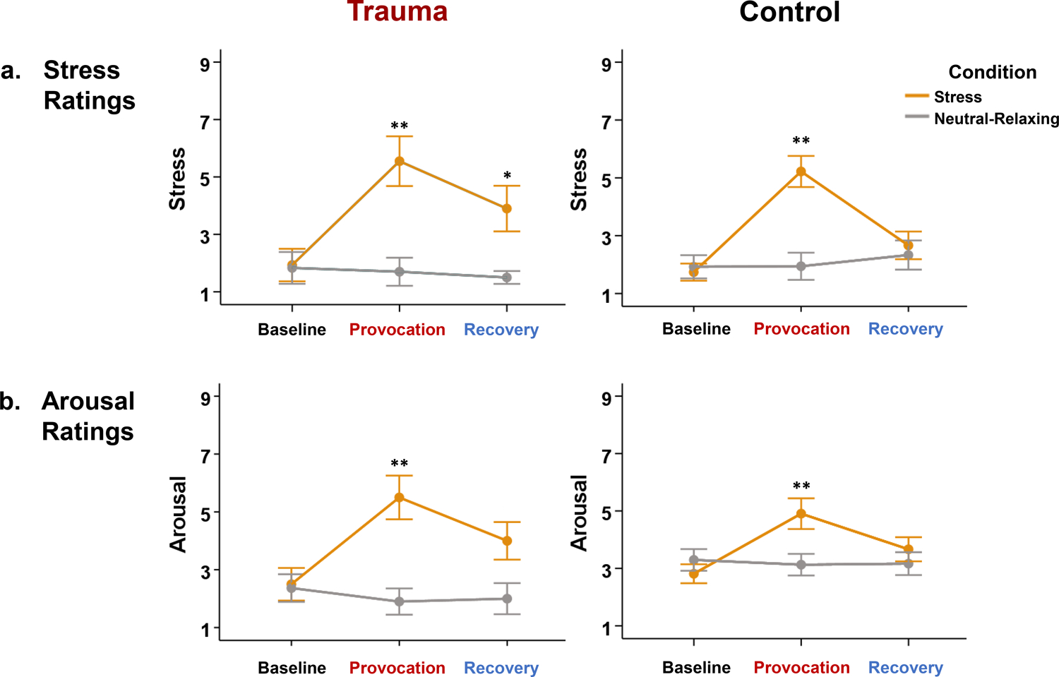
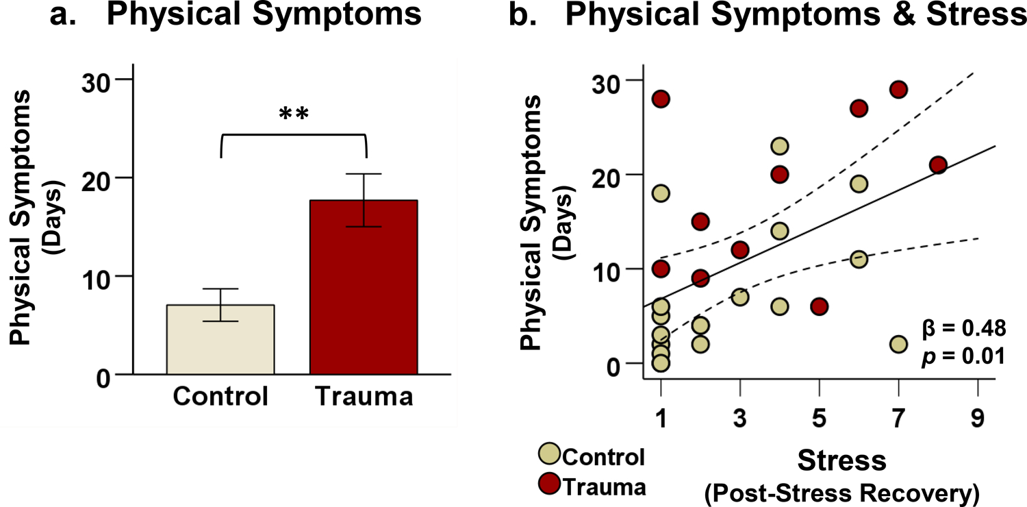
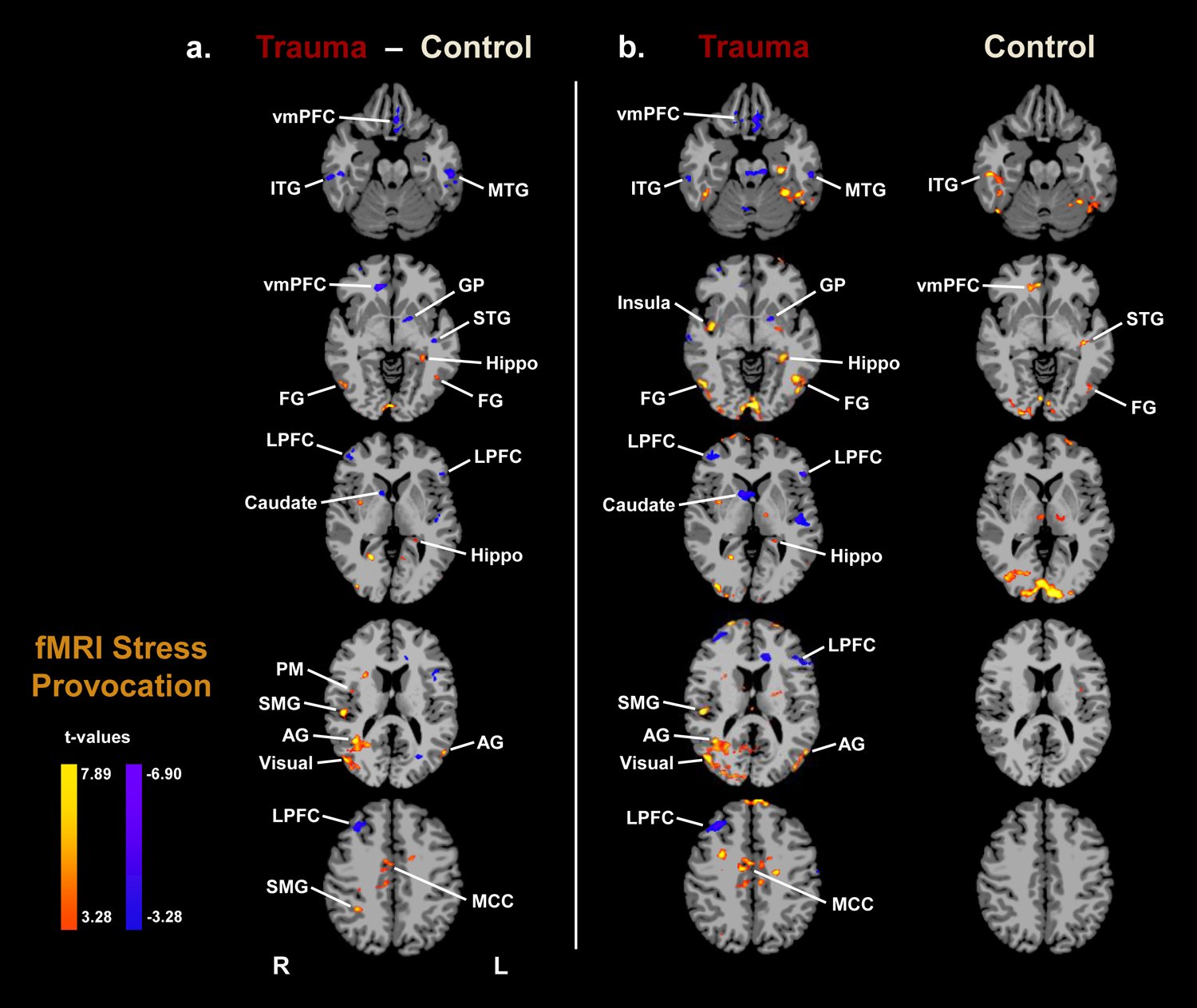
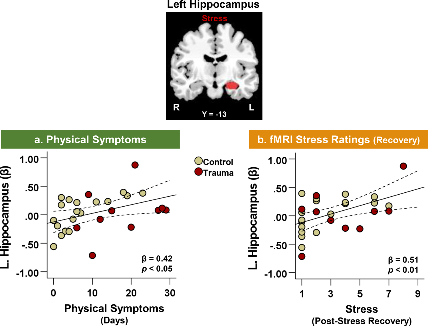
Trauma and chronic pain frequently co-occur, but the underlying neurological mechanisms are poorly understood. The current study investigated the neural correlates of stress and physical symptoms in trauma patients using functional magnetic resonance imaging (fMRI) and follow-up smartphone surveys. Participants were 10 patients diagnosed with Trauma- and Stressor-Related Disorders and 18 demographically-matched healthy controls who completed a fMRI stress provocation task in which they viewed stressful and neutral-relaxing images. Subsequently, participants completed daily smartphone surveys which prospectively monitored their stress and physical symptoms for 30 days. The trauma group experienced a significantly higher frequency of physical symptoms than controls during the follow-up period. During stress, trauma patients exhibited increased activity in the hippocampus, insula, and sensorimotor areas, but decreased activity in the ventromedial prefrontal cortex (vmPFC), lateral prefrontal cortex (LPFC), and dorsal striatum relative to controls. In all participants, higher physical symptom frequency was significantly associated with a hyperactive left hippocampal response to stress. The current study reports that trauma is characterized by greater physical symptoms and decreased prefrontal but increased limbic responses to stress. Our findings suggest that trauma may increase physical health symptoms by compromising hippocampal function, which could also increase vulnerability to stress- and pain-related disorders.

Keywords: Hippocampus; Pain; Physical symptoms; Stress; Trauma; fMRI.


Conflict of interest statement

Declaration of Competing Interest All authors declare no conflicts of interest or financial relationships with commercial interests.

Figures

Figure 1.
Figure 1.. fMRI task design.
The fMRI stress provocation task (adapted from Sinha et al. (2016)) consisted of two blocks representing the stress and neutral-relaxing conditions. Condition order was counterbalanced across participants. Each block consisted of three phases: baseline, provocation, and recovery. During the baseline phase, blank gray images with central fixation cross were displayed for three runs (Runs 1–3). During the provocation phase, participants viewed either stressful or neutral-relaxing images for six runs (Runs 4–9). During each run, 11 images (or gray blanks) were shown in succession for 5 seconds each with a 1-second interstimulus interval (ISI), for a total of 66 seconds (1.1 minutes) per run. During the recovery phase (4 minutes), participants were asked to relax and were not shown any images. Participants were asked to rate their stress and arousal using two-button fMRI response pads. Stress (1 = not at all stressed to 9 = extremely stressed) and arousal (1 = calm/relaxed to 9 = aroused/excited) ratings were collected after each baseline and provocation run and after each recovery phase.
Figure 2.
Figure 2.. fMRI task ratings.
(a) Stress Ratings: Both trauma patients and healthy controls reported significantly higher stress ratings during stress provocation than at baseline (p < 0.01). However, only trauma patients exhibited elevated stress ratings after stress recovery relative to baseline (p < 0.05). (b) Arousal Ratings: Both trauma patients and healthy controls reported significantly higher arousal ratings during stress provocation than at baseline (p < 0.01). Note: *p < 0.05, **p < 0.01. Error bars represent ± 1 standard error (SE) of the mean.
Figure 3.
Figure 3.. Physical symptoms and stress.
After the fMRI scan, participants completed daily smartphone surveys monitoring their stress and physical symptoms over a 30-day follow-up period. (a) Trauma patients experienced a higher frequency of physical symptoms (indexed by the number of days on which at least one physical symptom was reported) compared to healthy controls, t(26) = 3.57, p < 0.01. (b) In all participants, higher fMRI stress ratings after stress recovery were associated with a greater number of days with physical symptoms in the follow-up period (β = 0.48, p = 0.01). Note: **p < 0.01. Error bars represent ± 1 standard error (SE) of the mean.
Figure 4.
Figure 4.. fMRI results.
Whole-brain voxel-based analysis indicated significant group differences in the fMRI BOLD response to stress. (a) Group differences during stress provocation (Trauma − Control): Trauma patients, relative to controls, showed decreased activity in the ventromedial prefrontal cortex (vmPFC), lateral prefrontal cortex (LPFC), temporal lobe (superior, middle, and inferior temporal gyri), and dorsal striatum (caudate and globus pallidus) but increased activity in the left hippocampus, right insula, occipital cortex (visual association areas), parietal lobe (angular and supramarginal gyri), fusiform gyrus, primary motor cortex, and midcingulate cortex (p < 0.001, α < 0.05, whole-brain family-wise error corrected). A blue/purple color denotes decreased brain activity (Trauma < Control), while a yellow/red color denotes increased brain activity (Trauma > Control). (b) The fMRI BOLD response to stress provocation relative to baseline are displayed by group (p < 0.001, α < 0.05, whole-brain family-wise error corrected). Note: R = Right; L = Left; vmPFC = ventromedial prefrontal cortex; LPFC = lateral prefrontal cortex; Hippo = hippocampus; GP = globus pallidus; ITG = inferior temporal gyrus; MTG = middle temporal gyrus; STG = superior temporal gyrus; FG = fusiform gyrus; SMG = supramarginal gyrus; AG = angular gyrus; Visual = visual association areas; PM = primary motor cortex; MCC = midcingulate cortex.
Figure 5.
Figure 5.. Neural correlates of physical symptoms and stress.
Left hippocampal activity during fMRI stress provocation was positively associated with (a) number of days with physical symptoms over the 30-day follow-up period (β = 0.42, p < 0.05) and (b) fMRI stress ratings after stress task recovery (β = 0.51, p < 0.01).

References

    1. Abdallah CG, & Geha P (2017). Chronic Pain and Chronic Stress: Two Sides of the Same Coin? Chronic Stress (Thousand Oaks), 1. 10.1177/2470547017704763 - DOI - PMC - PubMed
    1. Agustin-Pavon C, Braesicke K, Shiba Y, Santangelo AM, Mikheenko Y, Cockroft G, Asma F, Clarke H, Man MS, & Roberts AC (2012). Lesions of ventrolateral prefrontal or anterior orbitofrontal cortex in primates heighten negative emotion. Biol Psychiatry, 72(4), 266–272. 10.1016/j.biopsych.2012.03.007 - DOI - PubMed
    1. Ahmadian AJ, Neylan TC, Metzler T, & Cohen BE (2019). Longitudinal association of PTSD symptoms and self-reported physical functioning among Veterans. J Affect Disord, 250, 1–8. 10.1016/j.jad.2019.02.048 - DOI - PubMed
    1. Alba-Delgado C, Llorca-Torralba M, Horrillo I, Ortega JE, Mico JA, Sanchez-Blazquez P, Meana JJ, & Berrocoso E (2013). Chronic pain leads to concomitant noradrenergic impairment and mood disorders. Biol Psychiatry, 73(1), 54–62. 10.1016/j.biopsych.2012.06.033 - DOI - PubMed
    1. Asmundson GJ, Coons MJ, Taylor S, & Katz J (2002). PTSD and the experience of pain: research and clinical implications of shared vulnerability and mutual maintenance models. Can J Psychiatry, 47(10), 930–937. 10.1177/070674370204701004 - DOI - PubMed

Publication types

MeSH terms