Review
. 2022 Nov 2;110(21):3597-3626.
doi: 10.1016/j.neuron.2022.10.017.

The sympathetic nervous system in the 21st century: Neuroimmune interactions in metabolic homeostasis and obesity

Affiliations
Review

The sympathetic nervous system in the 21st century: Neuroimmune interactions in metabolic homeostasis and obesity

Noelia Martinez-Sanchez et al. Neuron. .

Abstract

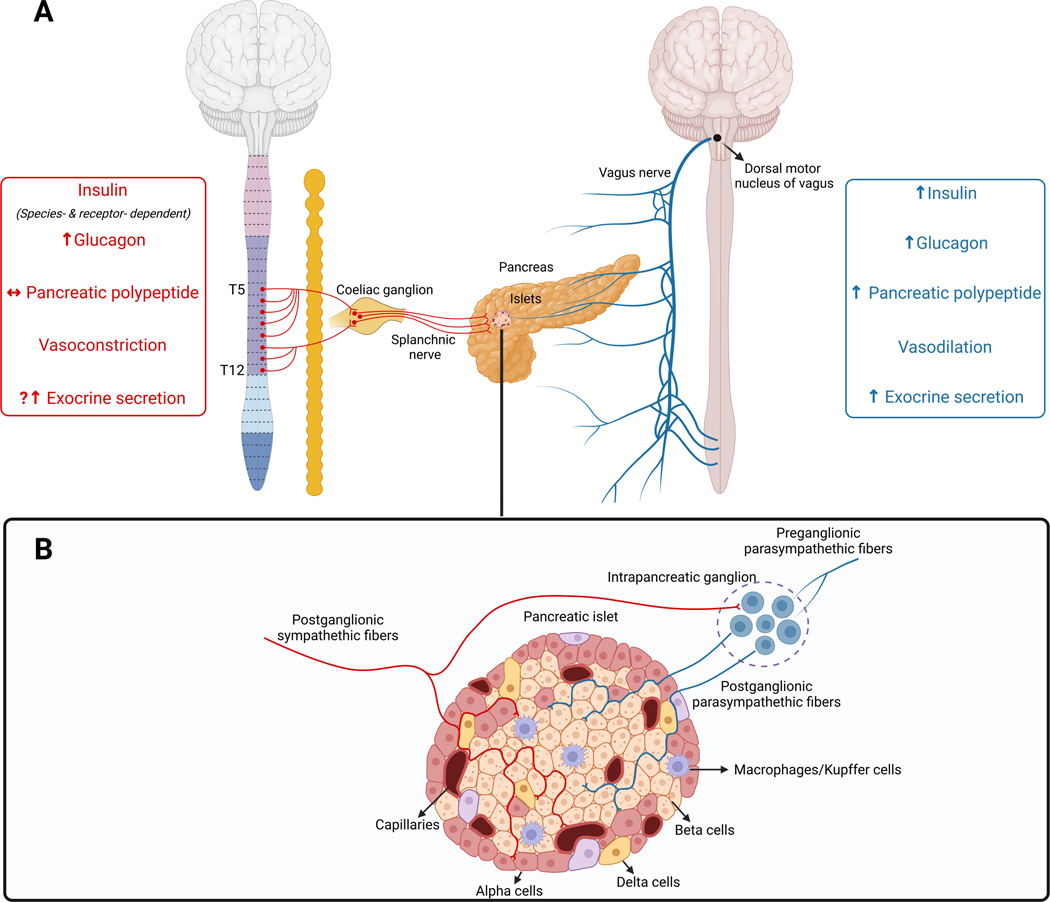
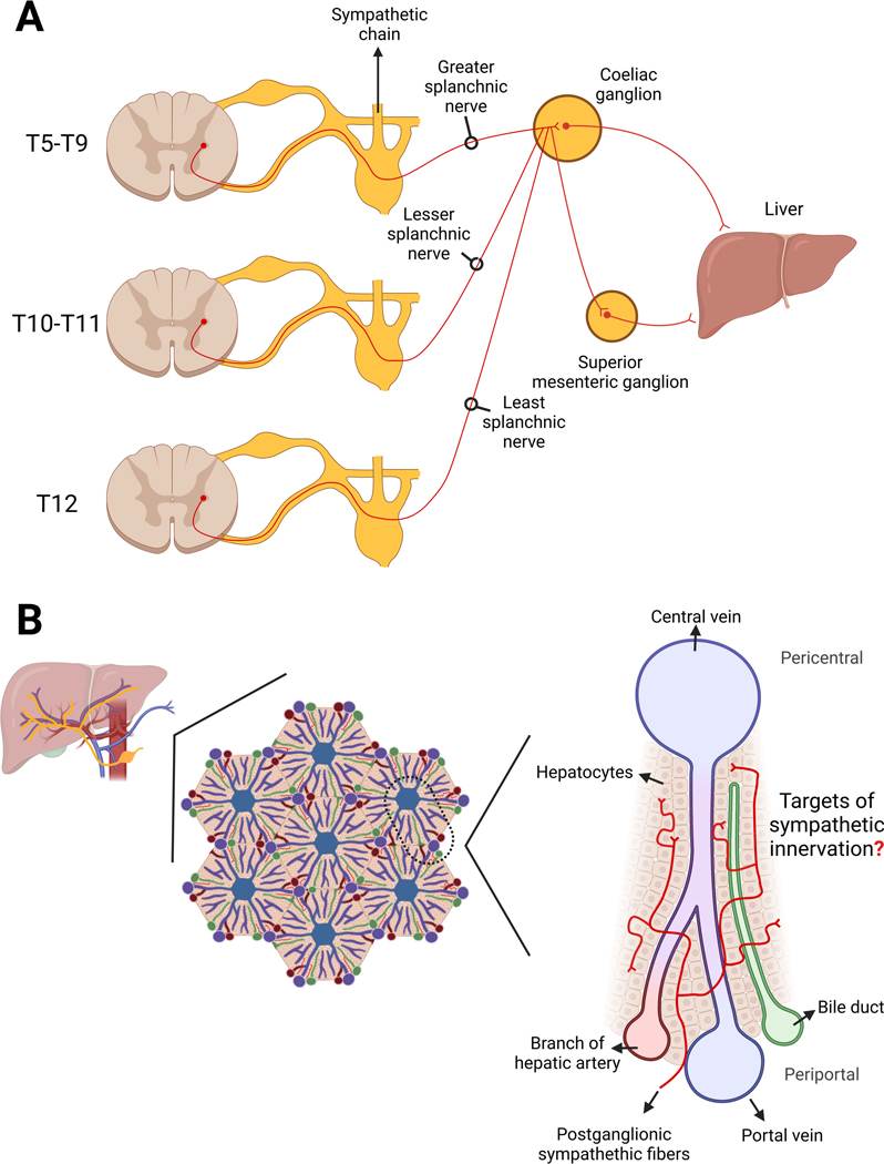
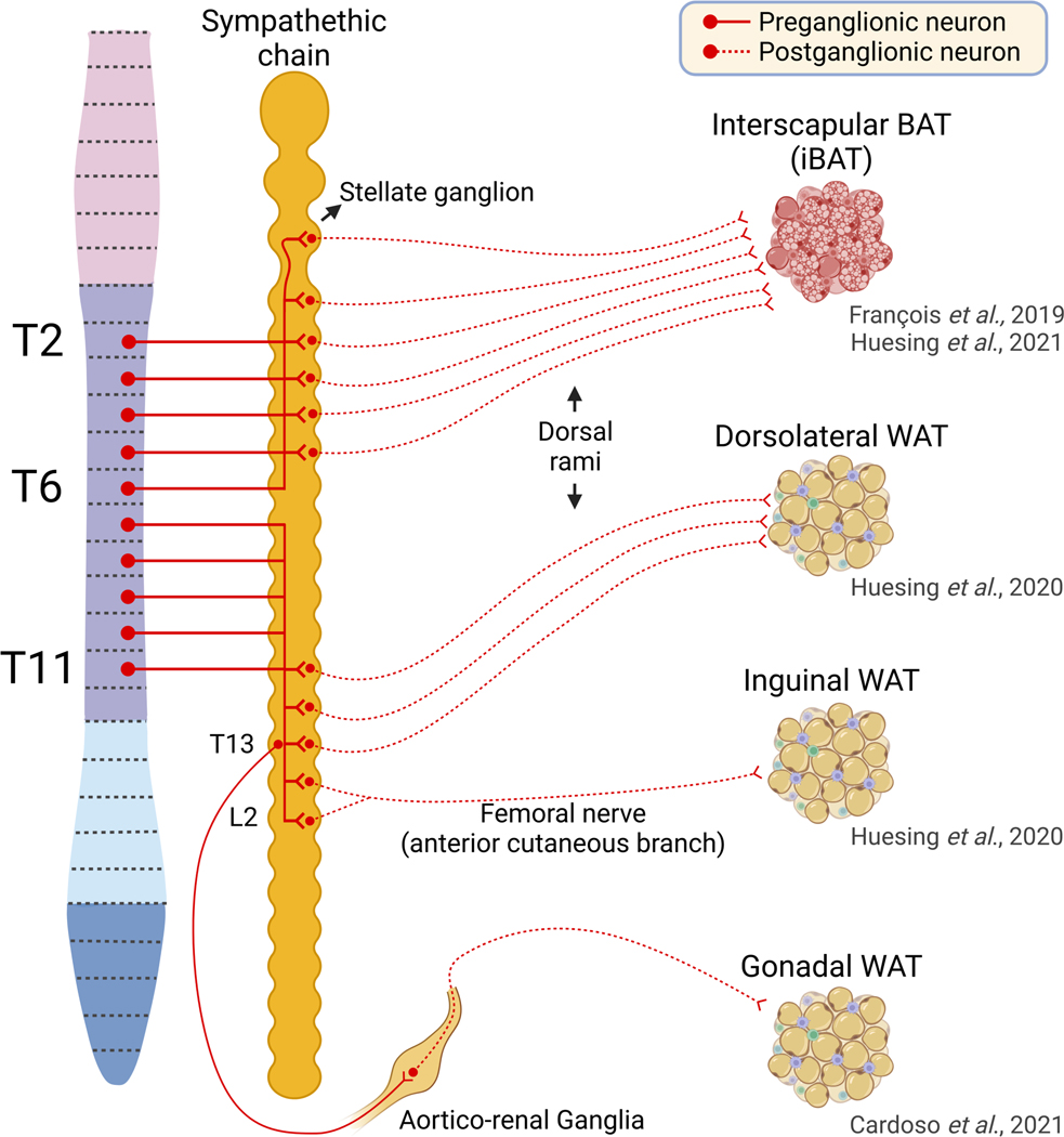
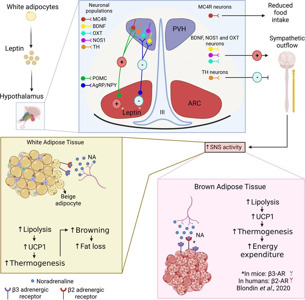
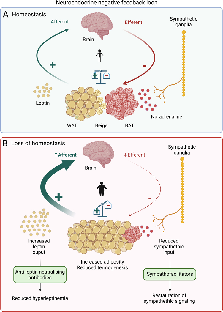
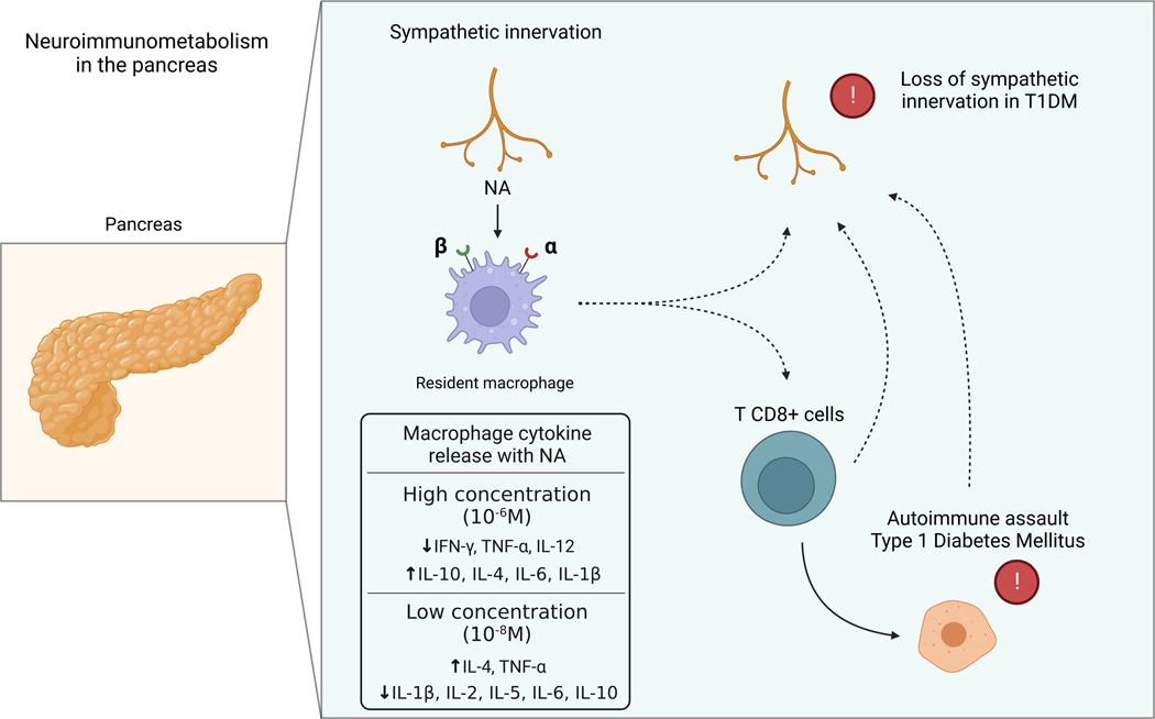
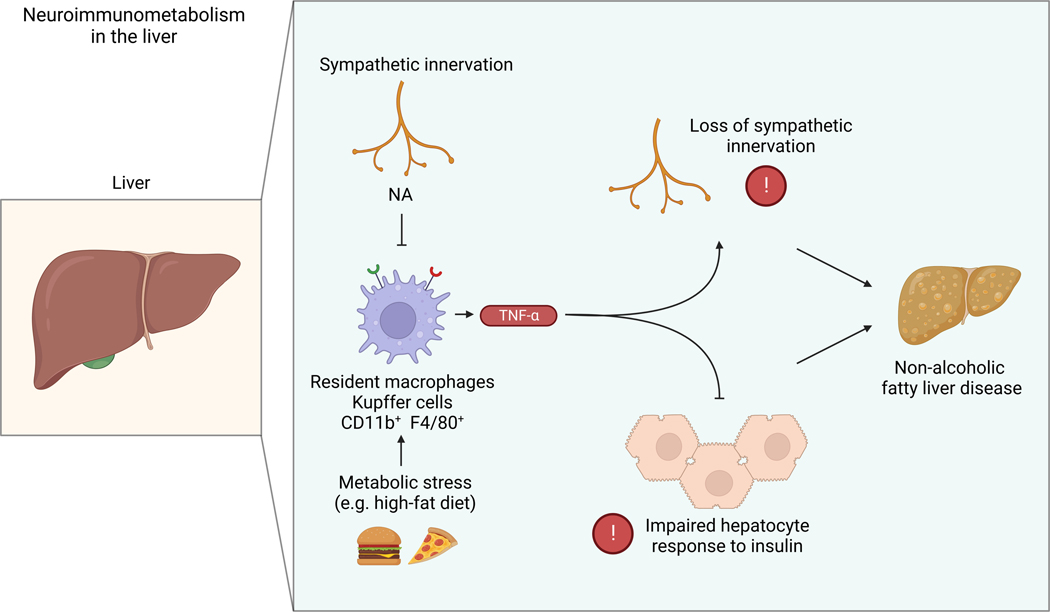
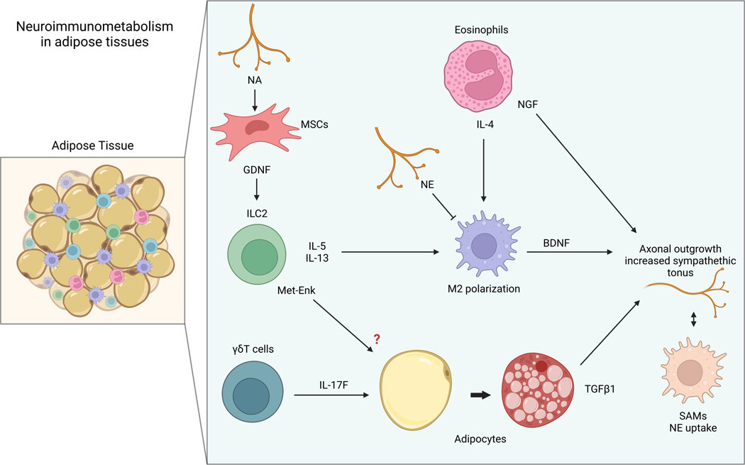
The sympathetic nervous system maintains metabolic homeostasis by orchestrating the activity of organs such as the pancreas, liver, and white and brown adipose tissues. From the first renderings by Thomas Willis to contemporary techniques for visualization, tracing, and functional probing of axonal arborizations within organs, our understanding of the sympathetic nervous system has started to grow beyond classical models. In the present review, we outline the evolution of these findings and provide updated neuroanatomical maps of sympathetic innervation. We offer an autonomic framework for the neuroendocrine loop of leptin action, and we discuss the role of immune cells in regulating sympathetic terminals and metabolism. We highlight potential anti-obesity therapeutic approaches that emerge from the modern appreciation of SNS as a neural network vis a vis the historical fear of sympathomimetic pharmacology, while shifting focus from post- to pre-synaptic targeting. Finally, we critically appraise the field and where it needs to go.

Keywords: adipose tissue; leptin; liver; metabolism; neuroimmunology; pancreas; sympathetic neurons.


Conflict of interest statement

Declaration of interests The authors declare no competing interests.

Figures

Figure 1.
Figure 1.. Efferent pancreatic innervation.
A) The pancreas receives efferent sympathetic and parasympathetic innervation. Preganglionic sympathetic inputs arise from spinal levels T5-T12 projecting to the coeliac ganglia. Preganglionic parasympathetic inputs from the dorsal motor nucleus of the vagus project to the pancreas via the vagus nerve. The main reported results of sympathetic and parasympathetic stimuli are summarized in the left and right boxes. B) Postganglionic sympathetic fibres with en-passant boutons and terminal fibres to innervate pancreatic vasculature, intrapancreatic ganglia, exocrine and endocrine cells. Preganglionic parasympathetic fibres innervate a network of intrapancreatic ganglia. Postganglionic parasympathetic fibres innervate pancreatic vasculature, exocrine and endocrine cells.
Figure 2.
Figure 2.. Sympathetic Innervation of Liver.
A) Postganglionic neurons projecting to the main lobe of the liver were identified in medial ganglia including the celiac-superior mesenteric complex. B) Sympathetic nerve fibres enter the liver along the portal vein. Nerve plexuses were identified in the hepatic triads, particularly around the branches of the hepatic arteries. From these nerve plexuses, branches enter the hepatic lobule in the space of Disse and extend towards the central vein to outline the hepatic sinusoids. These nerve fibres run primarily around the portal vein, hepatic artery and bile duct and it is debated whether they make direct contact with hepatocytes in the peripheral zone of the lobule, parenchymal cells, sinusoidal cells, and Kupffer cells.
Figure 3:
Figure 3:. Neuroanatomy of sympathetic innervation of Adipose tissues.
Retrograde poly-synaptic tracing of sympathetic circuity with pseudorabies virus (PRV) GFP, visualised after clearing of whole murine mediastini of TH-CreR26dtTomato reporter mice showed that interscapular BAT (iBAT) receives preganglionic input originating from spinal levels T2-T6, and post-ganglionic fibres from the stellate ganglia and T1–5 paravertebral ganglia. The continuous white fat pad that extends from the dorsolateral to the ventromedial inguinal region (inguinal WAT) is anatomically divided into ‘dorsolateral’ WAT (dlWAT) and ‘inguinal’ WAT (iWAT) by the superficial ilium circumflex vein. Studies analysing this fat depot using a similar retrograde tracing approach show preganglionic innervation to the iWAT arising from spinal levels T7 to T11. dlWAT receives postganglionic innervation from T11-T13 paravertebral ganglia, whereas iWAT receives innervation possibly from the anterior cutaneous branch of the femoral nerve, originating from L1–2 sympathetic chain neurons that merge onto the lumbar plexus. Retrograde tracing with GFP-labelled adeno-associated virus shows that gonadal WAT (gWAT) receives sympathetic innervation from the aortic renal ganglion at the level of T13, but the spinal level from which these fibres originate is not known. (Solid line represents preganglionic innervation and dashed line represents postganglionic innervation).
Figure 4.
Figure 4.. Pre-Sympathetic brain outputs to White and Brown Adipose Tissues.
Central hypothalamic nuclei detect circulating leptin. Within arcuate (ARC), leptin activates anorexigenic pro-opiomelanocortin (POMC) neurons and inhibits orexigenic neuropeptide Y/agouti-related protein (NPY/AGRP) neurons and this increases sympathetic outflow to adipose tissues. Both populations project to different areas in the hypothalamus, especially to the paraventricular nucleus (PVH) that receives a dense input of these neurons. Melanocortin 4 receptor (MC4R), one of the key players in the regulation of energy expenditure, is densely expressed in the PVH where it has an essential role in regulating food intake (Garfield et al., 2015) but not directly in energy expenditure (Balthasar et al., 2005). However, MC4Rs in other sites rather than in the PVH, like the median preoptic nucleus, dorsomedial or sub zona incerta, seem to mediate melanocortin effects on the energy expenditure (Monge-Roffarello et al., 2014, Vaughan et al., 2011, Voss-Andreae et al., 2007). Non-MC4R-expressing neurons in PVH, like oxytocin (OXT) neurons or nitric oxide synthase-1 (Nos1) expressing neurons, can regulate thermogenesis increasing energy expenditure (Sutton et al., 2014). Projections from ARC NPY neurons to PVH tyrosine hydroxylase (TH)-expressing neurons are important to modulate energy expenditure by reducing sympathetic activity in brown adipose tissue (BAT) (Shi et al., 2013). Through brainderived neurotropic factor-expressing (BDNF) neurons in the PVH, leptin exerts actions in the sympathetic innervation in the adipose tissue (Wang et al., 2020). In addition (and not represented in the figure), LepR+ dorsomedial hypothalamus (DMH) neurons regulate brown adipose tissue (BAT) thermogenesis and browning. Similarly, median preoptic nucleus (MPO) LepR+ and ventromedial nucleus (VMH) LepR+ neurons are also implicated in regulating both energy expenditure and thermogenesis (not represented). In BAT, sympathetic noradrenaline (NA) acts on ß3-adrenoceptors (ß2 in humans) to increase lipolysis and lipid metabolism. The mitochondrial H+ gradient established by substrate oxidation is then dissipated across the inner membrane via UCP-1, causing energy to be dissipated as heat rather than stored chemically. In white adipose tissue (WAT), sympathetic acts on ß3-adrenoceptors to increase lipolysis, liberating FFAs to be metabolised elsewhere. In addition, adrenergic signalling can encourage a phenotype change, causing WAT to adapt for nonshivering thermogenesis, beige adipose tissue (BeAT). Like BAT, BeAT expresses UCP-1 to dissipate electrochemical energy as heat. All three mechanisms reduce fat mass, thus closing a homeostatic feedback loop of leptin action.
Figure 5:
Figure 5:. Working models for reversing loss of homeostasis of the neuroendocrine loop of leptin action.
Leptin is produced by the adipose tissue and exerts its main actions through the brain, mainly in the hypothalamus’ arcuate and indirectly in the paraventricular nuclei. In health, increased leptin results in an increase in sympathetic signalling to the adipose tissues, increasing lipolysis and thus reducing plasma leptin. In chronic metabolic disease, persistently elevated leptin can cause insensitivity to signal, so-called ‘Leptin Resistance, which is concomitant with a decrease in sympathetic tone onto the adipose tissues, ultimately leading to sympathetic neuropathy. Together, the afferent and efferent arms of the metabolic homeostatic feedback loop are impeded, and a state of metabolic dysregulation emerges, causing obesity and other metabolic syndromes. Sympathofacilitators aim to selectively supplement peripheral sympathetic signalling and encourage lipolysis. Hyperleptinemia driving leptin resistance can be reduced with anti-leptin neutralising antibodies, allowing sensitisation to leptin. In tandem, the neuroendocrine negative feedback loop governing metabolism can be restored back to homeostasis.
Figure 6:
Figure 6:. Neuroimmunometabolism in the pancreas.
Noradrenaline (NA) is released by sympathetic nerve terminals within the pancreas. RNA sequencing of isolated islet macrophages incubated with high (10−6 M) or low (10−8 M) concentrations of NA indicates different patterns of cytokine activation. Treatment with high concentrations, assumed to mimic the concentrations experienced near nerve terminals, has anti-inflammatory effects via activation of low affinity β2 adrenergic receptors. High concentrations of NA reduced the expression of INF-γ, TNF-α and IL-12, and increased the expression of IL-10, IL-4, IL-6 and IL-1B. Conversely, treatment with lower concentrations of NA, which bind high affinity alpha adrenoreceptors, increased the expression of IL-4 and TNF-α, and reduced the expression of IL-10, IL- 6, IL-5, IL-2 and IL-1B. Decreased sympathetic activation through pharmacological blockade and denervation prevents infiltration of T CD8+ cells and is associated with the autoimmune assault observed in T1DM, possibly through macrophage signalling. The loss of sympathetic innervation observed in T1DM is associated with altered macrophage signalling, increased T CD8+ infiltration and autoimmune assault, but the precise etiology is unclear. Solid lines represent known interactions and dashed lines represent putative interactions.
Figure 7:
Figure 7:. Neuroimmunometabolism in the liver.
Noradrenaline (NA) is released by sympathetic nerve terminals within the liver. Under metabolic stress (e.g. consumption of a high-fat diet), impaired sympathetic activity is associated with increased production of TNF-α by macrophages/Kupffer cells, which is followed by impaired hepatocyte response to insulin and ultimately sympathetic neuropathy. These responses have been widely associated with the development of non-alcoholic fatty liver disease (NAFLD). Administration of anti-TNF-α neutralizing antibodies was sufficient to completely reverse the loss of sympathetic axons, highlighting the interplay between sympathetic signalling and inflammatory signalling in metabolic liver disease.
Figure 8:
Figure 8:. Neuroimmunometabolism in adipose tissue.
Noradrenaline (NA) is released by sympathetic nerve terminals within adipose depots. Adipose mesenchymal stromal cells (MSCs) neighbouring these terminals are activated via the β2 adrenergic receptor to produce glial-derived neurotrophic factor (GDNF), which controls the activity of ILC2s in visceral fat. In turn, ILC2s produce type 2 innate cytokines and Met-enkephalin (Met-Enk), whose direct role in browning is debated. Type 2 innate cytokines act on macrophages to induce M2 polarization, which is also induced by eosinophil-derived IL-4 and inhibited by direct NA signalling to macrophages. M2 polarization is associated with BDNF secretion by macrophages, which leads to axonal outgrowth and increased sympathetic tone. Eosinophils producing nerve growth factor (NGF) can also stimulate sympathetic axonal outgrowth. T cells stimulate beiging via IL-17F signalling to adipocytes, which produce transforming growth-factor beta-1 (TGF) to stimulate axonal outgrowth. Sympathetic neuron-associated macrophages (SAMs) contribute to obesity and negatively control sympathetic tone by importing and metabolizing NA.

References

    1. ADORI C, DARAIO T, KUIPER R, BARDE S, HORVATHOVA L, YOSHITAKE T, IHNATKO R, VALLADOLID-ACEBES I, VERCRUYSSE P, WELLENDORF AM, GRAMIGNOLI R, BOZOKY B, KEHR J, THEODORSSON E, CANCELAS JA, MRAVEC B, JORNS C, ELLIS E, MULDER J, UHLEN M, BARK C. & HOKFELT T. 2021. Disorganization and degeneration of liver sympathetic innervations in nonalcoholic fatty liver disease revealed by 3D imaging. Sci Adv, 7. - PMC - PubMed
    1. AHREN B, ERICSON LE, LUNDQUIST I, LOREN I. & SUNDLER F. 1981. Adrenergic innervation of pancreatic islets and modulation of insulin secretion by the sympatho-adrenal system. Cell Tissue Res, 216, 15–30. - PubMed
    1. AHREN B. & TABORSKY GJ JR. 1986. The mechanism of vagal nerve stimulation of glucagon and insulin secretion in the dog. Endocrinology, 118, 1551–7. - PubMed
    1. AHREN B, VEITH RC & TABORSKY GJ JR. 1987. Sympathetic nerve stimulation versus pancreatic norepinephrine infusion in the dog: 1). Effects on basal release of insulin and glucagon. Endocrinology, 121, 323–31. - PubMed
    1. ALMACA J, WEITZ J, RODRIGUEZ-DIAZ R, PEREIRA E. & CAICEDO A. 2018. The Pericyte of the Pancreatic Islet Regulates Capillary Diameter and Local Blood Flow. Cell Metab, 27, 630–644 e4. - PMC - PubMed

Publication types

LinkOut - more resources