. 2022 Dec;298(12):102660.
doi: 10.1016/j.jbc.2022.102660. Epub 2022 Oct 31.

A noncanonical RNA-binding domain of the fragile X protein, FMRP, elicits translational repression independent of mRNA G-quadruplexes

Affiliations

A noncanonical RNA-binding domain of the fragile X protein, FMRP, elicits translational repression independent of mRNA G-quadruplexes

MaKenzie R Scarpitti et al. J Biol Chem. 2022 Dec.

Abstract

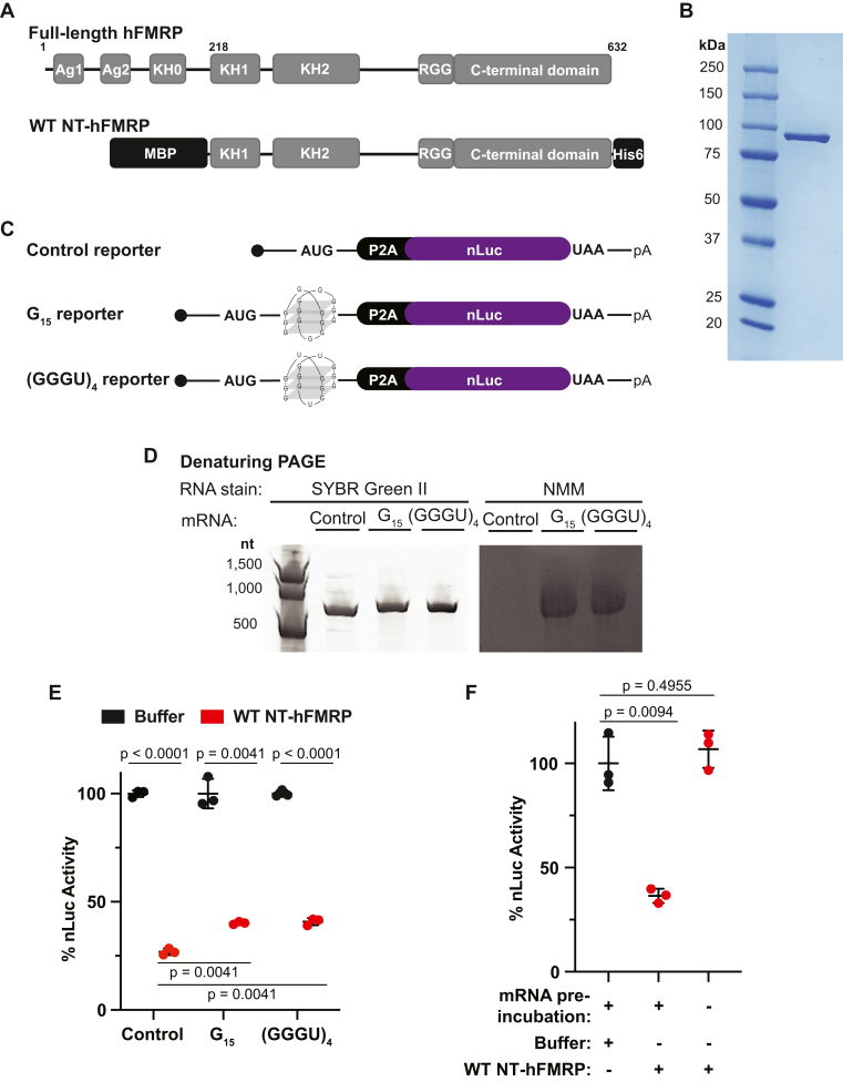
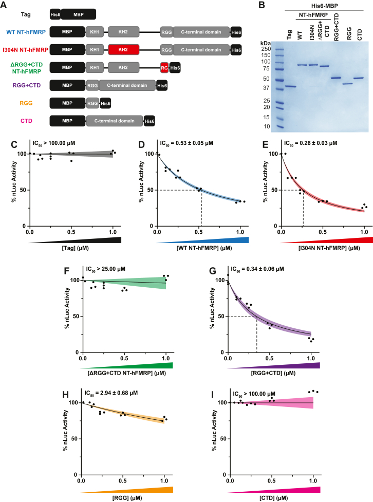
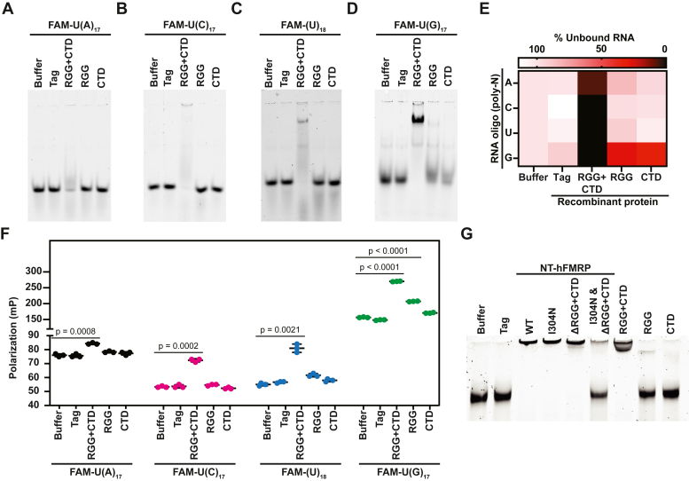
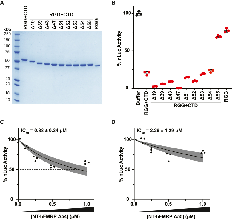
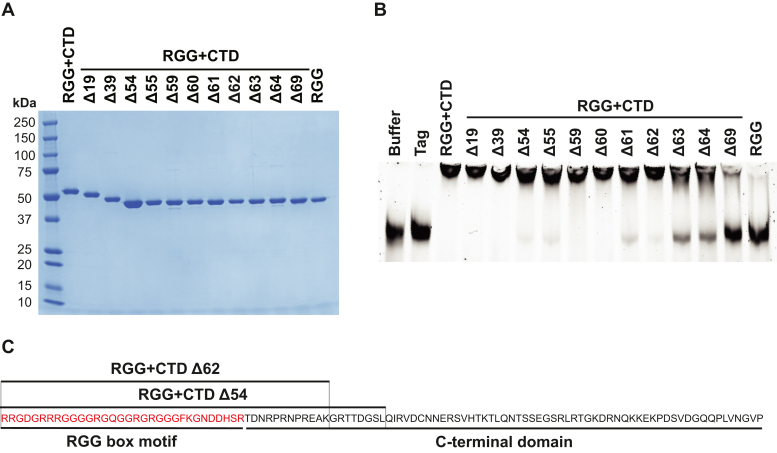
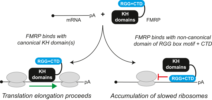
Loss of functional fragile X mental retardation protein (FMRP) causes fragile X syndrome, the leading form of inherited intellectual disability and the most common monogenic cause of autism spectrum disorders. FMRP is an RNA-binding protein that controls neuronal mRNA localization and translation. FMRP is thought to inhibit translation elongation after being recruited to target transcripts via binding RNA G-quadruplexes (G4s) within the coding sequence. Here, we directly test this model and report that FMRP inhibits translation independent of mRNA G4s. Furthermore, we found that the RGG box motif together with its natural C-terminal domain forms a noncanonical RNA-binding domain (ncRBD) that is essential for translational repression. The ncRBD elicits broad RNA-binding ability and binds to multiple reporter mRNAs and all four homopolymeric RNAs. Serial deletion analysis of the ncRBD identified that the regions required for mRNA binding and translational repression overlap but are not identical. Consistent with FMRP stalling elongating ribosomes and causing the accumulation of slowed 80S ribosomes, transcripts bound by FMRP via the ncRBD cosediment with heavier polysomes and were present in puromycin-resistant ribosome complexes. Together, this work identifies a ncRBD and translational repression domain that shifts our understanding of how FMRP inhibits translation independent of mRNA G4s.

Keywords: RNA-binding protein; mRNA; protein synthesis; ribosome; translation control.


Conflict of interest statement

Conflict of interest The authors declare that they have no conflicts of interest with the contents of this article.

Figures

Figure 1
Figure 1
FMRP inhibits translation independent of mRNA G-quadruplexes in the CDS.A, schematic of full-length (residues 1–632) and MBP- and His6-tagged WT N-terminally truncated human FMRP isoform 1 (NT-hFMRP). The Agenet 1 (Ag1), Agenet 2 (Ag2), and KH0 domains are absent in WT NT-hFRMP. Ag1 and Ag2 are also referred to as Tudor domains in some previous literature. WT NT-hFRMP harbors residues 218 to 632 of full-length human FMRP isoform 1. B, Coomassie stain of recombinant WT NT-hFMRP. C, schematic of custom nLuc reporters either lacking a G4 (control reporter) or harboring a G4 in the coding sequence (G15 and (GGGU)4 reporters). A P2A ribosome skipping motif was included immediately upstream of the nLuc coding sequence to ensure equal nLuc function between reporters. D, denaturing PAGE of control, G15, and (GGGU)4 reporters stained for total RNA with SYBR Green II or for G4 structures with NMM. E, in vitro translation of control, G15, and (GGGU)4 reporter mRNA preincubated with protein buffer or 1 μM WT NT-hFMRP. Data are shown as mean ± SD. n = 3 biological replicates. Comparisons were made using a two-tailed unpaired t test with Welch’s correction. F, in vitro translation of control nLuc reporter mRNA with protein storage buffer as a negative control, with 1 μM WT NT-hFMRP and nLuc mRNA preincubated together and with 1 μM WT NT-hFMRP without a preincubation step. Data are shown as mean ± SD. n = 3 biological replicates. Comparisons were made using a two-tailed unpaired t test with Welch’s correction. CDS, coding sequence; KH, K homology.
Figure 2
Figure 2
The RGG box motif and CTD together are essential and sufficient to inhibit translation.A, schematic of recombinant WT and mutant NT-hFMRP. Mutated/truncated domains are highlighted in red. B, Coomassie stain of recombinant proteins. CI, in vitro translation of nLuc mRNA with a titration of recombinant WT and mutant NT-hFMRP isoforms. IC50 values were determined for the His6-MBP tag (negative control; 633.50 ± infinity μM) (C), WT NT-hFMRP (D), I304N KH2 domain patient-derived mutant NT-hFMRP (E), ΔRGG + CTD mutant NT-hFMRP (25.72 ± infinity μM) (F), the RGG box motif + CTD fusion (G), the RGG box motif alone (H), and the CTD alone (5.75 × 1033 ± infinity μM) (I). n = 3 biological replicates. A nonlinear regression was used to calculate the IC50 and is shown as the line with the 95% confidence interval (CI) included as a watermark. The IC50 is reported ± 95% CI. CTD, C-terminal domain.
Figure 3
Figure 3
The RGG + CTD noncanonical RBD binds all four homopolymeric RNAs and mRNA. AD, EMSAs of 5ʹ FAM-labeled homopolymeric RNA oligonucleotides with the indicated recombinant proteins. Due to the suspected net neutral charge of the RNA oligo–FMRP complex causing the RNP to not enter the gel, the intensity of unbound RNA was quantified and set relative to the protein storage buffer only sample (E). F, fluorescence polarization of 5ʹ FAM-labeled homopolymeric RNA with the indicated recombinant protein. n = 3 biological replicates. Data are shown as mean ± SD. n = 3 biological replicates. Comparisons were made using a two-tailed unpaired t test with Welch’s correction (p-values for each comparison are listed in Table S2). G, EMSA of control reporter mRNA incubated with the indicated recombinant protein and stained with SYBR Green II. CTD, C-terminal domain; EMSA, electrophoretic mobility shift assay; RBD, RNA-binding domain.
Figure 4
Figure 4
The RGG + CTD Δ54 is required for translational repression.A, Coomassie stain of recombinant proteins with serial truncations to the CTD. The number of amino acids truncated from the CTD is indicated. B, in vitro translation of nLuc mRNA with 1 μM CTD truncated recombinant RGG + CTD proteins and protein storage buffer as a negative control. Data are shown as mean ± SD. n = 3 biological replicates. All recombinant protein tested was statistically significant compared to buffer negative control using a two-tailed unpaired t test with Welch’s correction (each comparison had p < 0.01). C and D, in vitro translation of nLuc mRNA with a titration of recombinant NT-hFMRP Δ54 (C) and NT-hFMRP Δ55 (D). n = 3 biological replicates. A nonlinear regression was used to calculate the IC50 value for each truncation and is shown as the line with the 95% confidence interval (CI) included as a watermark. The IC50 is reported ± 95% CI. CTD, C-terminal domain.
Figure 5
Figure 5
The RGG + CTD Δ62 is the key region of the ncRBD required for robust mRNA binding.A, Coomassie stain of recombinant proteins with C-terminal truncations to the ncRBD (RGG + CTD). The number of amino acids truncated from the CTD is indicated. B, EMSA of control reporter mRNA incubated with recombinant protein and stained with SYBR Green II. C, amino acid composition of overlapping regions of the ncRBD that are required for mRNA binding (RGG + CTD Δ62) and translational repression (RGG + CTD Δ54). CTD, C-terminal domain, ncRBD, noncanonical RNA-binding domain.
Figure 6
Figure 6
Human FMRP inhibits translation post-initiation when binding mRNA via the ncRBD.A, in vitro translation of the control reporter mRNA that harbors the 50 nt human β-globin 5ʹ UTR and a long 5ʹ UTR reporter mRNA that harbors three β-globin 5ʹ UTRs in tandem (150 nt total). mRNPs were formed with protein storage buffer or 1 μM WT NT-hFMRP. Data are shown as mean ± SD. n = 3 biological replicates. Comparisons were made using a two-tailed unpaired t test with Welch’s correction. B, distribution of nLuc reporter mRNA across sucrose gradients to assess polysome formation when preincubated with 1 μM ΔRGG + CTD (Control) or 1 μM WT NT-hFMRP. Abundance of reporter mRNA in each gradient fraction was determined by RT-qPCR. C, cumulative nLuc abundance in heavy polysomes in fractions 10 to 12 from 1 μM ΔRGG + CTD (Control) and 1 μM WT NT-hFMRP samples. D, fold change of nLuc mRNA abundance in heavy polysomes. Data are shown as mean ± SD. n = 3 biological replicates. Comparisons were made using a two-tailed unpaired t test with Welch’s correction. E, relative quantification of nLuc reporter mRNA pelleted through a 35% (w/v) sucrose cushion after a low-speed centrifugation (see Fig. S10). Lane 1 is a negative control lacking nLuc mRNA. Lane 2 is a negative control containing mRNA in RRL but not incubated at 30 °C to start translation. Lane 3 is nLuc mRNA in RRL translated for 15 min at 30 °C. Lane 4 is nLuc mRNA in RRL translated for 15 min at 30 °C and then incubated with 0.1 mM puromycin (final) for 30 min at 30 °C. Lanes 5 and 6 are negative control untranslated reactions of nLuc•FMRP mRNPs formed with 1 μM ΔRGG + CTD (Control) and 1 μM WT NT-hFMRP, respectively, in RRL kept on ice demonstrating poor pelleting without active translation through the low-speed sucrose cushion as described in the Experimental procedures and outlined in Fig. S10). Data are shown as mean ± SD. n = 3 biological replicates. Comparisons were made using a two-tailed unpaired t test with Welch’s correction. F, relative quantification of nLuc reporter mRNA pelleted through a 35% (w/v) sucrose cushion after a low-speed centrifugation. nLuc•ΔRGG + CTD (Control) and nLuc•WT NT-hFMRP mRNPs were translated and treated with 0.1 mM puromycin (final) before being overlayed on the cushion and low-speed centrifugation. Final concentration of recombinant protein was 1 μM. Data are shown as mean ± SD. n = 3 biological replicates. Comparisons were made using a two-tailed unpaired t test with Welch’s correction. CTD, C-terminal domain; ncRBD, noncanonical RNA-binding domain; RRL, rabbit reticulocyte lysate; RT-qPCR, reverse transcription quantitative PCR.
Figure 7
Figure 7
Model of FMRP-mediated translational repression via a ncRBD. FMRP can bind mRNA through canonical and noncanonical RBDs. When bound to mRNA through its canonical KH domain(s), FMRP does not robustly inhibit translation. The RGG box motif and CTD of FMRP together form a ncRBD that allows FMRP to bind multiple mRNAs and subsequently inhibit translation to cause accumulation of slowed/stalled ribosomes. CTD, C-terminal domain; KH, K homology; ncRBD, noncanonical RNA-binding domain.

References

    1. Kremer E.J., Pritchard M., Lynch M., Yu S., Holman K., Baker E., et al. Mapping of DNA instability at the fragile X to a trinucleotide repeat sequence p(CCG)n. Science. 1991;252:1711–1714. - PubMed
    1. Oberle I., Rousseau F., Heitz D., Kretz C., Devys D., Hanauer A., et al. Instability of a 550-base pair DNA segment and abnormal methylation in fragile X syndrome. Science. 1991;252:1097–1102. - PubMed
    1. Verkerk A.J., Pieretti M., Sutcliffe J.S., Fu Y.H., Kuhl D.P., Pizzuti A., et al. Identification of a gene (FMR-1) containing a CGG repeat coincident with a breakpoint cluster region exhibiting length variation in fragile X syndrome. Cell. 1991;65:905–914. - PubMed
    1. Yu S., Pritchard M., Kremer E., Lynch M., Nancarrow J., Baker E., et al. Fragile X genotype characterized by an unstable region of DNA. Science. 1991;252:1179–1181. - PubMed
    1. Niu M., Han Y., Dy A.B.C., Du J., Jin H., Qin J., et al. Autism symptoms in fragile X syndrome. J. Child Neurol. 2017;32:903–909. - PubMed

Publication types

MeSH terms

Substances

LinkOut - more resources