. 2022 Nov 3;13(1):6592.
doi: 10.1038/s41467-022-34469-1.

JAK2V617F mutation drives vascular resident macrophages toward a pathogenic phenotype and promotes dissecting aortic aneurysm

Affiliations

JAK2V617F mutation drives vascular resident macrophages toward a pathogenic phenotype and promotes dissecting aortic aneurysm

Rida Al-Rifai et al. Nat Commun. .

Erratum in

Abstract

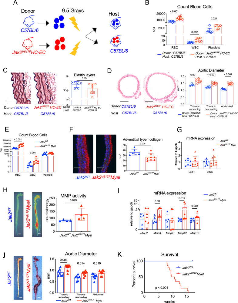
JAK2V617F mutation is associated with an increased risk for athero-thrombotic cardiovascular disease, but its role in aortic disease development and complications remains unknown. In a cohort of patients with myeloproliferative neoplasm, JAK2V617F mutation was identified as an independent risk factor for dilation of both the ascending and descending thoracic aorta. Using single-cell RNA-seq, complementary genetically-modified mouse models, as well as pharmacological approaches, we found that JAK2V617F mutation was associated with a pathogenic pro-inflammatory phenotype of perivascular tissue-resident macrophages, which promoted deleterious aortic wall remodeling at early stages, and dissecting aneurysm through the recruitment of circulating monocytes at later stages. Finally, genetic manipulation of tissue-resident macrophages, or treatment with a Jak2 inhibitor, ruxolitinib, mitigated aortic wall inflammation and reduced aortic dilation and rupture. Overall, JAK2V617F mutation drives vascular resident macrophages toward a pathogenic phenotype and promotes dissecting aortic aneurysm.


Conflict of interest statement

The authors declare no competing interests

Figures

Fig. 1
Fig. 1. JAK2V167F mutation leads to aortopathy in both human and mice.
A Adjusted risk factor for aorta dilation in a cohort of JAK2V167F + patients (n = 157) and age-gender-matched controls (N = 157), scale bar 2 cm. B CT-scan 3D-reconstruction of JAK2V167F + patient (right) and control (left) showing ascending and descending thoracic aorta. C Paw pictures of 7-week-old Jak2WTcontrol and Jak2V617F HC-EC mice showing palmar erythema in mutant mice, scale bar 1 cm. D Quantitative analysis and representative photomicrographs of spleen size in 7-week-old control Jak2WT(N = 11) and Jak2V617F HC-EC mice (N = 11) (scale bar 1 cm). E red blood cell, platelet, and myeloid cell count of 7-week-old control Jak2WT(N = 5) and Jak2V617F HC-EC mice (N = 9). F Representative photomicrographs and quantitative analysis of the mean aortic diameter in 7-week-old control Jak2WT(N = 11) and Jak2V617F HC-EC mice (N = 7) in the thoracic (ascending and descending) and the abdominal aorta (scale bar 1 mm). G Quantification of the number of elastin layers in the aortic wall by orcein staining in 7-week-old animals (N = 10–12/group), scale bar 50 μm. H Quantification of the collagen content (Cola1 immunostaining) in the aortic wall in 7-week-old animals (N = 9/group), scale bar 50 μm. I Fluorescent Molecular Tomography quantification of matrix metalloproteinase (MMP-2, −3, −9 and −13) activity in aorta of 10-week-old animals (N = 4/group), scale bar 1 mm. J Survival curve of control Jak2WT (N = 15) and Jak2V617F HC-EC mice (N = 25). K Representative photomicrographs of descending thoracic aorta after Orcein staining showing dissecting aortic aneurysm in Jak2V617F HC-EC mouse (representative of 10 aortas/group), scale bar 100 µm. Data are presented as mean values ± SD. [Two-tailed Mann-Whitney test, *P < 0.05, **P < 0.01, ***P < 0.001]. Difference in survival was evaluated using log-rank test. CT computed tomography, HC-EC hematopoietic cells-endothelial cells, MMP matrix metalloprotease. Source data are provided as a Source Data file.
Fig. 2
Fig. 2. JAK2V167F mutation in hematopoietic cells promoted dissecting aortopathy.
A Experimental protocol of WT or Jak2V617F HC-EC bone marrow cell transplantation in lethally irradiated C57BL/6 mice (7/Group). B Red blood cell, white blood cell and platelet count of 20-week-old chimeric mice (N = 7/group). C Quantification of the number of elastin layers in the aortic wall by Orcein staining at sacrifice (N = 7/group), scale bar 50 μm. D Representative photomicrographs and quantitative analysis of the mean aortic diameter in WT or Jak2V617F HC-EC chimeric mice (N = 7/group) in the thoracic (ascending and descending) and the abdominal aorta, scale bar 0.5 mm. E Red blood cell, white blood cell and platelet count of 7-week-old Jak2WT and Jak2V617F Myel mice (N = 7/group). F Quantification of the collagen content (Cola1 immunostaining) in the aortic wall in 7-week-old Jak2WT(N = 6) and Jak2V617F Myel animals (N = 8), scale bar 50 μm. G Quantification of Cola1 and Cola3 mRNA expression in aorta of 7-week-old Jak2WT (N = 7) and Jak2V617F Myel mice (N = 8). H Fluorescent Molecular Tomography quantification of matrix metalloproteinase (MMP-2, −3, −9, and −13) activity in aorta of 9-week old Jak2WT and Jak2V617F Myel mice (N = 4/group), scale bar 1 mm. I, quantification of Mmp2, Mmp3, Mmp9, Mmp12, Mmp13 mRNA expression in aorta of 7-week-old Jak2WT (N = 5) and Jak2V617F Myel mice (N = 7). J Representative photomicrographs and quantitative analysis of the mean aortic diameter in 7-week-old Jak2WT (N = 7) and Jak2V617F Myel (N = 8) mice in the thoracic (ascending and descending) and the abdominal aorta, scale bar 1 mm. K Survival curve of Jak2WT (N = 11) and Jak2V617F Myel mice (N = 14). Data are presented as mean values ± SD. [Two-tailed Mann-Whitney test, *P < 0.05, **P < 0.01, ***P < 0.001]. Difference in survival was evaluated using log-rank test. HC-EC, hematopoietic cells-endothelial cells; MMP, matrix metalloprotease. Source data are provided as a Source Data file.
Fig. 3
Fig. 3. JAK2V167F mutation induces pro-inflammatory signature in the aortic wall.
A Difference in gene expression in the aorta of 7-week-old Jak2WT (N = 4) and Jak2V617F HC-EC mice (N = 3). B Top Gene Ontology terms mostly enriched among genes upregulated in the aorta of Jak2V617F HC-EC mice when compared to Jak2WT mice. C Flow cytometry quantification of CD45 + CD11clowCMHIIlowCD11b + Ly6G-F4/80lowCD64lowLy6C + monocytes and CD45 + CD11clowCMHIIlowCD11b + Ly6G+ neutrophils in the aorta of 7-week-old Jak2WT (N = 10) and Jak2V617F HC-EC animals (N = 8). D Flow quantification and representative dot plot of CD45 + CD11clowCMHIIlowCD11b + Ly6G-F4/80highCD64high macrophage count in the aorta of 7-week-old Jak2WT (N = 10) and Jak2V617F HC-EC animals (N = 8). E Quantification and representative photomicrographs of CD68 positive macrophages in the aorta of 7-week-old mice (N = 11 in control group and N = 8 in Jak2V617F HC-EC group), scale bar 50 µm. F Quantification of Il6, Tnfα, Il1β, and Il10 mRNA expression by RT-qPCR in the aorta of 7-week-old Jak2WT(N = 8) and Jak2V617F HC-EC animals (N = 9). G Immunofluorescence staining in the aortic wall of Jak2WT and Jak2V617F HC-EC mice, DAPI (Blue), CD68 (Red), LYVE-1 (Green), scale bar 50 μm. resident macrophages were defined as CD68 + LYVE-1+ cells (N = 5/group). H GSEA for genes related to a gene set signature for tissue resident macrophage in the aorta of 7-week-old Jak2WT and Jak2V617F HC-EC animals (N = 4/group). Data are presented as mean values ± SD. [Two-tailed Mann-Whitney test, *P < 0.05, **P < 0.01, ***P < 0.001]. HC-EC, hematopoietic cells-endothelial cells; Il, interleukin; Tnf, tumor necrosis factor; GSEA, gene set enrichment analysis. Source data are provided as a Source Data file.
Fig. 4
Fig. 4. Jak2V617FMyel aortas harbor pro-inflammatory Lyve1 + tissue resident macrophages.
A Experimental design, B UMAP visualization of scRNA-seq profiles of CD45 + cells extracted from Jak2WT and Jak2V617F Myel aortas. C Single-cells annotated as macrophages on the UMAP displayed in B were in silico extracted, reanalyzed with dimensional reduction (UMAP) and clustered identifying 7 major populations (Annotated A to G). D Expression intensity of transcripts (top) and cell surface markers (bottom) characteristic of tissue resident macrophages (Lyve1, Mrc1, TIMD4, CD169) and recruited monocytes/macrophages (Ccr2, CD14) projected on the macrophage UMAP plot. E Expression of selected marker transcripts in the 7 macrophage populations. F Macrophage UMAP plot split according to experimental condition (color code as in B). G Proportions of macrophage populations among total aortic macrophages. H Proportions of tissue resident macrophage subsets among total aortic tissue resident macrophages. I Normalized expression of the indicated transcripts in tissue resident macrophage subpopulations. J Average expression of the indicated cell surface markers (measured by CITE-seq) in tissue resident macrophage subpopulations. UMAP, Uniform Manifold Approximation and Projection for Dimension Reduction, sc single cell, CITE-seq Cellular Indexing of Transcriptomes and Epitopes by Sequencing.
Fig. 5
Fig. 5. JAK2 mutation in vascular tissue resident macrophage cells promotes dissecting aortopathy.
A Left, cells corresponding to monocytes/macrophages/dendritic cells were extracted from murine healthy and atherosclerotic plaques and separately reanalyzed with dimensional reduction (UMAP). Right, expression of Pf4 transcript projected on the vascular monocytes/macrophages/dendritic cells UMAP plot. B Left, cells corresponding to macrophages were extracted from murine aorta and separately reanalyzed with dimensional reduction (UMAP). Right, expression of Pf4 transcript projected on the vascular tissue resident macrophage subsets UMAP plot. C Jak2V617F WT/Flex mice were backcrossed with Pf4 Cre ± mice to generate control Pf4 Cre- Jak2WT and Pf4 Cre + Jak2V617F (Called Jak2V617F Pf4) mice. D Red blood cell, white blood cell and platelet count of 7-week-old Jak2WT and Jak2V617F Pf4 mice (N = 5/group). E, F representative photomicrographs and quantitative analysis of the mean aortic diameter in control 7-week-old Jak2WT (N = 7) and Jak2V617F Pf4 mice (N = 8) in the thoracic (ascending and descending) and the abdominal aorta, scale bar 1 mm. G Quantification of the collagen content (Cola1 immunostaining) in the aortic wall in 7-week-old Jak2WTand Jak2V617F Pf4 animals (N = 5/group), scale bar 50 µm. H Quantification of Cola1 and Cola3 mRNA expression in aorta of 7-week-old Jak2WT (N = 8) and Jak2V617F Pf4 mice (N = 9). I Quantification of Mmp2, Mmp3, Mmp9, Mmp12, Mmp13 mRNA expression in aorta of 7-week-old Jak2WT (N = 8) and Jak2V617F Pf4 mice (N = 9). J Quantification of LYVE-1 + CD68 + tissue resident macrophages in the aorta of 7-week-old mice (Immunostaining, N = 5/group), scale bar 50 µm. K Quantification of Il6, Il1β and Tnfα mRNA expression in aorta of Jak2WT (N = 8) and Jak2V617F Pf4 mice (N = 9). L Survival curve (N > 30/group). Data are presented as mean values ± SD. [Two-tailed Mann-Whitney test, *P < 0.05, **P < 0.01]. Difference in survival was evaluated using log-rank test. UMAP Uniform Manifold Approximation and Projection for Dimension Reduction, Pf Platelet factor, MMP matrix metalloprotease, Il interleukin, Tnf tumor necrosis factor. Source data are provided as a Source Data file.
Fig. 6
Fig. 6. Pharmacological blockade of vascular tissue resident macrophage cells attenuates JAK2V617F-mediated dissecting aortopathy.
A Experimental protocol of Ki20227 treatment in C57BL/6 male mice. B Immunostaining of CD68 + LYVE-1+ macrophages (Yellow) in the aorta of Ki20227- or PBS-treated mice (N = 5/group), scale bar 50 μm. C Representative dot plot of CD45 + CD11b + Ly6G-Ly6C-CD64 + F4/80+ macrophage count in the aorta of Ki20227- or PBS-treated C57BL/6 mice. D Red blood cell, white blood cell and platelet count of Ki20227- or PBS-treated C57BL/6 mice. E experimental protocol of PBS or Ki20227 treatment in Jak2WT and Jak2V617F Myel mice. Jak2WT (blue, N = 8) and Jak2V617F Myel (Green, N = 8) mice received Ki20227 and a third group of Jak2V617F Myel (Red, N = 12) only received control diet without pharmacological drug. F survival curve of treated mice. G Immunostaining of CD68 + LYVE-1+ macrophages (Yellow) in the aorta of surviving Ki20227-treated Jak2WT (blue) and Jak2V617F Myel (green) mice (N = 8/group), scale bar 50 μm. H Quantification of Il6, Tnfα, Il1β and Ccl2 mRNA expression by RT-qPCR in the aorta of surviving Ki20227-treated Jak2WT (blue) (N = 8) and Jak2V617F Myel (green) (N = 7) mice. I Quantitative analysis of the mean aortic diameter of surviving Ki20227-treated Jak2WT (blue) and Jak2V617F Myel (green) mice (N = 8/group) in the thoracic ascending (Asc.), thoracic descending (Desc.) and the abdominal (Abd.) aorta. J Experimental protocol describing ruxolitinib (60 mg/kg, daily during 5 weeks) or PBS treatment in Jak2V617F HC-EC mice. K quantification of Il6, Tnfα, Il1β and Ccl2 mRNA expression by RT-qPCR in the aorta of Jak2V617F HC-EC mice treated with PBS (Red) (N = 10) or Ruxolitinib (Green) (N = 8). L Quantitative analysis of the mean aortic diameter of PBS (Red, N = 7) or ruxolitinib (Green, N = 8) -treated Jak2V617F HC-EC mice using ultrasonography in the thoracic ascending (Asc.), thoracic descending (Desc.) and the abdominal (Abd.) aorta. M experimental protocol describing Ruxolitinib (60 mg/kg, daily during 10 weeks) or PBS treatment in Jak2V617F Myel mice. N survival curve of treated mice (N = 11 PBS and N = 7 Ruxolitinib). Data are presented as mean values ± SD. [Two-tailed Mann-Whitney test or Kruskal-Wallis H, I, *P < 0.05, **P < 0.01, ***P < 0.001]. Difference in survival was evaluated using log-rank test. HC-EC, hematopoietic cells-endothelial cells; Il, interleukin; Tnf, tumor necrosis factor. Source data are provided as a Source Data file.

References

    1. Nordon IM, Hinchliffe RJ, Loftus IM, Thompson MM. Pathophysiology and epidemiology of abdominal aortic aneurysms. Nat. Rev. Cardiol. 2011;8:92–102. doi: 10.1038/nrcardio.2010.180. - DOI - PubMed
    1. Mehta CK, et al. Management of acute aortic syndromes from initial presentation to definitive treatment. Am. J. Emerg. Med. 2022;51:108–113. doi: 10.1016/j.ajem.2021.10.016. - DOI - PubMed
    1. Davis FM, Rateri DL, Daugherty A. Abdominal aortic aneurysm: novel mechanisms and therapies. Curr. Opin. Cardiol. 2015;30:566–573. doi: 10.1097/HCO.0000000000000216. - DOI - PMC - PubMed
    1. Mellak S, et al. Angiotensin II mobilizes spleen monocytes to promote the development of abdominal aortic aneurysm in Apoe−/− mice. Arterioscler Thromb. Vasc. Biol. 2015;35:378–388. doi: 10.1161/ATVBAHA.114.304389. - DOI - PubMed
    1. Ye P, et al. GM-CSF contributes to aortic aneurysms resulting from SMAD3 deficiency. J. Clin. Invest. 2013;123:2317–2331. doi: 10.1172/JCI67356. - DOI - PMC - PubMed

Publication types