. 2022 Nov 3;5(1):1176.
doi: 10.1038/s42003-022-04120-2.

ATP-binding and hydrolysis of human NLRP3

Affiliations

ATP-binding and hydrolysis of human NLRP3

Rebecca Brinkschulte et al. Commun Biol. .

Abstract

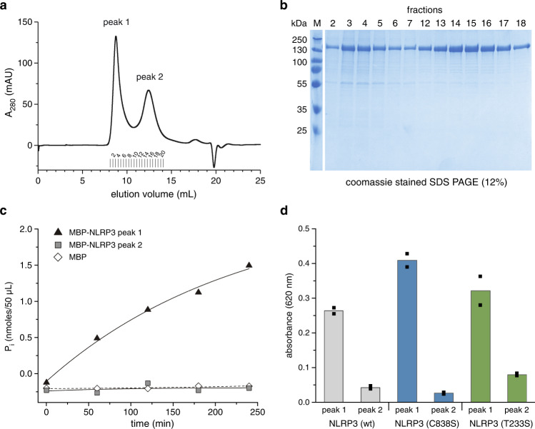
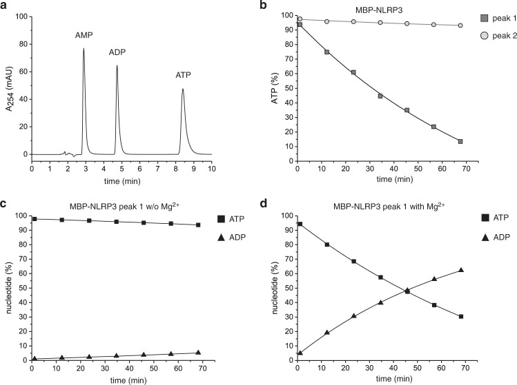
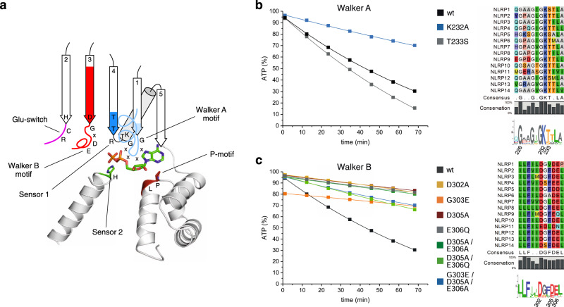
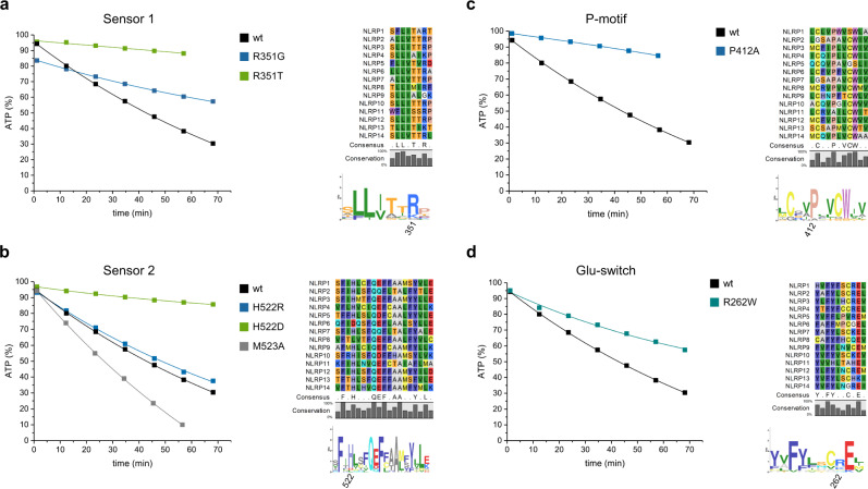
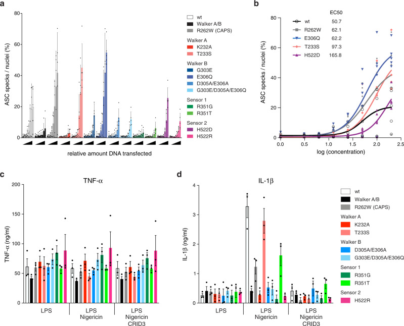
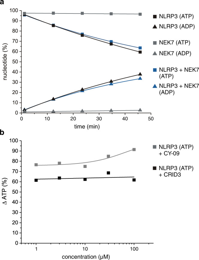
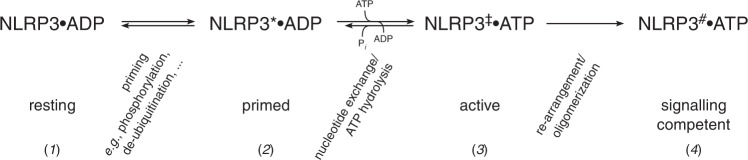
The innate immune system uses inflammasomal proteins to recognize danger signals and fight invading pathogens. NLRP3, a multidomain protein belonging to the family of STAND ATPases, is characterized by its central nucleotide-binding NACHT domain. The incorporation of ATP is thought to correlate with large conformational changes in NLRP3, leading to an active state of the sensory protein. Here we analyze the intrinsic ATP hydrolysis activity of recombinant NLRP3 by reverse phase HPLC. Wild-type NLRP3 appears in two different conformational states that exhibit an approximately fourteen-fold different hydrolysis activity in accordance with an inactive, autoinhibited state and an open, active state. The impact of canonical residues in the nucleotide binding site as the Walker A and B motifs and sensor 1 and 2 is analyzed by site directed mutagenesis. Cellular experiments show that reduced NLRP3 hydrolysis activity correlates with higher ASC specking after inflammation stimulation. Addition of the kinase NEK7 does not change the hydrolysis activity of NLRP3. Our data provide a comprehensive view on the function of conserved residues in the nucleotide-binding site of NLRP3 and the correlation of ATP hydrolysis with inflammasome activity.


Conflict of interest statement

The authors declare the following competing interests: M.G. and E.L. are co-founders and consultants of IFM Therapeutics. The other authors declare no competing interests.

Figures

Fig. 1
Fig. 1. The two conformers of NLRP3 exhibit different ATP hydrolysis activity.
a Human MBP-NLRP3, subjected to gel filtration analysis (Superose 6 10/300 GL), elutes in two distinct peaks, peak 1 and peak 2, respectively. b SDS-PAGE (12%) analysis of untreated MBP-NLRP3, according to gel filtration fractions shown in a. The protein band at about 165 kDa corresponds to the theoretical MW of MBP-NLRP3. c The malachite green phosphate assay was used to determine the ATP hydrolysis activity of MBP-NLRP3. The amount of Pi was measured in a time course experiment over 240 min. MBP-NLRP3 peak 1 and 2 (0.4 µM) were incubated at 20 °C in the presence of 1 mM ATP and 5 mM MgCl2. MBP was included as control; n = 1. d The ATP-hydrolysis activity of MBP-NLRP3 wt, C838S and T233S (0.4 µM), each showing peak 1 and peak 2, is compared. The amount of Pi generated in the presence of 1 mM ATP and MgCl2 was determined using the malachite green phosphate assay after 90 min incubation at 22 °C. Bars represent mean of technical duplicates shown as individual data points.
Fig. 2
Fig. 2. ATP-hydrolysis activity of NLRP3 determined by reverse-phase HPLC.
a Separation of adenosine nucleotides by HPLC on a RP-18 column. Nucleotides elute at about 3 mL (AMP), 4.5 mL (ADP) and 8.5 mL (ATP). b MBP-NLRP3 peak 1 or peak 2 (3 µM) were incubated at 25 °C for 68 min in the presence of 5 mM MgCl2 and 100 µM ATP. The amount of AMP, ADP and ATP was determined in 10 min intervals using RP-HPLC. The eluted peaks were evaluated by integration and the total integral adjusted to 100%. The relative ATP concentration is shown on the y-axis and the time on the x-axis (min). Shown is one representative measurement of n > 5 biologically independent experiments. c The hydrolysis activity of MBP-NLRP3 peak 1 depends on MgCl2. MBP-NLRP3 peak 1 (3 µM) was incubated at 25 °C for 68 min in the presence of 100 µM ATP, but in the absence of MgCl2. Shown is one representative experiment of n = 2 independent experiments. d ATP is hydrolyzed by NLRP3 peak 1 to ADP. MBP-NLRP3 peak 1 at 3 µM concentration was incubated at 25 °C for 68 min in the presence of 5 mM MgCl2 and 100 µM ATP. The relative amount of nucleotide is shown for ATP and ADP. Shown is one representative measurement of n > 5 biologically independent experiments.
Fig. 3
Fig. 3. Mutational analysis of Walker A/B nucleotide-binding motifs in NLRP3.
a A cartoon of the nucleotide-binding site of NLRP3 including ADP is shown based on the structure of inactive NLRP3 (PDB: 7PZC). The Walker A and B motifs as well as the Glu-switch, Sensor 1, Sensor 2 and the P-motif are indicated. b MBP-NLRP3 wild type and Walker A motif point-mutants were analyzed by RP-HPLC. Sequence alignments and sequence logos of the labeled motifs and residues are shown for human NLRP1-14. Protein samples (3 µM) were incubated at 25 °C in the presence of 100 µM ATP and 5 mM MgCl2. The amount of ATP (%) is shown on the y-axis and the time (min) on the x-axis. The amount of ATP was determined in 10 min intervals using RP-HPLC, integrated and normalized to 100% nucleotide. c MBP-NLRP3 wild type and Walker B motif point-mutants were analyzed by RP-HPLC using the same set-up as in b. All mutant significantly reduced the hydrolysis activity.
Fig. 4
Fig. 4. Mutational analysis of sensory ATP-binding motifs in NLRP3.
a MBP-NLRP3 wild type and two different Sensor 1 mutants were analyzed by RP-HPLC. The protein samples (3 µM) were incubated at 25 °C in the presence of 100 µM ATP and 5 mM MgCl2. The relative amount of ATP (%) is shown on the y-axis and the time (min) on the x-axis. The amount of ATP was determined in 10 min intervals, integrated and normalized to 100% nucleotide. Sequence alignments and sequence logos of the labeled motifs and residues are shown for human NLRP1-14. b Hydrolysis analysis of MBP-NLRP3 Sensor 2 mutants performed similarly as in a. c ATP hydrolysis analysis of the P412A mutant. d ATP hydrolysis analysis of the R262W CAPS/Glu-switch mutant.
Fig. 5
Fig. 5. Cellular activity assays for NLRP3 mutants targeting the AAA+ ATPase fold.
a Increasing amounts of different NLRP3 variants targeting the AAA+ ATPase fold were transiently over-expressed in HeLa cells stably over-expressing ASC-mTurquoise. ASC specks and nuclei were quantified using microscopy, and the ratio ASC speck/nuclei was plotted as a measure of inflammasome activation. Bars represent mean and SD of 5 independent experiments. b Dose response curves for the active NLRP3 mutants described in a. The half maximal effective concentration (EC50) was calculated for each of the NLRP3 variants. Data points of 5 independent experiments are shown. c, d NLRP3-deficient immortalized macrophages were reconstituted with wildtype NLRP3-mCitrine or indicated variants. c TNF-α and d IL-1β secretion of LPS primed immortalized macrophages stimulated with nigericin in the presence or absence of CRID3. Means ±SEM of pooled data from 3 independent experiments are presented.
Fig. 6
Fig. 6. Effects of NEK7 and antagonists CRID3 (MCC950) or CY-09 on the ATP-hydrolysis activity of NLRP3.
a The hydrolysis activity of MBP-NLRP3 peak 1 is unaffected by NEK7. MBP-NLRP3 peak 1 (3 µM) was incubated with NEK7 at 25 °C for 60 min in the presence of 100 µM ATP. The amount of ADP and ATP (%) is shown for MBP-NLRP3 peak 1 alone and addition of NEK7 to MBP-NLRP3. As a control, the kinase NEK7 exhibits no residual ATPase activity in the absence of substrate (gray symbols). b The hydrolysis activity of MBP-NLRP3 peak 1 is unchanged by CRID3 (MCC950) or CY-09. MBP-NLRP3 peak 1 (3 µM) was incubated at 25 °C for 60 min in the presence of 100 µM ATP. Increasing concentrations of CRID3 or CY-09 were added in a dose-response measurement from 1 to 100 µM and the relative concentration of ATP after 60 min measured.
Fig. 7
Fig. 7. Schematic representation of the proposed activation mechanism of human NLRP3.
From an inactive, resting state (1) the protein is turned, for example, by reversible PTMs such as phosphorylation, into a primed state (2) characterized by loss of autoinhibition. This state is capable of ATP uptake, which converts the protein to an active state (3). The ability of intrinsic ATP hydrolysis allows the protein to relapse to its resting or primed state. The active protein is eventually able to assemble into a quaternary structure, state (4), competent for downstream signaling, such as the induction of ASC filament formation.

References

    1. Akira S, Uematsu S, Takeuchi O. Pathogen recognition and innate immunity. Cell. 2006;124:783–801. - PubMed
    1. Beutler B, et al. Genetic analysis of host resistance: Toll-like receptor signaling and immunity at large. Annu Rev. Immunol. 2006;24:353–389. - PubMed
    1. Takeuchi O, Akira S. Pattern recognition receptors and inflammation. Cell. 2010;140:805–820. - PubMed
    1. Swanson KV, Deng M, Ting JP. The NLRP3 inflammasome: molecular activation and regulation to therapeutics. Nat. Rev. Immunol. 2019;19:477–489. - PMC - PubMed
    1. Dick MS, Sborgi L, Rühl S, Hiller S, Broz P. ASC filament formation serves as a signal amplification mechanism for inflammasomes. Nat. Commun. 2016;7:11929. - PMC - PubMed

Publication types

MeSH terms