Review
. 2022 Nov 3;22(1):632.
doi: 10.1186/s12887-022-03671-x.

Advances and limitations for the treatment of spinal muscular atrophy

Affiliations
Review

Advances and limitations for the treatment of spinal muscular atrophy

John W Day et al. BMC Pediatr. .

Abstract

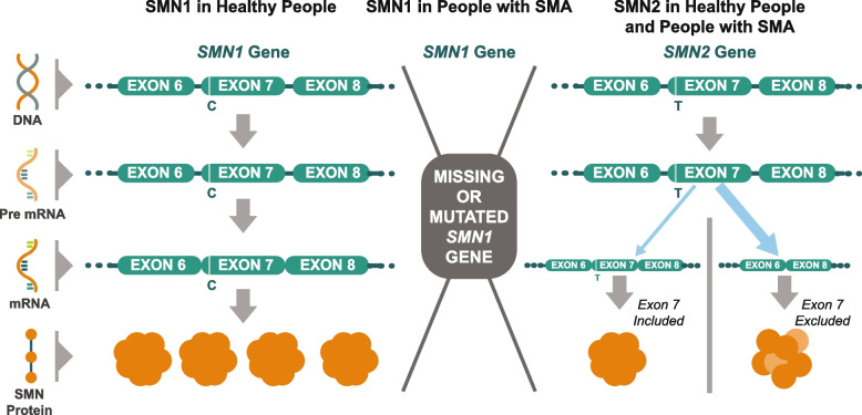
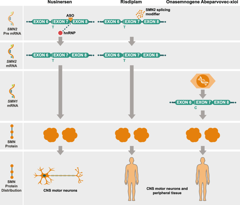
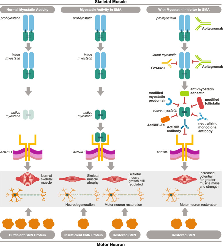
Spinal muscular atrophy (5q-SMA; SMA), a genetic neuromuscular condition affecting spinal motor neurons, is caused by defects in both copies of the SMN1 gene that produces survival motor neuron (SMN) protein. The highly homologous SMN2 gene primarily expresses a rapidly degraded isoform of SMN protein that causes anterior horn cell degeneration, progressive motor neuron loss, skeletal muscle atrophy and weakness. Severe cases result in limited mobility and ventilatory insufficiency. Untreated SMA is the leading genetic cause of death in young children. Recently, three therapeutics that increase SMN protein levels in patients with SMA have provided incremental improvements in motor function and developmental milestones and prevented the worsening of SMA symptoms. While the therapeutic approaches with Spinraza®, Zolgensma®, and Evrysdi® have a clinically significant impact, they are not curative. For many patients, there remains a significant disease burden. A potential combination therapy under development for SMA targets myostatin, a negative regulator of muscle mass and strength. Myostatin inhibition in animal models increases muscle mass and function. Apitegromab is an investigational, fully human, monoclonal antibody that specifically binds to proforms of myostatin, promyostatin and latent myostatin, thereby inhibiting myostatin activation. A recently completed phase 2 trial demonstrated the potential clinical benefit of apitegromab by improving or stabilizing motor function in patients with Type 2 and Type 3 SMA and providing positive proof-of-concept for myostatin inhibition as a target for managing SMA. The primary goal of this manuscript is to orient physicians to the evolving landscape of SMA treatment.

Keywords: Apitegromab; Myostatin; Nusinersen; Onasemnogene abeparvovec-xioi; Risdiplam; SRK-015; Spinal muscular atrophy; Survival motor neuron; Survival motor neuron-1 gene.


Conflict of interest statement

Drs. George Nomikos, Nathalie Kertesz and Jose Rossello are employees of Scholar Rock, Inc. and are shareholders. Drs. Amy Place and Kimberly Long are former employees of Scholar Rock. Dr. John Day reports grants from: AMO Pharma; Audentes; Avidity; Biogen; Cytokinetics; Genentech; Ionis Pharmaceuticals; Novartis Gene Therapies; Roche Pharmaceuticals; Sanofi-Genzyme; Sarepta Therapeutics; Scholar Rock, and has received support from participating on advisory boards or having consulted with: Affinia Therapeutics; AMO Pharmaceuticals; Avidity Biosciences; Biogen; Cytokinetics; Epirium Bio; Ionis Pharmaceuticals; Kate Therapeutics; Novartis Gene Therapies; Roche/Genentech Pharmaceuticals; Sarepta Therapeutics; Scholar Rock; Shift Therapeutics; and Vertex Pharmaceuticals. Dr Kelly Howell has nothing to disclose.

Figures

Fig. 1
Fig. 1
Normal SMN protein expression and in patients with spinal muscular atrophy. Survival motor neuron gene 1 (SMN1) encodes full length SMN protein needed to ensure survival of motor neurons and normal muscle growth and function (left). The nearly identical SMN2 gene differs by only two nucleotides, a CT base change inside exon 7 that affects gene splicing and leads to exon 7 skipping in the majority of SMN2 mRNA (messenger ribonucleic acids) (right). SMN2 mRNA transcripts with exon 7 included provide a supplementary source of normal SMN protein; SMN2 mRNA lacking exon 7 encodes truncated, rapidly degraded SMN protein. In patients with SMA, there is a homozygous deletion or loss of function of the SMN1 gene, eliminating the body’s main source of SMN protein (center). The functional protein made by the SMN2 gene is identical to that produced by the SMN1 gene but is produced in insufficient quantity to support normal motor neuron functioning, muscle growth, and development. SMN1 codes for full length functional SMN1 protein which is the primary source of the SMN protein. SMN2 with exon 7 included is a full length, functional SMN protein (10–20%). SMN2 with Exon 7 excluded is an unstable rapidly degraded SMN protein (80–90%). Patients with SMN may have up to eight copies of the SMN2 gene, all of which can produce limited quantities of SMN protein. Patients with more SMN2 gene copies generally have less severe SMA [–15]. Figure property of Scholar Rock, Inc
Fig. 2
Fig. 2
SMN correcting therapy, mechanism of action. The antisense oligonucleotide (ASO) nusinersen is an intrathecally-delivered splicing modifier that binds to the exon 7 silencer region on SMN2 pre-mRNA (Pre mRNA) (left). By displacing the splicing repressor protein hnRNP, nusinersen promotes inclusion of exon 7 and boosts production of full-length SMN2 mRNA. Functional SMN protein in central nervous system motor neurons is increased. Risdiplam is an orally available, selective small molecule that modifies SMN2 pre-mRNA (Pre mRNA) splicing (center). Risdiplam increases exon 7 inclusion in SMN2 mRNA transcripts and production of full-length SMN protein in the brain. This leads to increased production of functional SMN protein in the brain and throughout peripheral tissues. Onasemnogene abeparvovec-xioi is an adeno-associated virus 9 (AAV9)-based therapy that delivers a fully functional copy of SMN complementary deoxyribonucleic acid (cDNA) (right). Administered intravenously as a single-dose, the SMN transgene passes the blood–brain barrier and is introduced directly into target motor neuron cells throughout the CNS. Transduced cells produce full-length SMN mRNA transcripts, which enable continuous production of SMN protein in motor neurons and peripheral tissue over time [–46]. Figure property of Scholar Rock, Inc
Fig. 3
Fig. 3
Myostatin inhibition MOA as add-on to SMN correctors in SMA. SMN protein promotes normal motor neuron function, which in turn provides the signals that activate and sustain muscle tissue (left). In SMA, insufficient SMN protein leads to degeneration of motor neurons and subsequent skeletal muscle atrophy (center). SMN correctors help to increase SMN protein production, stabilize neurodegeneration, and improve or maintain motor function, but may not return muscle to its normal size and function (right) Myostatin circulates as a complex of inhibitory prodomains. When the prodomains are proteolytically cleaved, the active myostatin dimer can bind to its receptor ActRIIB, The heterocomplex translocates to the nucleus where it regulates transcription. Several inhibitors of this signaling pathway have been developed including modified myostatin prodomain, modified follistatin, neutralizing monoclonal antibody and adnectin, ActRIIB-Fc, and ActRIIB blocking antibody. These strategies all lead to blocking myostatin binding to its receptor. Myostatin inhibition in combination with SMN correctors may directly address muscle atrophy and further restore motor function [, , –84]. Apitegromab is a monoclonal antibody that selectively blocks the precursor, or inactive form of myostatin, blocking its activation in skeletal muscle. Myostatin is a negative regulator of skeletal muscle growth. Apitegromab specifically targets the upstream pro and latent forms of myostatin, which avoids cross-reactivity with other TGF-ß ligands and inhibits activation of myostatin. Apitegromab improves muscle mass and strength with fewer off-target effects and related toxicities than possible with less selective myostatin inhibitors [82, 85]. Figure property of Scholar Rock, Inc

References

    1. Strunk A, Abbes A, Stuitje AR, Hettinga C, Sepers EM, Snetselaar R, et al. Validation of a fast, robust, inexpensive, two-tiered neonatal screening test algorithm on dried blood spots for spinal muscular atrophy. Int J Neonatal Screen. 2019;5:21. - PMC - PubMed
    1. Cure SMA. Elk Grove Village, IL. https://www.curesma.org/about-sma/. Accessed 18 July 2021.
    1. Lally C, Jones C, Farwell W, Reyna SP, Cook SF, Flanders WD. Indirect estimation of the prevalence of spinal muscular atrophy Type I, II, and III in the United States. Orphanet J Rare Dis. 2017;12:175. - PMC - PubMed
    1. Verhaart IEC, Robertson A, Wilson IJ, Aartsma-Rus A, Cameron S, Jones CC, et al. Prevalence, incidence and carrier frequency of 5q-linked spinal muscular atrophy - a literature review. Orphanet J Rare Dis. 2017;12:124. - PMC - PubMed
    1. Sugarman EA, Nagan N, Zhu H, Akmaev VR, Zhou Z, Rohlfs EM, et al. Pan-ethnic carrier screening and prenatal diagnosis for spinal muscular atrophy: clinical laboratory analysis of >72,400 specimens. Eur J Hum Genet. 2012;20:27–32. - PMC - PubMed