. 2022 Sep 13;9(3):395-422.
doi: 10.3934/Neuroscience.2022022. eCollection 2022.

Chronic treatment with escitalopram and venlafaxine affects the neuropeptide S pathway differently in adult Wistar rats exposed to maternal separation

Affiliations

Chronic treatment with escitalopram and venlafaxine affects the neuropeptide S pathway differently in adult Wistar rats exposed to maternal separation

Miłosz Gołyszny et al. AIMS Neurosci. .

Abstract

Neuropeptide S (NPS), which is a peptide that is involved in the regulation of the stress response, seems to be relevant to the mechanism of action of antidepressants that have anxiolytic properties. However, to date, there have been no reports regarding the effect of long-term treatment with escitalopram or venlafaxine on the NPS system under stress conditions. This study aimed to investigate the effects of the above-mentioned antidepressants on the NPS system in adult male Wistar rats that were exposed to neonatal maternal separation (MS). Animals were exposed to MS for 360 min. on postnatal days (PNDs) 2-15. MS causes long-lasting behavioral, endocrine and neurochemical consequences that mimic anxiety- and depression-related features. MS and non-stressed rats were given escitalopram or venlafaxine (10mg/kg) IP from PND 69 to 89. The NPS system was analyzed in the brainstem, hypothalamus, amygdala and anterior olfactory nucleus using quantitative RT-PCR and immunohistochemical methods. The NPS system was vulnerable to MS in the brainstem and amygdala. In the brainstem, escitalopram down-regulated NPS and NPS mRNA in the MS rats and induced a tendency to reduce the number of NPS-positive cells in the peri-locus coeruleus. In the MS rats, venlafaxine insignificantly decreased the NPSR mRNA levels in the amygdala and a number of NPSR cells in the basolateral amygdala, and increased the NPS mRNA levels in the hypothalamus. Our data show that the studied antidepressants affect the NPS system differently and preliminarily suggest that the NPS system might partially mediate the pharmacological effects that are induced by these drugs.

Keywords: antidepressants; maternal separation; neuropeptide S; neuropeptide S receptor.


Conflict of interest statement

Conflict of interest: The authors do not declare any conflict of interest.

Figures

Figure 1.
Figure 1.. Neuropeptide S (NPS) pathways in the rat brain. NPS is expressed in the brainstem and reaches the limbic system and anterior olfactory nucleus (AON). In the brainstem, NPS is synthesized in the peri-locus coeruleus (peri-LC) (blue squares). The link between the peri-LC and hypothalamic nuclei, e.g., the paraventricular nucleus (PVN), is a pathway that is involved in the expanded regulation of the limbic system and neuroendocrine response to stress, according to Nauta's conceptualization. The neuropeptide S receptors (NPSR) are localized in the ventral tegmental area, hypothalamic nuclei, amygdaloid complex, hippocampus and AON (red circles), according to previous studies ,.
Abbreviations: AMY—amygdala; AON—anterior olfactory nucleus; ARC—arcuate nucleus; BLA—basolateral amygdala; BMA—basomedial amygdala;CeA—central amygdala; CA1-CA3—areas of hypothalamus; HT—hypothalamus; LA—lateral amygdala; LC—locus coeruleus; LH—lateral hypothalamus;MeA—medial amygdala; NPS—neuropeptide S; NPSR—neuropeptide S receptor; OB—olfactory bulb; PAG—periaqueductal grey area; PFC—prefrontal cortex; PVN—paraventricular nucleus; VMH—ventromedial hypothalamus; VTA—ventral tegmental area; 4V—4th ventricle.
Figure 2.
Figure 2.. The effects of maternal separation (MS) and/or the long-term administration of escitalopram (10 mg/kg IP) or venlafaxine (10 mg/kg IP) on the relative NPS mRNA expression in the brainstem and NPS-immunopositive cells in peri-LC of adult male Wistar rats. (A) Values are the relative expression ± SEM (n = 8–10 per group). # p < 0.05 vs. MS (post hoc Dunn's test). (B) NPS-immunopositive cells (% of control ±) (n = 4 per group); (C) A representative photomicrograph showing the evaluated area – peri-LC in the cresyl violet stain (magnification 4 x). $ p < 0.05 vs. MS + Esci (post hoc Dunn's test).
Abbreviations: LPB—lateral parabrachial nucleus; peri-LC—peri-locus coeruleus; Contr—non-stressed rats receiving saline (IP); Esci—non-stressed rats receiving escitalopram (IP); Ven—non-stressed rats receiving venlafaxine (IP); MS—rats subjected to MS on PNDs 2–15 and receiving saline (IP); MS + Esci—rats subjected to MS on PND 2–15 and receiving escitalopram (IP); MS + Ven—rats subjected to MS on PNDs 2–15 and receiving venlafaxine (IP).
Figure 3.
Figure 3.. The effects of maternal separation (MS) and/or the long-term administration of escitalopram (10 mg/kg IP) or venlafaxine (10 mg/kg IP) on the relative NPSR mRNA expression in the brainstem, hypothalamus and amygdala of adult male Wistar rats. Values are the relative expression ± SEM (n = 8–10 per group). $ p < 0.05 vs. MS + Esci (post hoc Dunn's test); ## p < 0.01 vs. MS (post hoc Dunn's test). Abbreviations are explained in the legend for Fig. 2.
Figure 4.
Figure 4.. Neuropeptide S in the peri-LC – the representative photomicrographs.
Figure 5.
Figure 5.. The effects of maternal separation (MS) and/or long-term administration of escitalopram (10 mg/kg IP) or venlafaxine (10 mg/kg IP) on the number of immunopositive NPSR cells in the ventromedial hypothalamus (VMH), lateral hypothalamus (LH), arcuate nucleus (ARC) and paraventricular nucleus (PVN). Values are the percentage of control ± SEM (n = 4 per group). Representative photomicrographs of NPSR immunostaining in the studied subregions in control group. Abbreviations are explained in the legends for Fig. 1 and Fig. 2.
Figure 6.
Figure 6.. The effects of maternal separation (MS) and/or the long-term administration of escitalopram (10mg/kg IP) or venlafaxine (10mg/kg IP) on the number of immunopositive NPSR cells and optical density in the basolateral amygdala (BLA). (A) A representative photomicrograph of the evaluated area in the amygdaloid complex (magnification 4x) (coronal plane −2.04 from the bregma); (B-C) The NPS-immunopositive cells and optical density (% of control); (D) Representative photomicrographs showing the NPSR immunopositive cells in the BLA (magnification 20×).
Abbreviations: CeC—central amygdaloid nucleus, capsular, CeM—central amygdaloid nucleus, medial division, LaDL—lateral amygdaloid nucleus, dorsolateral. The otherabbreviations are explained in the legends of Fig. 1 and Fig. 2.
Figure 7.
Figure 7.. The NPSR-immunopositive cells in the rat anterior olfactory nucleus (AON). (A) Part of the brain including the anterior olfactory nucleus from which the slices were prepared according to the atlas by Paxinos and Watson (2006) (the coronal planes 4.20 to 6.12 from the bregma); (B) A representative photomicrograph showing NPSR expression in control rats (3,3′-Diaminobenzidine–immunostaining, magnification 4×).
Abbreviations: aci—anterior commissure, intrabulbar; E/OV—ependyma/olfactory ventricle
Figure 8.
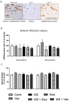
Figure 8.. The effects of maternal separation (MS) and/or long-term administration of escitalopram (10 mg/kg IP) or venlafaxine (10 mg/kg IP) on the number of immunopositive NPSR cells and optical density in the anterior olfactory nucleus (AON). (A) A representative microphotograph showing the result of NPSR-cytoplasmatic positive reaction. NPSR protein was not detected in cellular nucleus. Color deconvolution; (B) NPSR-immunopositive neurons in the ventrolateral and dorsomedial areas of the AON (% of control); (C) Optical density (% of control).

Similar articles

References

    1. Kormos V, Gaszner B. Role of neuropeptides in anxiety, stress, and depression: from animals to humans. Neuropeptides. 2013;47(6):401–419. doi: 10.1016/j.npep.2013.10.014. - DOI - PubMed
    1. Yamazoe M, Shiosaka S, Emson PC, et al. Distribution of neuropeptide Y in the lower brainstem: an immunohistochemical analysis. Brain Res. 1985;335(1):109–120. doi: 10.1016/0006-8993(85)90281-1. - DOI - PubMed
    1. Douglas FL, Palkovits M, Brownstein MJ. Regional distribution of substance P-like immunoreactivity in the lower brainstem of the rat. Brain Res. 1982;245(2):376–378. doi: 10.1016/0006-8993(82)90821-6. - DOI - PubMed
    1. Spirovski D, Li Q, Pilowsky PM. Brainstem galanin-synthesizing neurons are differentially activated by chemoreceptor stimuli and represent a subpopulation of respiratory neurons. J Comp Neurol. 2012;520(1):154–173. doi: 10.1002/cne.22723. - DOI - PubMed
    1. Liu C, Lovenberg TW. Relaxin-3, INSL5, and their receptors. Orphan G Protein-Coupled Receptors and Novel Neuropeptides, 46. Berlin, Heidelberg: Springer; 2008. pp. 213–237. - DOI - PubMed

LinkOut - more resources