Case Reports
. 2023 Sep;26(3):306-311.
doi: 10.5397/cise.2022.01151. Epub 2022 Nov 3.

Normal ossification of the glenoid mimicking a glenoid fracture in an adolescent patient: a case report

Affiliations
Case Reports

Normal ossification of the glenoid mimicking a glenoid fracture in an adolescent patient: a case report

María Galán-Olleros et al. Clin Shoulder Elb. 2023 Sep.

Abstract

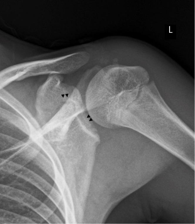
A 13-year-old male was diagnosed with a glenoid fracture following direct shoulder trauma, for which surgical treatment was considered. After referral to a center for pediatric orthopedic care, physical examination, contralateral shoulder X-ray, and detailed computed tomography examination ruled out the presence of fracture; these findings were later confirmed by magnetic resonance imaging. Normal ossification patterns in the adolescent shoulder may simulate a fracture in traumatic settings. To accurately diagnose and manage pediatric shoulder pathology, orthopedic surgeons must be aware of the normal anatomy of the growing shoulder, its secondary ossification centers, and growth plates.

Keywords: Child development; Physiologic ossification; Shoulder; Trauma; Glenoid cavity.


Conflict of interest statement

Conflict of interest

None.

Figures

Fig. 1.
Fig. 1.
Shoulder X-rays of the patient. (A) Anteroposterior radiograph of the right shoulder after trauma in a patient aged 13 years and 6 months. An irregular, radiolucent line is seen extending from the base of the coracoid to the articular surface of the glenoid and another trace appears to be directed posteriorly (▲▲). (B) Scapular Y-projection showing the same discontinuity in the anterior and posterior cortex (*) at the level of the middle third of the glenoid. First interpreted as fracture lines on presentation to the first health center, these images actually correspond to the subcoracoid ossification center and the glenoid growth plates.
Fig. 2.
Fig. 2.
Right shoulder computed tomography scan of the same patient. Despite being reported as a fracture of the glenoid surface extending to the coracoid ossification core with 3 mm thickness and discrete irregularity of the glenoid surface at the inferior margin with 2 millimetric bone fragments, the images are compatible with the subcoracoid ossification centers (↑↑↑) and normal inferior glenoid secondary ossification centers (*) of the shoulder of a 13-year-old boy in axial views (A-D), coronal view (E), and three-dimensional reconstruction (F). The undulating appearance of the subcoracoid growth plate and sclerosis of the inferior secondary glenoid ossification centers are in evidence.
Fig. 3.
Fig. 3.
Anteroposterior radiograph of the left shoulder of the same patient showing a radiolucent line extending from the base of the coracoid to the articular surface of the glenoid (▲▲), similar to the contralateral shoulder, which appears to correspond to the glenoid physeal area.
Fig. 4.
Fig. 4.
Magnetic resonance imaging (MRI) (A-C). Axial, coronal, and sagittal T1-weighted MRI in which the subcoracoid ossification center (*) can be seen extending toward the growth plate between the base of the coracoid c and the scapula s and presents the same intensity as the rest of the scapular bone. The hypointense undulations of the glenoid growth plate (▲▲) and the multilamellar appearance of the glenoid (◆◆) can be seen in coronal view (B). (D) Axial T2-fast field echo image showing the low-signal subcoracoid secondary ossification center (*), a bipolar growth plate prior to closure (▲▲), the residual unossified glenoid cartilage and hyperintense overlying articular cartilage (↑↑↑), and the hypointense glenoid labrum (■). H: humeral head. (E) Coronal proton density spectral attenuated inversion recovery image showing normal undulation and irregularity of the glenoid bone plate (◆◆), which may simulate a subchondral lesion, but is physiologic. (F) Axial PD-SPIR image showing a typically curved and lobulated hypointense interface (▲▲) between the subcoracoid ossification center and the rest of the glenoid. No signs of bone edema or physeal fracture are seen on long repetition time sequences (D-F).
Fig. 5.
Fig. 5.
Schematic illustration of the secondary ossification centers of the glenoid [1].

References

    1. Kothary S, Rosenberg ZS, Poncinelli LL, Kwong S. Skeletal development of the glenoid and glenoid-coracoid interface in the pediatric population: MRI features. Skeletal Radiol. 2014;43:1281–8. - PubMed
    1. Sidharthan S, Greditzer HG, 4th, Heath MR, Suryavanshi JR, Green DW, Fabricant PD. Normal glenoid ossification in pediatric and adolescent shoulders mimics bankart lesions: a magnetic resonance imaging-based study. Arthroscopy. 2020;36:336–44. - PubMed
    1. Chauvin NA, Jaimes C, Laor T, Jaramillo D. Magnetic resonance imaging of the pediatric shoulder. Magn Reson Imaging Clin N Am. 2012;20:327–47. - PubMed
    1. Zember JS, Rosenberg ZS, Kwong S, Kothary SP, Bedoya MA. Normal skeletal maturation and imaging pitfalls in the pediatric shoulder. Radiographics. 2015;35:1108–22. - PubMed
    1. Zember J, Vega P, Rossi I, Rosenberg ZS. Normal development imaging pitfalls and injuries in the pediatric shoulder. Pediatr Radiol. 2019;49:1617–28. - PubMed

Publication types

LinkOut - more resources