. 2022 Nov 30;136(22):1661-1681.
doi: 10.1042/CS20220607.

Cardiomyocyte BRAF is a key signalling intermediate in cardiac hypertrophy in mice

Affiliations

Cardiomyocyte BRAF is a key signalling intermediate in cardiac hypertrophy in mice

Hajed O Alharbi et al. Clin Sci (Lond). .

Abstract

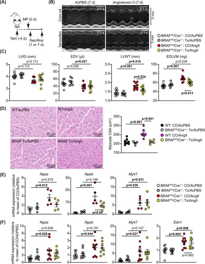
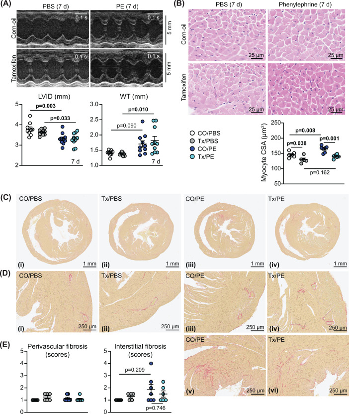
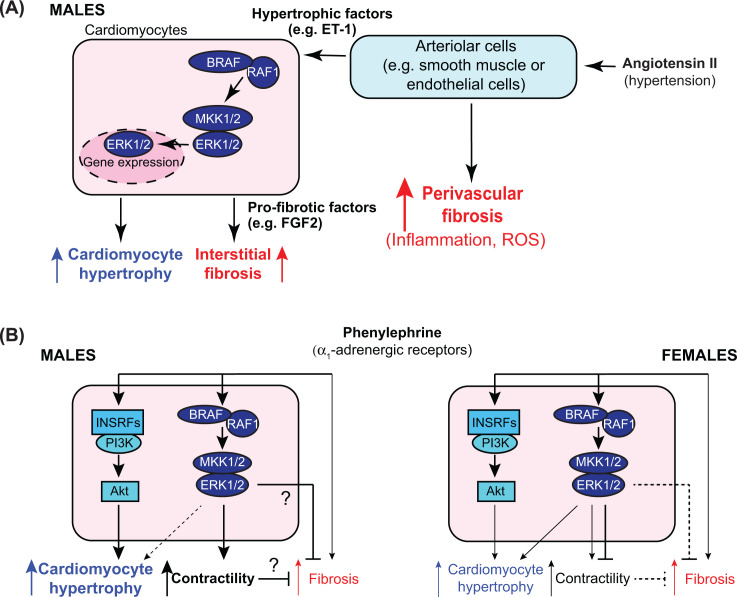
Cardiac hypertrophy is necessary for the heart to accommodate an increase in workload. Physiological, compensated hypertrophy (e.g. with exercise) is reversible and largely due to cardiomyocyte hypertrophy. Pathological hypertrophy (e.g. with hypertension) is associated with additional features including increased fibrosis and can lead to heart failure. RAF kinases (ARAF/BRAF/RAF1) integrate signals into the extracellular signal-regulated kinase 1/2 cascade, a pathway implicated in cardiac hypertrophy, and activation of BRAF in cardiomyocytes promotes compensated hypertrophy. Here, we used mice with tamoxifen-inducible cardiomyocyte-specific BRAF knockout (CM-BRAFKO) to assess the role of BRAF in hypertension-associated cardiac hypertrophy induced by angiotensin II (AngII; 0.8 mg/kg/d, 7 d) and physiological hypertrophy induced by phenylephrine (40 mg/kg/d, 7 d). Cardiac dimensions/functions were measured by echocardiography with histological assessment of cellular changes. AngII promoted cardiomyocyte hypertrophy and increased fibrosis within the myocardium (interstitial) and around the arterioles (perivascular) in male mice; cardiomyocyte hypertrophy and interstitial (but not perivascular) fibrosis were inhibited in mice with CM-BRAFKO. Phenylephrine had a limited effect on fibrosis but promoted cardiomyocyte hypertrophy and increased contractility in male mice; cardiomyocyte hypertrophy was unaffected in mice with CM-BRAFKO, but the increase in contractility was suppressed and fibrosis increased. Phenylephrine induced a modest hypertrophic response in female mice and, in contrast with the males, tamoxifen-induced loss of cardiomyocyte BRAF reduced cardiomyocyte size, had no effect on fibrosis and increased contractility. The data identify BRAF as a key signalling intermediate in both physiological and pathological hypertrophy in male mice, and highlight the need for independent assessment of gene function in females.

Keywords: BRAF; cardiac hypertrophy; cardiomyocytes; fibrosis; hypertension; protein-serine-threonine kinases.


Conflict of interest statement

The authors declare that there are no competing interests associated with the manuscript.

Figures

Figure 1
Figure 1. Male mice with tamoxifen-inducible cardiomyocyte-specific BRAF knockout
BRAFfl/fl/Cre+/− (homozygous for floxed BRAF; hemizygous for Cre) male mice were treated with corn-oil (CO) or tamoxifen in CO (Tx) for 24 h or 11 d as indicated. (A) Confirmation of BRAF knockout using cDNA prepared from RNA extracted from powdered tissues. PCR amplification used forward primers in exon 10 (upper image) or exon 9 (lower image) with reverse primers in exon 13. Deletion of exon 12 in cardiomyocytes resulted in the appearance of a smaller product following recombination with tamoxifen administration in heart (H), but not liver (L) or kidney (K). Representative images are shown. (B) Immunoblot analysis of RAF isoforms in samples of mouse hearts treated with vehicle or tamoxifen. Representative immunoblots are in the upper panels (positions of relative molecular mass markers are on the right) with densitometric analysis below. RAF expression was normalized to Gapdh and data presented relative to the means of the vehicle treated controls. Statistical analysis used unpaired two-tailed t tests. (C) mRNA expression in mouse hearts after 11 d treatment with tamoxifen. RNA was extracted and expression of selected genes assessed by qPCR. (D,E) Assessment of cardiomyocyte cross-sectional area (CSA) and fibrosis in wild-type (WT) mice from the same breeding stock as the BRAFfl/fl/Cre+/− mice in comparison with the BRAFfl/fl/Cre+/− mice. Mice were treated with Tx for 11 d, AngII (0.8 mg/kg/d, 7 d) in acidified PBS (AcPBS) or AcPBS only. Data for CSA are from haematoxylin and eosin stained sections. Data for fibrosis are from Masson’s trichrome and picrosirius red stained sections. Data are presented as individual values with means ± SEM. Statistical analysis used unpaired two-tailed t tests.
Figure 2
Figure 2. Cardiomyocyte BRAF knockout inhibits the hypertrophic response induced by AngII in male mouse hearts
Male BRAFfl/fl/Cre+/− (BRAF) or wild-type (WT) mice were treated with corn-oil vehicle (CO) or tamoxifen in CO (Tx) 4 days before minipumps (MP) were implanted to deliver acidified PBS (AcPBS) or 0.8 mg/kg/d AngII. Mice were sacrificed after 1 or 7 d. Echocardiograms were collected and hearts taken at 7 d. (A) Schematic of experimental protocol. (B) Representative M-mode echocardiograms taken from short axis views of the hearts of BRAFfl/fl/Cre+/− at 7 d. (C) Analysis of echocardiograms taken at 7 d to assess cardiac dimensions. Abbreviations: EDV, end diastolic volume; EDLVM, end diastolic left ventricular mass; LVID, left ventricle internal diameter; LVWT, left ventricle wall thickness (posterior plus anterior walls). LVID and LVWT were measured at diastole from M-mode images of short axis views of the heart. EDV and EDLVM were predicted from B-mode images of long axis views of the heart. (D) Haematoxylin and eosin staining of mouse heart sections (left panels) from hearts collected at 7 d, with assessment of cardiomyocyte cross-sectional area (CSA; right panel). Images and measurements are from the periphery of the left ventricle. (E,F) mRNA expression in mouse hearts after 24 h (E) or 7 d (F) treatment with AngII. RNA was extracted and expression of Nppa, Nppb, Myh7 and Edn1 were assessed by qPCR. Data are individual values with means ± SEM. Statistical analysis used two-way ANOVA with Holm-Sidak’s post-test. Statistically significant values (P<0.05) are in bold type.
Figure 3
Figure 3. Cardiomyocyte BRAF knockout inhibits interstitial but not perivascular fibrosis induced by AngII in male mouse hearts
Male BRAFfl/fl/Cre+/− (BRAF) or wild-type (WT) mice were treated with CO or tamoxifen in CO (Tx) 4 days before minipumps were implanted to deliver acidified PBS (AcPBS) or 0.8 mg/kg/d AngII in AcPBS (AngII) for 7 d. (A,B) Picrosirius red staining of mouse heart sections showing short axis views of the whole heart (A) with enlarged sections (B) from the same views (the red stain shows accumulation of fibrotic material). Representative (average) images are shown for mice treated with CO/AcPBS (i), Tx/AcPBS (ii), CO/AngII (iii) and Tx/AngII (v). Additional images are shown for the most severe degree of fibrosis with CO/AngII (iv) and Tx/AngII (vi). (C) Quantification of the degree of fibrosis. This was scored as: 1 = the least amount of fibrosis; 2 = low level fibrosis; 3 = high level fibrosis in at least one area of the myocardium; 4 = high level fibrosis throughout the myocardium (half scores were used). Statistical analysis used two-way ANOVA with Holm-Sidak’s post-test. Statistically significant values (P<0.05) are in bold type.
Figure 4
Figure 4. Effects of cardiomyocyte BRAF knockout on changes in mRNA expression induced by AngII in male mouse hearts
Male BRAFfl/fl/Cre+/− (BRAF) mice were treated with corn-oil (CO) or tamoxifen in CO (Tx) 4 days before minipumps were implanted to deliver acidified PBS (AcPBS) or 0.8 mg/kg/d AngII in AcPBS (AngII) for 1 or 7 d. RNA was extracted and expression of selected genes assessed by qPCR. (A) Fgf2 was assessed at 7 d. (B) CTGF and TGFB1 were assessed at 1 or 7 d as indicated. (C) Pro-fibrotic genes (Ddr2, Lox, Col1a1, Col4a1, Postn, Fn1) were assessed at 7 d. (D) Pro-inflammatory (IL1b, IL6) and oxidant (Hif1a, NOX1, NOX2, NOX4) genes were assessed at 7 d. Data are presented as individual values with means ± SEM. Statistical analysis used two-way ANOVA with Holm-Sidak's post-test. Statistically significant values (P<0.05) are in bold type.
Figure 5
Figure 5. Cardiomyocyte BRAF knockout does not inhibit cardiomyocyte hypertrophy induced by phenylephrine in male mouse hearts but increases cardiac fibrosis
Male BRAFfl/fl/Cre+/− mice were treated with corn-oil (CO) or tamoxifen in CO (Tx) 4 days before minipumps were implanted to deliver PBS or 40 mg/kg/d phenylephrine in PBS (PE) for 7 d. (A) Representative M-mode echocardiograms taken from short axis views of the heart (upper panels) with analysis of echocardiograms to assess cardiac dimensions (lower panels). Abbreviations: LVID, left ventricle internal diameter; WT, left ventricle wall thickness (posterior plus anterior walls). Diastolic measurements are shown. (B) mRNA expression in mouse hearts after 7 d treatment with phenylephrine. (C) Haematoxylin and eosin staining of mouse heart sections (left panels) with assessment of cardiomyocyte cross-sectional area (CSA; right panel). Images and measurements are from the periphery of the left ventricle. (D,E) Picrosirius red staining of mouse heart sections showing short axis views of the whole heart (D) with enlarged sections (E) from the same views (the red stain shows accumulation of fibrotic material). Representative (average) images are shown for mice treated with CO/PBS (i), Tx/PBS (ii), CO/PE (iii) and Tx/PE (v). Additional images are shown for the most severe degree of fibrosis with CO/PE (iv) and Tx/PE (vi). (F) Quantification of fibrosis. This was scored as: 1 = the least amount of fibrosis; 2 = low level fibrosis; 3 = high level fibrosis in at least one area of the myocardium; 4 = high level fibrosis throughout the myocardium (half scores were used). Data are presented as individual values with means ± SEM. Statistical analysis used two-way (A,C,F) or one-way (B) ANOVA with Holm-Sidak’s post-test. Statistically significant values (P<0.05) are in bold type.
Figure 6
Figure 6. Comparison of cardiac function/dimensions and confirmation of recombination in female BRAFfl/fl/Cre+/− mice compared with male littermates
(A–D) Data were gathered from mice at the time of the first baseline echocardiogram taken from male and female BRAFfl/fl/Cre+/− littermates (males: 7–8 weeks; females: 9–10 weeks). (A) Body weights. (B) Heart rate, ejection fraction and fractional shortening, global longitudinal strain (GLS) and global circumferential strain (GCS) were measured from B-mode images of long-axis views using VevoStrain speckle-tracking software. (C,D) Stroke volume, cardiac output, end diastolic volume (EDV), end systolic volume (ESV) and end diastolic LV mass (EDLVM) were measured from B-mode images of long-axis views using VevoStrain speckle-tracking software. Diastolic (d) and systolic (s) left ventricle (LV) internal diameter (ID) and wall thickness (WT: anterior plus posterior walls) measurements were measured from M-mode images of short axis views using VevoLab software. (E) Confirmation of recombination using cDNA prepared from RNA extracted from the hearts of male (upper image) and female (lower image) littermates 11 d post-tamoxifen treatment. PCR amplification used forward primers in exon 9 with reverse primers in exon 13. Deletion of exon 12 in cardiomyocytes resulted in the appearance of a smaller product in heart (H) but not kidney (K) of mice treated with tamoxifen (Tx) in corn-oil (CO) but not CO alone. Representative images are shown. (F–H) Immunoblot analysis of RAF isoforms (F) or phosphorylated and total ERK1/2 (G,H) in relation to Gapdh in samples of female or male (as indicated) mouse hearts treated with CO or Tx 4 days before administration of phenylephrine (PE) in PBS or PBS alone for 7 d. Representative immunoblots are shown. Densitometric analysis of the blots in H are in the right panels. Individual data points are shown with the mean and range.
Figure 7
Figure 7. Assessment of the effects of cardiomyocyte BRAF knockout on the response of female mouse hearts to phenylephrine
Female BRAFfl/fl/Cre+/− mice were treated with corn-oil (CO) or tamoxifen in CO (Tx) 4 days before minipumps were implanted to deliver PBS or 40 mg/kg/d phenylephrine in PBS (PE) for 7 d. (A) Representative M-mode echocardiograms taken from short axis views of the heart (upper panels) with analysis of echocardiograms to assess cardiac dimensions (lower panels). Abbreviations: LVID, left ventricle internal diameter; WT, left ventricle wall thickness (posterior plus anterior walls). Measurements were taken at diastole. (B) Haematoxylin and eosin staining of mouse heart sections (upper panels) with assessment of cardiomyocyte cross-sectional area (CSA; lower panel). Images and measurements are from the periphery of the left ventricle. (C,D) Picrosirius red staining of mouse heart sections showing short axis views of the whole heart (C) with enlarged sections (D) from the same views (the red stain shows accumulation of fibrotic material). Representative (average) images are shown for mice treated with CO/PBS (i), Tx/PBS (ii), CO/PE (iii) and Tx/PE (iv). Additional images are shown for the most severe degree of fibrosis with CO/PE (v) and Tx/PE (vi). (E) Quantification of fibrosis. This was scored as: 1 = the least amount of fibrosis; 2 = low level fibrosis; 3 = high level fibrosis in at least one area of the myocardium; 4 = high level fibrosis throughout the myocardium (half scores were used). Data are presented as individual values with means ± SEM. Statistical analysis used two-way ANOVA with Holm-Sidak’s post-test. Statistically significant values (P<0.05) are in bold type.
Figure 8
Figure 8. Comparison of effects of cardiomyocyte BRAF knockout on cardiac function in male and female hearts
Male and female BRAFfl/fl/Cre+/− mice were treated with CO or tamoxifen in CO (Tx) 4 days before minipumps were implanted to deliver PBS (PBS) or 40 mg/kg/d phenylephrine in PBS (PE) for 7 d. (A,B) Representative images are shown for short axis (left two panels) and long axis (right two panels) views in diastole or systole in male (A) or female mice (B). (C–J) B-mode images were analysed with VevoStrain speckle-tracking software. (C,D) Heart rate (HR) and stroke volume (SV). (E,F) End diastolic volume (EDV) and end diastolic left ventricle mass (EDLVM). (G,H) Ejection fraction (EF) and fractional shortening (FS). (I,J) Global longitudinal strain (GLS) and global circumferential strain (GCS). (C,E,G,I) Data for male mice; (D,F,H,J) Data for female mice. All parameters except GCS were measured from long axis views; GCS was taken from short axis views. Data are individual values with means ± SEM. Statistical analysis used paired two-way ANOVA with Holm-Sidak’s post-test. Statistically significant values (P<0.05) are in bold type.
Figure 9
Figure 9. Schematic representations of the conclusions from this study
(A) AngII causes hypertension and directly stimulates cells in the walls of cardiac arterioles (e.g. endothelial cells or smooth muscle cells). Acutely (over 7 d), this resulted in perivascular fibrosis around arterioles, along with markers of inflammation and increased ROS. These cells also produced hypertrophic factors such as endothelin-1 (ET-1) that stimulate cardiomyocyte hypertrophy via BRAF/RAF1, MKK1/2 and ERK1/2. Cardiomyocyte hypertrophy was associated with production of pro-fibrotic factors such as fibroblast growth factor 2 (FGF2) downstream of BRAF/RAF1→ERK1/2 signalling, and these increased interstitial fibrosis, probably acting on resident fibroblasts. Loss of BRAF resulted in decreased cardiomyocyte hypertrophy and interstitial, but not perivascular, fibrosis. (B) Phenylephrine acts directly on cardiomyocytes in the heart but has some additional systemic effects that may lead to limited cardiac fibrosis. In male mice (left), phenylephrine causes cardiomyocyte hypertrophy acting primarily via insulin receptor family members (INSRFs) and Akt (as described in [29]). BRAF/RAF1→ERK1/2 signalling increased contractility and this increase was lost with cardiomyocyte BRAF knockout. In addition, cardiomyocyte BRAF knockout resulted in increased fibrosis, possibly due to loss of a direct inhibitory signal or because of an imbalance between hypertrophy and contractility. In female mice (right), phenylephrine had a modest effect on cardiomyocyte hypertrophy, possibly with some signal from the ERK1/2 cascade, but did not significantly affect contractility. Cardiomyocyte BRAF knockout increased contractility, possibly due to loss of an inhibitory signal, but there was no effect on fibrosis.

References

    1. Dorn G.W. II, Robbins J. and Sugden P.H. (2003) Phenotyping hypertrophy: eschew obfuscation. Circ. Res. 92, 1171–1175 10.1161/01.RES.0000077012.11088.BC - DOI - PubMed
    1. Tarone G., Balligand J.L., Bauersachs J., Clerk A., de Windt L., Heymans S.et al. (2014) Targeting myocardial remodelling to develop novel therapies for heart failure: A position paper from the Working Group on Myocardial Function of the European Society of Cardiology. Eur. J. Heart Fail. 16, 494–508 10.1002/ejhf.62 - DOI - PubMed
    1. Sugden P.H. and Clerk A. (1998) Cellular mechanisms of cardiac hypertrophy. J. Mol. Med. (Berl.) 76, 725–746 10.1007/s001090050275 - DOI - PubMed
    1. Rose B.A., Force T. and Wang Y. (2010) Mitogen-activated protein kinase signaling in the heart: angels versus demons in a heart-breaking tale. Physiol. Rev. 90, 1507–1546 10.1152/physrev.00054.2009 - DOI - PMC - PubMed
    1. Kehat I. and Molkentin J.D. (2010) Extracellular signal-regulated kinase 1/2 (ERK1/2) signaling in cardiac hypertrophy. Ann. N.Y. Acad. Sci. 1188, 96–102 10.1111/j.1749-6632.2009.05088.x - DOI - PMC - PubMed

Publication types