. 2022 Nov 11;7(77):eabm7200.
doi: 10.1126/sciimmunol.abm7200. Epub 2022 Nov 4.

Oxidized thioredoxin-1 restrains the NLRP1 inflammasome

Affiliations

Oxidized thioredoxin-1 restrains the NLRP1 inflammasome

Daniel P Ball et al. Sci Immunol. .

Abstract

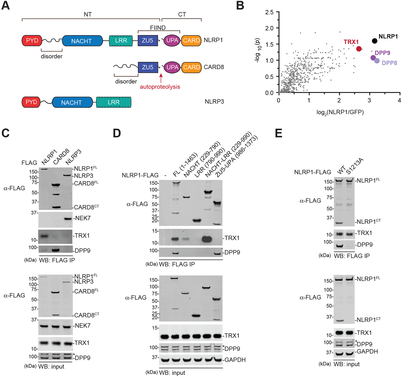
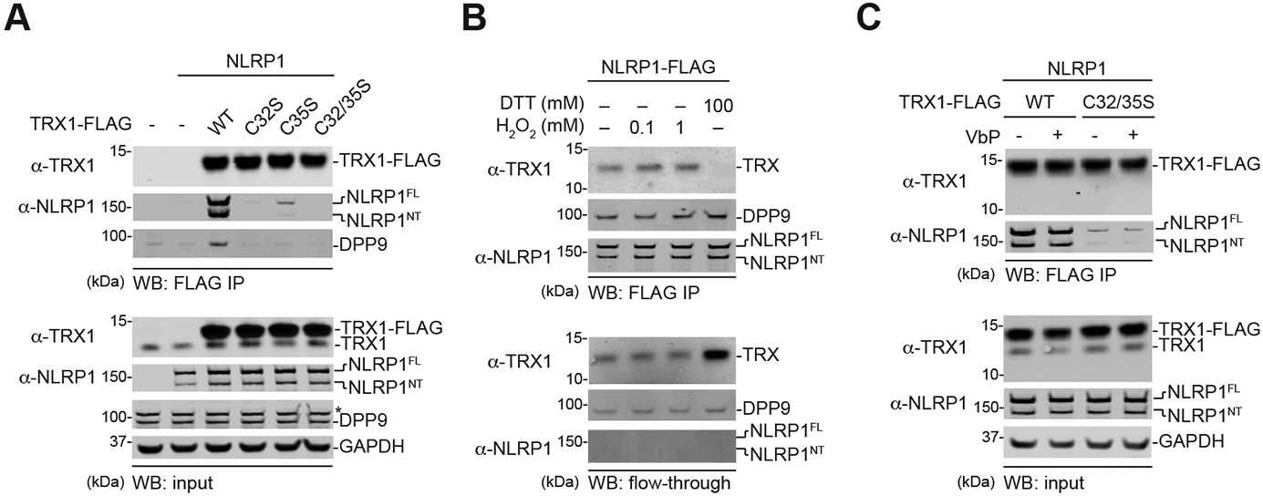
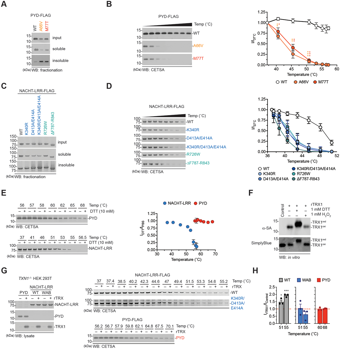
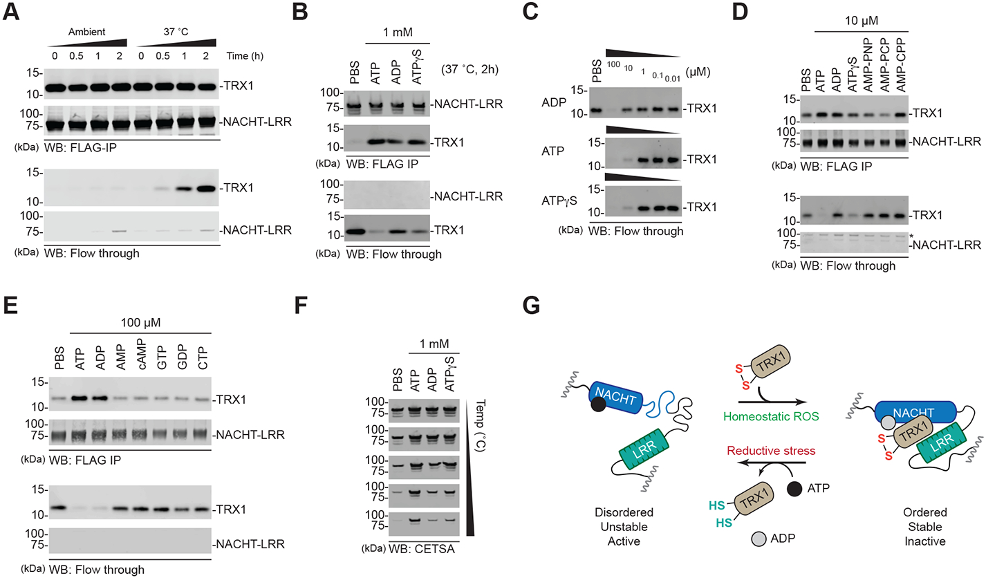
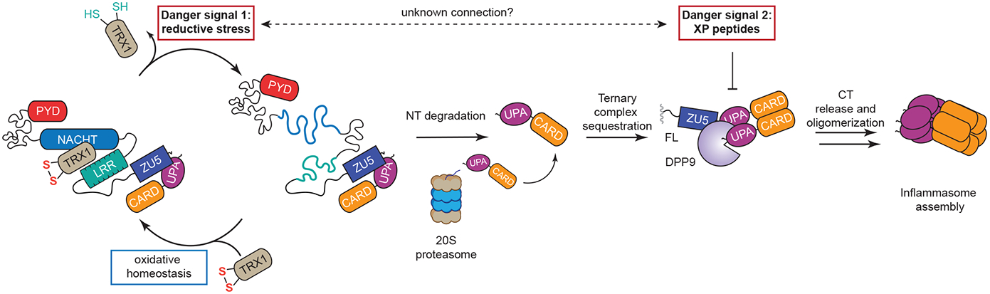
The danger signals that activate the NLRP1 inflammasome have not been established. Here, we report that the oxidized, but not the reduced, form of thioredoxin-1 (TRX1) binds to NLRP1. We found that oxidized TRX1 associates with the NACHT-LRR region of NLRP1 in an ATP-dependent process, forming a stable complex that restrains inflammasome activation. Consistent with these findings, patient-derived and ATPase-inactivating mutations in the NACHT-LRR region that cause hyperactive inflammasome formation interfere with TRX1 binding. Overall, this work strongly suggests that reductive stress, the cellular perturbation that will eliminate oxidized TRX1 and abrogate the TRX1-NLRP1 interaction, is a danger signal that activates the NLRP1 inflammasome.


Figures

Fig. 1.
Fig. 1.. The NACHT-LRR region of NLRP1 associates with TRX1.
(A) Domain organization of the human NLRP1, CARD8, and NLRP3 proteins. The FIINDs of NLRP1 and CARD8 undergo autoproteolysis between the ZU5 and UPA subdomains. NT, N-terminus. CT, C-terminus. (B) HEK 293T cells were transfected with GFP- or NLRP1-FLAG, subjected to anti-FLAG IP, and analyzed by quantitative mass spectrometry. The volcano plot depicts proteins enriched in the NLRP1-FLAG IP. (C-E) HEK 293T cells were transiently transfected with the indicated FLAG-tagged constructs, subjected to anti-FLAG IP, and analyzed by immunoblotting. In D, numbers indicate amino acid residues. Asterisks (*) denote background bands.
Fig. 2.
Fig. 2.. Only oxidized TRX1 binds NLRP1.
(A-C) HEK 293T cells were transfected with the indicated constructs before lysates were harvested, subjected to anti-FLAG IP, and immunoblotted. Anti-FLAG beads were treated with the indicated concentrations of (anti)oxidants in B and VbP (10 μM) in C. The flow-through in B is the unbound fraction after (anti)oxidant treatment. Note that cells were transfected with untagged NLRP1 in A and C. An asterisk (*) denotes a background band.
Fig. 3.
Fig. 3.. Thioredoxin restrains NLRP1 activation.
(A-D) N/TERT-1 keratinocytes were electroporated with control or TXN1 RNPs before being treated with VbP (10 μM) for 6 h or 16 h. Supernatants were analyzed for levels of LDH (A), IL-18 (B), and IL-1β (C) release, and both lysates and supernatants were subjected to immunoblotting analyses (D). (E,F) WT and NLRP1−/− N/TERT-1 keratinocytes were electroporated with control or TXN1 RNPs and treated with VbP (10 μM, 8 h). Supernatants were assessed for IL-1β release (E), and lysates and supernatants were evaluated by immunoblotting (F). Data in A-C and E are means ± SEM of three biological replicates. (G) Lysates from BMDMs from the indicated mouse strains were evaluated by immunoblotting. (H,I) The indicated mice were treated with vehicle or VbP (1 mg/kg, i.p.). The levels or serum cytokines were evaluated after 6 h by ELISA (H) or Luminex (I). Data in H are means ± SEM. Data in I are the column sum normalization of each cytokine (all data in Fig. S4D) depicted using Morpheus Software (Broad Institute). n = 8 and 11 for vehicle-treated WT and Txn1−/− mice, respectively, and n = 10 and 12 for VbP-treated WT and Txn1−/− mice, respectively. *p < 0.05, **p < 0.01,***p < 0.001 by two-sided Student’s t-test.
Fig. 4.
Fig. 4.. Hyperactive NLRP1 NACHT-LRR mutations weaken TRX1 binding.
(A) Diagram of NLRP1 mutations. Patient-derived mutations are shown above the protein. ATPase- and FIIND-inactivating mutations are shown below the protein. (B-D) NLRP1−/− N/TERT-1 keratinocytes were complemented with the indicated NLRP1 mutant behind a tetracycline-inducible promotor. Cells were stimulated with doxycycline (1 μg/mL) in the presence or absence of VbP (10 μM, 24 h) before analysis of inflammasome activation by LDH release (B), IL-1β ELISA (C), or immunoblotting (D). (E,F) HEK 293T cells were transiently transfected with the indicated FLAG-tagged NLRP1 constructs before lysates were harvested, subjected to anti-FLAG IP, and immunoblotted. Data in B and C are means ± SEM of three biological replicates. *p < 0.05, **p < 0.01, ***p < 0.001 by two-sided Student’s t-test. Asterisks (*) denote background bands.
Fig. 5.
Fig. 5.. TRX1 stabilizes the NACHT-LRR region.
(A-E) HEK 293T cells were transfected with constructs encoding isolated FLAG-tagged WT or mutant PYD and NACHT-LRR domains as indicated. Lysates were harvested, fractionated by centrifugation (A,C) or subjected to CETSA analysis (B,D, and E) and probed by immunoblotting. In E, CETSA was performed in the presence or absence of 10 mM DTT. The graphs in B and D are the quantifications of immunoblot densitometry. The graph in E is the ratio of the immunoblot densitometry in the presence of DTT over PBS alone. (F) Recombinant TRX1 was incubated with DTT or H2O2 (1 mM) for 30 min at 37°C before the addition of iodoacetamide-biotin (20 mM, 2 h, 37°C) and immunoblotting analysis. (G) Constructs encoding isolated FLAG-tagged PYD, NACHT-LRR (WT or K340R/D413A/E414A) were transiently transfected into TXN1-deficient HEK 293T cells. Lysates were subjected to CETSA analysis in the presence or absence of recombinant oxidized TRX1 protein (1.58 μM). (H) Gel band intensities from G were standardized relative to lane 1 (no rTRX1, 37°C). The graph depicts the ratio of immunoblot density in the presence or absence of rTRX1 (IrTRX1/IControl) at 51°C and 55°C for the NACHT-LRRs and 60°C and 68°C for the PYD constructs (the hashed red line depicts no change in stabilization). Graphs in B, D, E, and H are means ± SEM of three biological replicates. *p < 0.05, **p < 0.01, ***p < 0.001 by a two-sided Student’s t-test.
Fig. 6.
Fig. 6.. ATP binding and hydrolysis is coupled to TRX1 binding.
(A-E) HEK 293T cells were transfected with constructs encoding the isolated FLAG-tagged NLRP1 NACHT-LRR region. Protein was immobilized on anti-FLAG beads, washed with PBS, and incubated for the indicated times under the specified conditions. Dissociated (flow through) and complexed (FLAG-IP) TRX1 levels were assessed by immunoblotting. (F) Eluates from B were subjected to CETSA analysis. (G) Proposed model for the nucleotide-dependent states of NLRP1’s NACHT-LRR region.
Fig. 7.
Fig. 7.. Proposed mechanism for NLRP1 activation.
During homeostasis, oxidized TRX1 is complexed with NLRP1, stabilizing it in an inactive conformation. In the presence of reductive stress (“Danger Signal 1”), the levels of oxidized TRX1 are lowered, causing it to dissociate from NLRP1. The TRX1-free form of NLRP1 is more unstable, leading to accelerated degradation by the proteasome. The CT fragments released by this mechanism are initially sequestered in a ternary complex with DPP9 and NLRP1FL. The accumulation of XP-containing peptides (“Danger Signal 2”) disrupts this ternary complex and cause inflammasome activation. It remains unclear if these two danger signals are in some way related.

Similar articles

Cited by

References

    1. D'Osualdo A et al., CARD8 and NLRP1 undergo autoproteolytic processing through a ZU5-like domain. PLoS One 6, e27396 (2011). - PMC - PubMed
    1. Finger JN et al., Autolytic proteolysis within the function to find domain (FIIND) is required for NLRP1 inflammasome activity. J. Biol. Chem 287, 25030–25037 (2012). - PMC - PubMed
    1. Frew BC, Joag VR, Mogridge J, Proteolytic processing of Nlrp1b is required for inflammasome activity. PLoS Pathog. 8, e1002659 (2012). - PMC - PubMed
    1. Sandstrom A et al., Functional degradation: A mechanism of NLRP1 inflammasome activation by diverse pathogen enzymes. Science, (2019). - PMC - PubMed
    1. Chui AJ et al., N-terminal degradation activates the NLRP1B inflammasome. Science 364, 82–85 (2019). - PMC - PubMed

Publication types