. 2022 Nov 4;8(44):eabm4089.
doi: 10.1126/sciadv.abm4089. Epub 2022 Nov 4.

Insights into the evolution of enzymatic specificity and catalysis: From Asgard archaea to human adenylate kinases

Affiliations

Insights into the evolution of enzymatic specificity and catalysis: From Asgard archaea to human adenylate kinases

Apoorv Verma et al. Sci Adv. .

Abstract

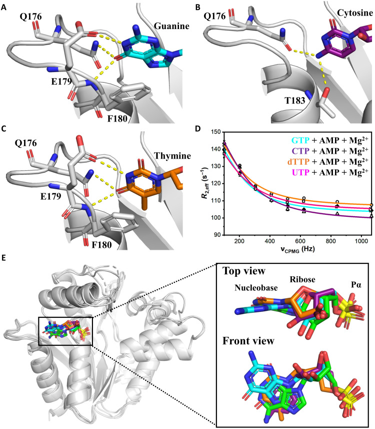
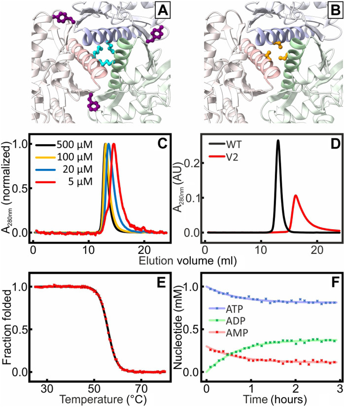
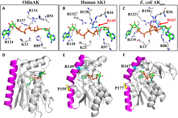
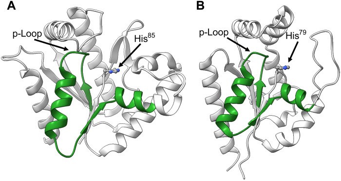
Enzymatic catalysis is critically dependent on selectivity, active site architecture, and dynamics. To contribute insights into the interplay of these properties, we established an approach with NMR, crystallography, and MD simulations focused on the ubiquitous phosphotransferase adenylate kinase (AK) isolated from Odinarchaeota (OdinAK). Odinarchaeota belongs to the Asgard archaeal phylum that is believed to be the closest known ancestor to eukaryotes. We show that OdinAK is a hyperthermophilic trimer that, contrary to other AK family members, can use all NTPs for its phosphorylation reaction. Crystallographic structures of OdinAK-NTP complexes revealed a universal NTP-binding motif, while 19F NMR experiments uncovered a conserved and rate-limiting dynamic signature. As a consequence of trimerization, the active site of OdinAK was found to be lacking a critical catalytic residue and is therefore considered to be "atypical." On the basis of discovered relationships with human monomeric homologs, our findings are discussed in terms of evolution of enzymatic substrate specificity and cold adaptation.


Figures

Fig. 1.
Fig. 1.. Multiple sequence alignment of archaeal AK enzymes.
Shown is a multiple sequence alignment between the two AK enzymes from Odinarchaeota (i.e., AdkA1, hereafter named OdinAK, and AdkA2) and three other archaeal AKs for which the crystallographic structure has been solved. The three other AK sequences are from Sulfolobus acidocaldarius (PDB ID: 1NKS), M. thermolithotrophicus (PDB ID: 1KI9), and Methanococcus voltae (PDB ID: 1KHT). The location of the ATP- and AMP-binding domains (ATPlid and AMPbd) is indicated, together with the location of the selectivity loop (7). The alignment was generated with Clustal Omega. Identical residues are colored green, while similar residues are colored yellow.
Fig. 2.
Fig. 2.. Structure of hyperthermophilic OdinAK.
(A) The thermal stability of OdinAK was followed with DSC. The Tm was determined from the inflection point of the thermogram and was found to be 95°C. The data have been corrected with the solvent background, and the normalized partial heat capacity is displayed. (B) Crystal structure of trimeric apo OdinAK showing trimer contacts between trimerization helices (magenta), β-hairpins (teal), and β strands (red) of adjacent monomers. (C) Structure of a single subunit of closed OdinAK in complex with Ap5A. The specific recognition of the adenosine base of ATP by the side chain of Q176 is highlighted. (D) Zoom-in on the selectivity loop showing recognition of the ATP base by the side chains of Q176 and T183. (E and F) Superimposition of 5 known trimeric and archaeal (E) and 30 monomeric (F) AK structures from the PDB illustrating that OdinAK has a unique and significantly longer selectivity loop (shown in black) relative to all other AKs.
Fig. 3.
Fig. 3.. OdinAK substrate-binding affinity and enzyme kinetics.
(A) ITC binding isotherm between OdinAK and ATP at 25°C. The best fitted isotherm is shown as a green line, and fitted parameters are displayed in Table 1. (B) Representative 31P NMR spectra showing the real-time evolution of mono-, di-, and trinucleoside phosphates from AMP, ADP, and ATP, respectively, over time. The experiment was initiated by mixing ATP, AMP, and Mg2+ together with catalytic amounts of OdinAK. (C and D). Quantification of enzymatic activity by a real-time 31P NMR assay at 25°C (C) and 65°C (D). The rate equations (16) were fitted to the evolution of nucleotide concentrations to obtain kcat (Table 2).
Fig. 4.
Fig. 4.. OdinAK conformational dynamics during catalysis.
(A) Selection of suitable probe sites for 19F NMR by comparing the structures of OdinAK in open (substrate-free) and closed (Ap5A-bound) states. Wild-type W174 (magenta) located in the selectivity loop yielded flat dispersion profiles and therefore Y44, V50, and G132 (cyan) were tested for insertion of 19F probe. (B) One-dimensional (1D) 19F NMR spectra of apo (black) and Ap5A-bound (red) OdinAK Y44W at 65°C, showing chemical shifts of mutant Y44W and wild-type W174 peaks as fingerprints for open and closed states of the enzyme. (C) Titration of OdinAK Y44W with ADP at 65°C showing that the enzyme adopts the closed conformation only in the presence of both binding site substrates and Mg2+. (D) 19F NMR spectra of substrate-bound OdinAK Y44W at 65°C. The green and blue spectra correspond to saturating concentrations [ATP (1:10) + AMP (1:10)] and ADP (1:20), respectively. Under these conditions, the signals from both tryptophans (44 and 174) are overlapping. (E) 19F NMR spectra of OdinAK Y44W under turnover conditions, i.e., substrate-bound states supplemented with 5 mM Mg2+ at 65°C. Addition of Mg2+ shifts the enzyme toward the closed state as deduced from the chemical shift of Y44W and indicated with the dashed line. (F) Quantification of dynamics during catalysis from 19F RD experiments under turnover conditions at 65°C. The observed R2 relaxation rate (R2,eff) obtained from Y44W is plotted against the applied CPMG field for samples generated from ATP, AMP, and Mg2+ (green) or ADP and Mg2+ (blue). The fitted parameters are shown in table S2, and rate constants for enzyme closing (kclose) and opening (kopen) are shown in Table 2.
Fig. 5.
Fig. 5.. Structural basis for universal NTP binding by OdinAK.
(A to C) Crystal structures of OdinAK in complex with NTPs demonstrate that the key determinant of selective binding of the nucleobases is the interactions formed by the side chain of Q176. In addition, the interactions formed by other important side chains are indicated in the different complexes. (A) Structure of OdinAK in complex with GTP. (B) Structure of OdinAK in complex with CTP. (C) Structure of OdinAK in complex with dTTP. (D) 19F CPMG RD profiles obtained from fluorinated Y44W for different substrates under turnover conditions (compare to Fig. 4F). The resulting fits from the data (table S2 and Table 2) show that OdinAK is rate-limited by slow conformational dynamics for all NTPs. (E) A key factor for the broad substrate specificity of OdinAK is harnessing of the endogenous flexibility of NTP molecules. Shown is a superimposition of NTP substrates bound to OdinAK. The color coding is as follows: ATP (green), GTP (cyan), CTP (purple), and dTTP (orange). The overlay was generated by superimposing the carbon α atoms of OdinAK, and for simplicity, only the base, ribose, and α phosphate are shown for the NTPs.
Fig. 6.
Fig. 6.. Formation of monomeric OdinAK by disruption of trimerization interfaces.
The trimerization interfaces were destabilized by replacing key amino acid residues identified from structural analysis. In V1, M154 (cyan) and Y166 (purple) (A) were mutated into glutamic acid. In V2, Met154, Tyr166, and Ser158 (orange) (B) were mutated into arginines. (C) SEC of different concentration samples of V1 showed that the elution for samples with decreasing concentration samples was shifted toward higher volumes, indicating the presence of an equilibrium between different oligomeric states. (D) The SEC profile for V2 (red) indicated a fully monomeric state that elutes at a volume that is significantly larger than that of a wild-type (WT) reference trimer (black). (E) Thermal unfolding of V2 probed with CD spectroscopy at 222 nm; Tm was determined to 56°C. (F) The kcat value of monomeric V2 at 25°C is 0.5 s−1 as quantified from the 31P NMR activity assay.
Fig. 7.
Fig. 7.. Active site architecture in trimeric and monomeric AK enzymes.
(A to C) Annotated active sites in AK enzymes. (A) The active site of trimeric OdinAK in complex with Ap5A (PDB ID: 7OWE) is atypical because it is lacking a critical arginine and has one of the otherwise conserved arginine residues replaced with histidine (His85). (B) The typical AK active site of monomeric human AK1 in complex with Ap5A (PDB ID: 1Z83) with the critical arginine-149 present. (C) The active site of AKeco in complex with two ADP molecules (PDB ID: 7APU) is a representative of a bacterial and monomeric enzyme that has the critical arginine-167 present. (D) The straight and close to ideal conformation of the trimerization helix (magenta) in OdinAK occludes the possibility of a typical active site because the N terminus of the trimerization helix projects away from the active site. (E) The corresponding α helix in monomeric AK1 is kinked due to a conserved proline residue at position 159. The kink places the N terminus of the α helix in an optimal position for complementation of the active site by R149. (F) Analogous to AK1, the α helix of AKeco is kinked by the presence of P177, which results in complementation of the active site by R167.
Fig. 8.
Fig. 8.. Sequence similarities between OdinAK and nuclear hAK6 displayed on crystallographic structures.
The figure illustrates the sequence similarities obtained by interrogating the human proteome with a BLAST search using OdinAK as queries. The top hit for both OdinAK was hAK6. (A) The sequence similarity between OdinAK and hAK6 is confined to residues 4 to 39 in OdinAK, and this segment is colored in green on molecule A of the apo OdinAK structure (PDB ID: 7OWH). The p-loop and H85 are indicated. (B) The sequence similarity between OdinAK and hAK6 illustrated on the hAK6 structure (PDB ID: 1RKB). For hAK6, the segment is consists of amino acid residues 7 to 41. The p-loop together with His79 is indicated. The details of the sequence similarities obtained from the BLAST search are shown in fig. S16.

Similar articles

Cited by

References

    1. Deamer D., Singaram S., Rajamani S., Kompanichenko V., Guggenheim S., Self-assembly processes in the prebiotic environment. Philos. Trans. R. Soc. Lond. B Biol. Sci. 361, 1809–1818 (2006). - PMC - PubMed
    1. Rona P. A., Klinkhammer G., Nelsen T. A., Trefry J. H., Elderfield H., Black smokers, massive sulfides and vent biota at the Mid-Atlantic Ridge. Nature 321, 33–37 (1986).
    1. Spang A., Saw J. H., Jørgensen S. L., Zaremba-Niedzwiedzka K., Martijn J., Lind A. E., van Eijk R., Schleper C., Guy L., Ettema T. J. G., Complex archaea that bridge the gap between prokaryotes and eukaryotes. Nature 521, 173–179 (2015). - PMC - PubMed
    1. Zaremba-Niedzwiedzka K., Caceres E. F., Saw J. H., Bäckström D., Juzokaite L., Vancaester E., Seitz K. W., Anantharaman K., Starnawski P., Kjeldsen K. U., Stott M. B., Nunoura T., Banfield J. F., Schramm A., Baker B. J., Spang A., Ettema T. J. G., Asgard archaea illuminate the origin of eukaryotic cellular complexity. Nature 541, 353–358 (2017). - PubMed
    1. Fournier G. P., Poole A. M., A briefly argued case that Asgard archaea are part of the eukaryote tree. Front. Microbiol. 9, (2018). - PMC - PubMed

Substances