

Editorial
doi: 10.1038/s41419-022-05375-7.
High-potency PD-1/PD-L1 degradation induced by Peptide-PROTAC in human cancer cells
Affiliations
- PMID: 36333311
- PMCID: PMC9636179
- DOI: 10.1038/s41419-022-05375-7
Item in Clipboard
Editorial
High-potency PD-1/PD-L1 degradation induced by Peptide-PROTAC in human cancer cells
Cell Death Dis.
.
No abstract available
Conflict of interest statement
The authors declare no competing interests.
Figures
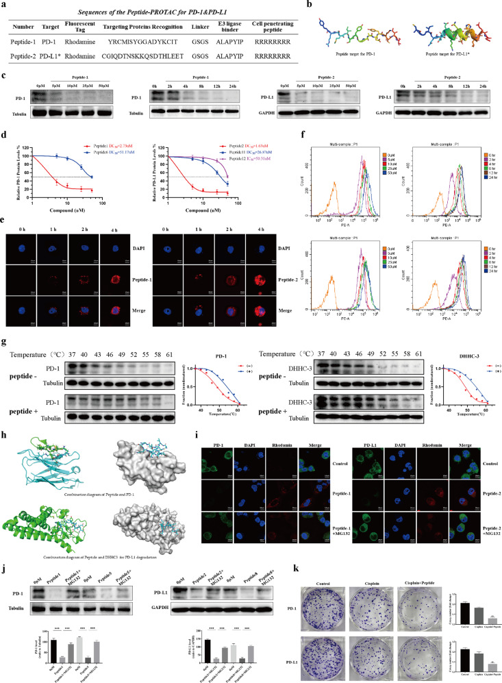
a The schematic illustrates the locations of the CPP sequence, TPR Peptide sequence, Peptide Linker, and ERP sequence. Peptide 1: the initial TPR sequence of Peptide 1 that target PD-1, and *Peptide 2 induced the decrease of PD-L1 dramatically by mean of the initial TPR sequence that target PD-L1 palmitoylation. b Schematic of peptides for PD-1 and PD-L1 palmitoylation. The AlphaFold 2 Artificial Intelligence system was used to generate three-dimensional structures of Peptide 1 (targeting PD-1) and Peptide 2 (targeting DHHC3, upstream of PD-L1) and visualizations were performed using the PyMOL software. c Western blotting analysis of C33A cells after treatment with the Peptide-PROTAC (Peptide 1 and Peptide 2) degraders at the indicated doses and timepoints. Values are presented as mean ± SEM. One-way ANOVA followed by Tukey’s post hoc test (n = 3): *P < 0.05, **P < 0.01, ***P < 0.001. d DC50 (50% protein degradation concentration) of Peptides targeting PD-1/PD-L1 in C33A cells. Both Peptide 1 and 6 target for PD-1, but Peptide 1 had a much better degradation effect than Peptide 6. Peptide 2 targeting for PD-L1 palmitoyltransferase ZDHHC3 (DHHC3) had a much better PD-L1 decrease effect than Peptide 11 targeting for PD-L1 directly and Peptide 12 which is the inhibition peptide without EPR sequence. e C33A cells were treated with 10 μM of rhodamine-labeled Peptide-PROTAC targeting PD-1 or PD-L1. After 4 h, the distribution of peptides in C33A cells was observed by confocal laser microscopy. Red; Peptide-PROTAC, blue; nucleus. Scale bar, 10 μM. f C33A cells were incubated with rhodamine-labeled Peptide-PROTAC targeting PD-1 or PD-L1 at the indicated concentrations and timepoints, then fluorescent cells were quantified by flow cytometry. Results are reported for 100,000 cells. n = 3. g CETSA-based determination of binding between Peptide (1 and 2) and PD-1/DHHC3. CETSA curves of PD-1/DHHC3 in C33A cells were determined in the absence and presence of 25 μM Peptide and analyzed by western blotting. GAPDH was used as an internal control. The band intensities of PD-1/DHHC3 were normalized with respect to the intensity at 40 °C. (−) without Peptide-PROTAC treatment, (+) with Peptide-PROTAC treatment. h Combination diagram of Peptide and PD-1/DHHC3. i Confocal microscopic images show the effects of Peptide-PROTAC (10 μM) and rescue by MG132 (4 mM) in C33A cells. Scale bars, 10 μM. j Western blotting analysis shows the PD-1 and PD-L1 levels after treating C33A cells with Peptide-PROTAC for 4 h. MG132 (4 mM) was added for 4 h before cell harvest. Values are presented as mean ± SEM. One-way ANOVA followed by Tukey’s post-hoc test (n = 3): ***P < 0.001. k C33A cells were treated with Peptide-PROTAC (10 μM) targeting either PD-1 or PD-L1 + cisplatin (15 μM). Colony formation assays were performed following treatment with cisplatin or cisplatin + Peptide-PROTAC treatments.
References
-
- Yao H, Lan J, Li C, Shi H, Brosseau JP, Wang H, et al. Inhibiting PD-L1 palmitoylation enhances T-cell immune responses against tumours. Nat Biomed Eng. 2019;3:306–17. - PubMed
Publication types
MeSH terms
Substances
LinkOut - more resources
Full Text Sources
Other Literature Sources
Medical
Research Materials
