. 2022 Nov 4;13(1):6648.
doi: 10.1038/s41467-022-34352-z.

Osteocytes directly regulate osteolysis via MYD88 signaling in bacterial bone infection

Affiliations

Osteocytes directly regulate osteolysis via MYD88 signaling in bacterial bone infection

Tetsuya Yoshimoto et al. Nat Commun. .

Abstract

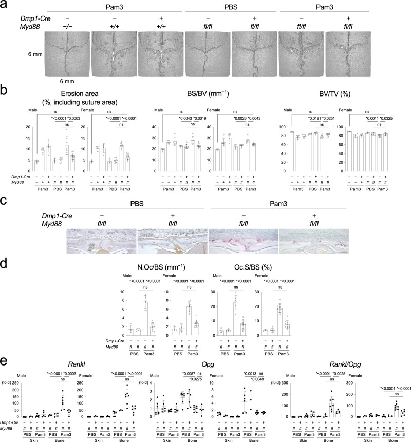
The impact of bone cell activation on bacterially-induced osteolysis remains elusive. Here, we show that matrix-embedded osteocytes stimulated with bacterial pathogen-associated molecular patterns (PAMPs) directly drive bone resorption through an MYD88-regulated signaling pathway. Mice lacking MYD88, primarily in osteocytes, protect against osteolysis caused by calvarial injections of bacterial PAMPs and resist alveolar bone resorption induced by oral Porphyromonas gingivalis (Pg) infection. In contrast, mice with targeted MYD88 restoration in osteocytes exhibit osteolysis with inflammatory cell infiltration. In vitro, bacterial PAMPs induce significantly higher expression of the cytokine RANKL in osteocytes than osteoblasts. Mechanistically, activation of the osteocyte MYD88 pathway up-regulates RANKL by increasing binding of the transcription factors CREB and STAT3 to Rankl enhancers and by suppressing K48-ubiquitination of CREB/CREB binding protein and STAT3. Systemic administration of an MYD88 inhibitor prevents jawbone loss in Pg-driven periodontitis. These findings reveal that osteocytes directly regulate inflammatory osteolysis in bone infection, suggesting that MYD88 and downstream RANKL regulators in osteocytes are therapeutic targets for osteolysis in periodontitis and osteomyelitis.


Conflict of interest statement

The authors declare no competing interests.

Figures

Fig. 1
Fig. 1. Lack of MYD88 in osteocytes and mature osteoblasts rescues calvarial osteolysis induced by Pam3CSK4 injection.
a MicroCT images of the calvaria. Representative images from each group of male mice (n ≥ 5/group). b Erosion area, BS/BV, and BV/TV of the calvaria (n = 5, 5, 5, 5, 8, 7, 11 in male, n = 4, 6, 4, 4, 7, 9, 10 in female). c TRAP staining of the calvarial bone. Representative images from each group of male mice (n ≥ 4/group). Scale bar = 100 μm. d Histomorphometric analysis of osteoclasts on the calvarial bone surface (n = 4, 7, 7, 10 in male, n = 4, 6, 9, 10 in female). e qPCR analysis of Rankl (n = 6, 3, 7, 5, 4, 4, 7, 5 in male, n = 6, 3, 6, 5, 6, 3, 7, 6 in female), Opg (n = 7, 6, 7, 5, 7, 6, 7, 5 in male, n = 6, 3, 7, 6, 6, 3, 7, 6 in female), and their ratio (n = 6, 3, 7, 5, 4, 4, 7, 5 in male, n = 6, 3, 6, 5, 6, 3, 7, 6 in female) in skin and calvarial bone tissues. ae Pam3 = Pam3CSK4. b, d, e Data are presented as mean ± SD. *p < 0.05 with one-way ANOVA with Tukey–Kramer test. ns = not significant. Each data point represents a biologically independent mouse. Source data are provided as a Source Data file.
Fig. 2
Fig. 2. The TLR2-MYD88 axis in osteocytes and mature osteoblasts regulates osteolysis in periodontitis induced by Porphyromonas gingivalis infection.
ac, f Top: MicroCT images of the right maxilla. Representative images from each group of male mice (n ≥ 5/group). Buccal side view. Bottom: The total CEJ-ABC distance of the right maxillary molars and alveolar BV/TV underneath the second molar of the right maxilla. a n = 8, 5, 5, 6 in male. n = 5, 5, 8, 6 in female. b n = 5, 5, 7, 6 in male. n = 4, 6, 7, 7 in female. c CEJ-ABC: n = 7, 9, 17, 13 in male. n = 5, 8, 4, 11 in female. BV/TV: n = 7, 8, 16, 10 in male. n = 5, 8, 4, 11 in female. f n = 5, 5, 5, 7 in male. n = 3, 6, 3, 8 in female. d, e Top: TRAP staining of the alveolar bone. Representative images from each group of male mice (n ≥ 4/group). Scale bar = 100 μm. Bottom: Histomorphometric analysis of osteoclasts on the alveolar bone surface. d n = 4, 5, 7, 6 in male. n = 4, 7, 7, 7 in female. e n = 5, 6, 15, 11 in male. n = 5, 6, 4, 9 in female. g Immunofluorescence images of the Pg component detected in the alveolar bone (green, indicated by arrows). Nuclei: DAPI (blue). AB = Alveolar bone. PDL = Periodontal ligament. Representative images from three independent experiments. Scale bar = 100 µm. ag Pg = Porphyromonas gingivalis. af Data are presented as mean ± SD. *p < 0.05 with one-way ANOVA with Tukey–Kramer test. ns = not significant. Each data point represents a biologically independent mouse. Source data are provided as a Source Data file.
Fig. 3
Fig. 3. Targeted activation of the MYD88 pathway in osteocytes and mature osteoblasts is sufficient to trigger calvarial osteolysis and alveolar bone loss.
a Top: MicroCT images of the calvaria. Representative images from each group of male mice injected with Pam3CSK4 (n ≥ 6/group). Bottom: Erosion area (n = 7, 10, 3, 8, 8, 6), BS/BV (n = 7, 9, 3, 8, 8, 6), and BV/TV (n = 7, 9, 3, 8, 8, 6) of the calvaria. b Top: MicroCT images of the right maxilla. Representative images from each group of male mice inoculated with Pg (n ≥ 3/group). Buccal side view. Bottom: The total CEJ-ABC distance of the right maxillary molars and alveolar BV/TV underneath the second molar of the right maxilla. n = 4, 4, 3, 4, 3, 5. c Top: TRAP staining of the calvarial bone. Representative images from each group of male mice injected with Pam3CSK4 (n ≥ 5/group). Scale bar = 100 μm. Bottom left: Histomorphometric analysis of osteoclasts on the calvarial bone surface. n = 5, 7, 3, 7, 5, 6. Bottom right: qPCR analysis of Rankl in calvarial bone tissue. n = 5, 7, 6, 8, 7, 7. d Top: TRAP staining of the alveolar bone. Representative images from each group of male mice inoculated with Pg (n ≥ 3/group). Scale bar = 100 μm. Bottom left: Histomorphometric analysis of osteoclasts on the alveolar bone surface. n = 3, 4, 3, 4, 3, 5. Bottom right: qPCR analysis of Rankl in alveolar bone tissue. n = 4, 5, 6, 4, 3, 5. e H&E staining of skin tissue on the intersection of the coronal and sagittal sutures. Representative images from each group of male mice injected with Pam3CSK4 (n ≥ 3/group). Scale bar = 100 μm. f qPCR analysis of inflammatory cytokines in skin tissue on the calvaria and gingival tissue. Skin Tnf and Il1b (n = 5, 3, 3, 9, 3, 4). Skin Il6 (n = 4, 3, 3, 9, 3, 4). Gingival Tnf, Il1b, and Il6 (n = 4, 3, 3, 4, 3, 3). g Left: Immunohistochemical staining of neutrophils and macrophages in skin tissue on the calvaria. Representative images from three independent experiments with similar results. Scale bar = 100 μm. Right: qPCR analysis of macrophage and neutrophil marker genes in skin tissue on the calvaria. Ly6g (n = 4, 3, 3, 8, 3, 4). Adgre1 (n = 5, 3, 3, 8, 3, 4). ag Data from male mice. Female mice showed the similar results. Pam3 = Pam3CSK4. PgPorphyromonas gingivalis. ad, f, g Data are presented as mean ± SD. *p < 0.05 with one-way ANOVA with Tukey–Kramer test. #p < 0.05 when two-tailed unpaired t-test was used (f, g). ns = not significant with ANOVA. Each data point represents a biologically independent mouse. Source data are provided as a Source Data file.
Fig. 4
Fig. 4. PAMPs induce more robust RANKL expression in the osteocyte-enriched cell population than the osteoblast-enriched cell population.
a qPCR analysis of Rankl in the osteoblast-enriched cell population (Ob) and osteocyte-enriched cell population (Ocy) stimulated with Pam3CSK4 (100 ng/mL), E. coli LPS (100 ng/mL), or PBS every 24 h. Average expression levels in Ob treated with PBS for 0 h were set as 1. Representative data from five independent experiments with similar results, each with three replicates (n = 3). b Western blotting of RANKL with cell lysates from Ob and Ocy stimulated with Pam3CSK4, E. coli LPS, or PBS. Representative images from five independent experiments with similar results. cf Co-culture of bone marrow-derived M-CSF-dependent macrophages from Myd88−/− male mice with Ob or Ocy isolated from wild-type (Myd88+/+) C57BL/6 J male mice. Cells were stimulated with Pam3CSK4, E. coli LPS, or PBS for 7 days. c, e Formation of TRAP + multinucleated cells (MNCs) and numbers of TRAP + MNCs per well. Representative images and data (n = 6/group) from three independent experiments with similar results. Scale bar = 100 μm. d, f Numbers of TRAP + MNCs per well in the presence and absence of RANKL neutralizing antibody for 7 days. Representative data from three independent experiments with similar results (n = 6/group). ad Pam3 = Pam3CSK4. a, cf Data are presented as mean ± SD. a #p < 0.05 with two-tailed unpaired t-test. c, e #p < 0.05 with two-tailed unpaired t-test. *p < 0.05 when one-way ANOVA with Tukey–Kramer test was used. ns = not significant with t-test. d, f *p < 0.05 with one-way ANOVA with Tukey–Kramer test. Source data are provided as a Source Data file.
Fig. 5
Fig. 5. Activation of the MYD88 pathway induces RANKL via the ERK-CREB/STAT3 axes in the osteocyte-enriched cell population.
a Western blotting of phosphorylated (p) and total ERK, JNK, p38, and NF-kB p65 proteins in Ocy stimulated with Pam3CSK4 or PBS. min. = minutes. b, c qPCR analysis of Rankl. Ocy was pretreated with increasing concentrations of inhibitors or vehicle (DMSO) for 2 h before stimulation with Pam3CSK4 or PBS for 8 h. Representative data from three independent experiments with similar results, each with three replicates (n = 3). d Western blotting of phosphorylated (p) and total CREB and STAT3 proteins in Ocy stimulated with Pam3CSK4 or PBS. min. = minutes. e Left: Western blotting of phosphorylated (p) and total CREB and STAT3 proteins. Ocy was pretreated with the MYD88 inhibitor T6167923 (20 µM) or vehicle (DMSO) for 2 h before stimulation with Pam3CSK4 or PBS for 8 h. Right: Densitometric analysis of the p-CREB/CREB and p-STAT3/STAT3 ratio using ImageJ. f Left: Western blotting of phosphorylated (p) and total CREB and STAT3 proteins. Ocy was pretreated with the MEK1/2 inhibitor U0126 (20 µM) or vehicle (DMSO) for 2 h before stimulation with Pam3CSK4 or PBS for 8 h. Right: Densitometric analysis of the p-CREB/CREB and p-STAT3/STAT3 ratio using ImageJ. e, f Graphs were created from the data of three independent experiments (n = 3). g CUT & RUN assays of CREB and STAT3 in Ocy stimulated with Pam3CSK4 or PBS for 8 h. Inductions of CREB and STAT3 binding to the Rankl promoter and enhancers were quantitated by qPCR relative to the isotype control IgGs (fold enrichment). Representative data from three independent experiments with similar results, each with three replicates (n = 3). The diagram shows the mouse Rankl promoter and enhancer loci and the binding sites of CREB and STAT3 in Ocy stimulated with Pam3CSK4. TSS = Transcription start site. ag Pam3 = Pam3CSK4. a, d, e, f Representative images from more than three independent experiments. b, c, e, f, g Data are presented as mean ± SD. b, c, e, f *p < 0.05 with one-way ANOVA with Tukey–Kramer test. ns = not significant. g #p < 0.05 with two-tailed unpaired t-test. Source data are provided as a Source Data file.
Fig. 6
Fig. 6. The MYD88 pathway regulates CREB, CBP, and STAT3 protein stability in the osteocyte-enriched cell population.
a Western blotting of CREB, CBP, and STAT3 proteins in Ocy isolated from wild-type (Myd88+l+) male mice. Ocy was treated with T6167923 or vehicle (DMSO) every 24 h. Graphs show the intensities of the protein bands relative to those of DMSO-treated Ocy at 0 h (%). Graphs were created from the data of three independent experiments (n = 3). Normalized by β -Actin. b qPCR analysis of Creb1, Cbp, and Stat3 in male Myd88+l+ Ocy treated with or without T6167923 (20 µM) for 48 h. T6167923 was added to the cultures every 24 h. Representative data from three independent experiments with similar results, each with three replicates (n = 3). c Western blotting of the indicated proteins in Ocy isolated from Dmp1-Cre;Myd88fl/fl and Myd88fl/fl male mice. Graphs show the intensities of the protein bands from Dmp1-Cre;Myd88fl/fl mice relative to those from Myd88fl/fl mice normalized by β-Actin (%). Graphs were created from the data of three independent experiments (n = 3). d qPCR analysis of Creb1, Cbp, and Stat3 in Ocy isolated from Dmp1-Cre;Myd88fl/fl and Myd88fl/fl male mice. Representative data from three independent experiments, each with three replicates (n = 3). e,f Western blotting of CREB, CBP, and STAT3 in Ocy isolated from Myd88+l+ male mice. Ocy was treated with a proteasomal inhibitor MG132 (1 μM) or a lysosomal inhibitor leupeptin (100 μM) for 3 h before treatment with T6167923 (20 μM) for 48 h. MG132, leupeptin, and T6167923 were added to the cultures every 24 h. a, b, c, d Data are presented as mean ± SD. #p < 0.05 with two-tailed unpaired t-test. ns = not significant. a, c, e, f Representative images from more than three independent experiments. Source data are provided as a Source Data file.
Fig. 7
Fig. 7. Activation of the MYD88 pathway induces RANKL by decreasing CREB, CBP, and STAT3 ubiquitination in the osteocyte-enriched cell population.
a Degradation kinetics of CREB, CBP, and STAT3 proteins in Ocy stimulated with Pam3CSK4 or PBS. Ocy was treated with cycloheximide (CHX, 10 μM) and Pam3CSK4/PBS every 24 h. Graphs show the intensities of the protein bands relative to those at 0 h (%). Normalized by β-Actin. bd Western blotting of K48-ubiquitinated proteins after immunoprecipitation (IP) of CREB, CBP, and STAT3. Ocy was stimulated with Pam3CSK4 or PBS for 48 h. e Western blotting of FBXL19 in Ocy stimulated with Pam3CSK4 or PBS. f IP of CREB and CBP followed by Western blotting for FBXL19. 48 h after Pam3CSK4 or PBS stimulation. g Western blotting of K48-ubiquitinated proteins after IP of CREB or CBP. Cell lysates of differentiated IDG-SW3 cells overexpressing FLAG-tagged mouse FBXL19 were used. Graphs show the relative intensities of CREB, CBP, and FBXL19 protein bands against β-Actin in whole cell lysates. h Left: Western blotting of RANKL using cell lysates from differentiated IDG-SW3 cells overexpressing FLAG-tagged mouse FBXL19. Right: Relative RANKL protein levels normalized by β-Actin. i qPCR analysis of Rankl in differentiated IDG-SW3 cells overexpressing FLAG-tagged mouse FBXL19 stimulated with Pam3CSK4 or PBS for 48 h. j Western blotting of PDLIM2 in Ocy stimulated with Pam3CSK4 or PBS. k IP of STAT3 followed by Western blotting for PDLIM2. 48 h after Pam3CSK4 or PBS stimulation. l Western blotting of K48-ubiquitinated proteins after IP of STAT3. Cell lysates of differentiated IDG-SW3 cells overexpressing MYC-tagged mouse PDLIM2 were used. Graphs show the relative intensities of STAT3 and PDLIM2 protein bands against β-Actin in whole cell lysates. m Left: Western blotting of RANKL using cell lysates from differentiated IDG-SW3 cells overexpressing MYC-tagged mouse PDLIM2. Right: Relative RANKL protein levels normalized by β-Actin. n qPCR analysis of Rankl in differentiated IDG-SW3 cells overexpressing MYC-tagged mouse FBXL19 stimulated with Pam3CSK4 or PBS for 48 h. af, ik, n Pam3 = Pam3CSK4. a, g, h, l, m Graphs were created from the data of three independent experiments (n = 3). i, n Representative data from three independent experiments with similar results, each with three replicates (n = 3). a, g, h, i, l, m, n Data are presented as mean ± SD. #p < 0.05 with two-tailed unpaired t-test. ah, jm Representative images from more than three independent experiments. Source data are provided as a Source Data file.
Fig. 8
Fig. 8. Administration of an MYD88 inhibitor protects against alveolar bone loss in periodontitis induced by Porphyromonas gingivalis infection.
a Left: MicroCT images of the right maxilla from Porphyromonas gingivalis (Pg)- or vehicle-inoculated wild-type C57BL/6 J male mice administered with T6167923 or DMSO. Buccal side view. Representative images from each group of male mice. Right: The total CEJ-ABC distance at the right maxillary molars. b Alveolar BV/TV underneath the right maxillary second molar. a, b Pg + DMSO (n = 5), Pg + T6167923 (n = 4), Vehicle + DMSO (n = 4), Vehicle + T6167923 (n = 4). c Left: TRAP staining of the alveolar bone. Representative images from each group of male mice (n = 5/group). Scale bar = 100 μm. Right: Histomorphometric analysis of osteoclasts on the alveolar bone surface. d qPCR analysis of osteoclast-associated genes in the alveolar bone (n = 5/group). ad Data are presented as mean ± SD. *p < 0.05 with one-way ANOVA with Tukey–Kramer test. ns = not significant. Each data point represents a biologically independent mouse. Source data are provided as a Source Data file.

References

    1. Ruff WE, Greiling TM, Kriegel MA. Host-microbiota interactions in immune-mediated diseases. Nat. Rev. Microbiol. 2020;18:521–538. - PubMed
    1. Robling AG, Bonewald LF. The osteocyte: new insights. Annu Rev. Physiol. 2020;82:485–506. - PMC - PubMed
    1. de Mesy Bentley KL, et al. Evidence of staphylococcus aureus deformation, proliferation, and migration in canaliculi of live cortical bone in murine models of osteomyelitis. J. Bone Miner. Res.: Off. J. Am. Soc. Bone Miner. Res. 2017;32:985–990. - PMC - PubMed
    1. Yang, D. Q. et al. Novel insights into staphylococcus aureus deep bone infections: the involvement of osteocytes. Mbio9, e00415–e00418 (2018). - PMC - PubMed
    1. de Mesy Bentley KL, MacDonald A, Schwarz EM, Oh I. Chronic osteomyelitis with staphylococcus aureus deformation in submicron canaliculi of osteocytes: a case report. JBJS Case Connect. 2018;8:e8. - PMC - PubMed

Publication types

MeSH terms

Substances