

Fabrication of an Au-doped Cu/Fe oxide-polymer core-shell nanoreactor with chemodynamic and photodynamic dual effects as potential cancer therapeutic agents
- PMID: 36333398
- PMCID: PMC9636373
- DOI: 10.1038/s41598-022-23002-5
Fabrication of an Au-doped Cu/Fe oxide-polymer core-shell nanoreactor with chemodynamic and photodynamic dual effects as potential cancer therapeutic agents
Abstract
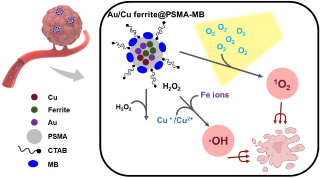
Nanoparticles are widely used in biomedical applications and cancer treatments due to their minute scale, multi-function, and long retention time. Among the various nanoparticles, the unique optical property derived from the localized surface plasmon resonance effect of metallic nanoparticles is a primary reason that metallic nanoparticles are researched and applied. Copper and Iron nanoparticles have the potential to generate hydroxyl radicals in excess H2O2 via Fenton or Fenton-like reactions. On the other hand, gold nanoparticles equipped with a photosensitizer can transfer the energy of photons to chemical energy and enhance the production of singlet oxygen, which is suitable for cancer treatment. With the actions of these two reactive oxygen species in the tumor microenvironment, cell apoptosis can further be induced. In this work, we first synthesized dual metal nanoparticles with poly[styrene-alt-(maleic acid, sodium salt)(Cu ferrite oxide-polymer) by a simple one-step hydrothermal reduction reaction. Then, gold(III) was reduced and doped into the structure, which formed a triple metal structure, Au-doped Cu ferrite nanoparticles (Au/Cu ferrite oxide-polymer NPs). The metal ratio of the product could be controlled by manipulating the Fe/Cu ratio of reactants and the sequence of addition of reactants. The core-shell structure was verified by transmission electron microscopy. Moreover, the hydroxyl radical and singlet oxygen generation ability of Au/Cu ferrite oxide-polymer was proved. The chemodynamic and photodynamic effect was measured, and the in vitro ROS generation was observed. Furthermore, the behavior of endocytosis by cancer cells could be controlled by the magnetic field. The result indicated that Au/Cu ferrite oxide-polymer core-shell nanoreactor is a potential agent for chemodynamic/photodynamic synergetic therapy.
© 2022. The Author(s).
Conflict of interest statement
The authors declare no competing interests.
Figures


References
-
- Kim D, Lee N, Park YI, Hyeon T. Recent advances in inorganic nanoparticle-based NIR luminescence imaging: Semiconductor nanoparticles and lanthanide nanoparticles. Bioconjug. Chem. 2017;28:115–123. - PubMed
-
- Wang Y, De S, Yan N. Rational control of nano-scale metal-catalysts for biomass conversion. Chem. Commun. 2016;52:6210–6224. - PubMed
-
- Liu T-M, Conde J, Lipiński T, Bednarkiewicz A, Huang C-C. Revisiting the classification of NIR-absorbing/emitting nanomaterials for in vivo bioapplications. Npg Asia Mater. 2016;8:e295.
