Review
. 2023 Feb;19(2):382-391.
doi: 10.1007/s12015-022-10467-0. Epub 2022 Nov 5.

Artificial Nerve Containing Stem Cells, Vascularity and Scaffold; Review of Our Studies

Affiliations
Review

Artificial Nerve Containing Stem Cells, Vascularity and Scaffold; Review of Our Studies

Ryosuke Kakinoki et al. Stem Cell Rev Rep. 2023 Feb.

Abstract

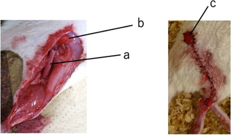
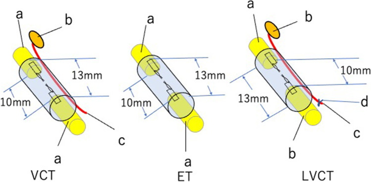
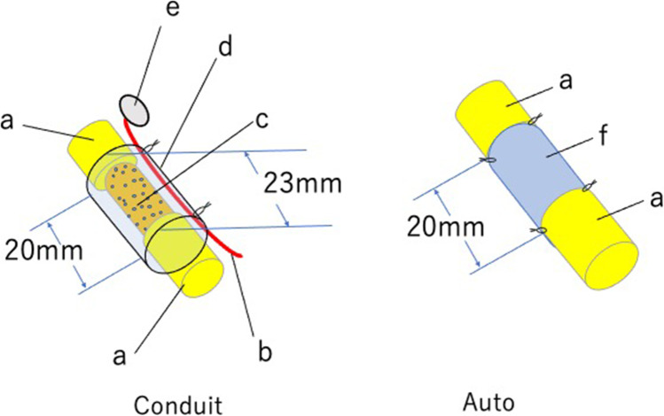
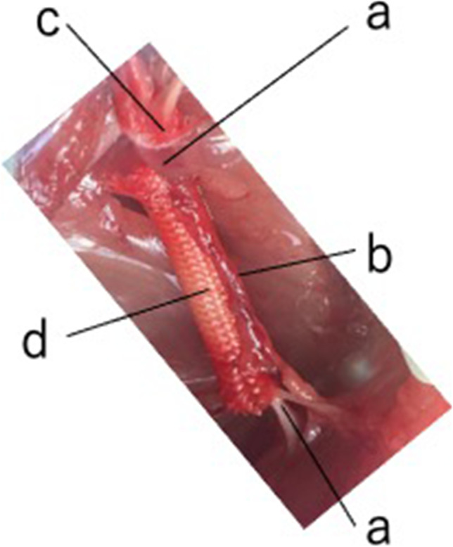
To promote nerve regeneration within a conduit (tubulation), we have performed studies using a tube model based on four important concepts for tissue engineering: vascularity, growth factors, cells, and scaffolds. A nerve conduit containing a blood vascular pedicle (vessel-containing tube) accelerated axon regeneration and increased the axon regeneration distance; however, it did not increase the number or diameter of the axons that regenerated within the tube. A vessel-containing tube with bone-marrow-derived mesenchymal stem cell (BMSC) transplantation led to the increase in the number and diameter of regenerated axons. Intratubularly transplanted decellularized allogenic nerve basal lamellae (DABLs) worked as a frame to maintain the fibrin matrix structure containing neurochemical factors and to anchor the transplanted stem cells within the tube. For the clinical application of nerve conduits, they should exhibit capillary permeability, biodegradability, and flexibility. Nerbridge® (Toyobo Co. Ltd., Osaka, Japan) is a commercially available artificial nerve conduit. The outer cylinder is a polyglycolic acid (PGA) fiber mesh and possesses capillary permeability. We used the outer cylinder of Nerbridge as a nerve conduit. A 20-mm sciatic nerve deficit was bridged by the PGA mesh tube containing DABLs and BMSCs, and the resulting nerve regeneration was compared with that obtained through a 20-mm autologous nerve graft. A neve-regeneration rate of about 70%-80% was obtained in 20-mm-long autologous nerve autografts using the new conduits.

Keywords: Artificial nerve; Axonal regeneration; Stem cells; Tissue engineering; Tubulation.


Conflict of interest statement

We have no competing interests with regard of this work.

Figures

Fig. 1
Fig. 1
Creation of a vessel-containing tube in a rat hind limb. A: A myocutaneous flap supplied by the sural vessels was elevated. B: The sural vascular pedicle was separated from the sural nerve and turned proximally. C: Through a longitudinal slit in the tube, the sural vascular pedicle was inserted into the tubular lumen. The sciatic nerve stumps were sutured to either end of the tube. a, Sural nerve; b, sural vascular pedicle; c, myocutaneous flap supplied by the sural vessels; d, sciatic nerve stump; e, tibial nerve; f, peroneal nerve; g, silicone tube; h, longitudinal slit (this was sealed with liquid silicone after vascular insertion)
Fig. 2
Fig. 2
Intraoperative (left) and postoperative (right) images of the vessel-containing tube model in rats. Left: Vessel-containing tube. a, vessel-containing silicone tube; b, myocutaneous flap. Right: A myocutaneous flap was used as a flap for monitoring sural vessel vascularity
Fig. 3
Fig. 3
Experimental groups: VCT, ET, and LVCT. VCT, a 13-mm-long sural vessel-containing silicone tube onto each end of which the sciatic nerve stumps were sutured, leaving a 10-mm interstump gap. ET, a 13-mm-long silicone tube without the vascular pedicle bridging the sciatic nerve stumps, leaving a 10-mm interstump gap. LVCT, a VCT tube with the vascular pedicle ligated at the popliteal fossa. a, sciatic nerve stump; b, myocutaneous flap; c, sural vessel pedicle; d, ligation of the sural vascular pedicle
Fig. 4
Fig. 4
Experimental groups: BMC, FIB, and VCT. BMC, an 18-mm-long sural-vessel-containing silicone tube, at each end of which the sciatic nerve stumps were sutured, leaving a 15-mm interstump gap, followed by the intratubular transplantation of 1 × 107 BMSCs. FIB, an 18-mm-long sural-vessel-containing silicone tube, at each end of which the sciatic nerve stumps were sutured, leaving a 15-mm interstump gap, followed by the intratubular transplantation of 1 × 107 fibroblasts. VCT, an 18-mm-long sural-vessel-containing silicone tube, at each end of which the sciatic nerve stumps were sutured, leaving a 15-mm interstump gap; no cells were transplanted into the tube. a, Sciatic nerve stump; b, myocutaneous flap; c, sural vessel pedicle; d, transplanted BMSCs; e, transplanted fibroblasts
Fig. 5
Fig. 5
Experimental groups: Conduit and Auto. Left: Conduit group: a 23-mm-long sural vessel-containing silicone tube with DABLs (prepared using the freeze–thaw technique) and 3 × 106 BMSCs intratubularly. Right: Auto group: a 20-mm-long sciatic nerve segment was transected. The segment was reversed and sutured between the proximal and distal sciatic nerve stumps
Fig. 6
Fig. 6
Nerbridge® (Toyobo, Osaka, Japan): the outer cylinder is a polyglycolic acid (PGA) fiber mesh, and the inner core is a collagen sponge. Images reproduced with permission from Toyobo Co Ltd
Fig. 7
Fig. 7
Experimental groups: Tube C + , Tube C–, and Auto. Tube C + : Two 20-mm-long DABLs (harvested from DA rats and prepared using chemosurfactants) seeded with 3 × 106 BMSCs were transplanted in a 23-mm-long polyglycolic acid mesh tube (the outer cylinder of Nerbridge®). A sural vascular pedicle was placed along the conduit. Tube C–: Two 20-mm-long DABLs (harvested from DA rats and prepared using chemosurfactants) without BMSC implantation were transplanted into a 23-mm-long polyglycolic acid mesh tube (the outer cylinder of Nerbridge®). A sural vascular pedicle was placed along the conduit. Auto: A 20-mm-long sciatic nerve segment was transected. The segment was reversed and sutured between the proximal and distal sciatic nerve stumps
Fig. 8
Fig. 8
Intraoperative photo of a conduit in the Tube C + group. Tube C + conduit. a, sciatic nerve stump; b, sural vascular pedicle; c, monitor flap; d, polyglycolic acid mesh tube
Fig. 9
Fig. 9
Transverse sections of the most distal part of the regenerated nerves in the Tube C + , Tube C–, and Auto groups. The vascular pedicles are indicated by arrows

References

    1. Lundborg G, Gelberman RH, Longo FM, et al. In vivo regeneration of cut nerves encased in silicone tubes: Growth across a six-millimeter gap. Journal Neuropatholgy and Experimental Neurolgy. 1982;41(4):412–422. doi: 10.1097/00005072-198207000-00004. - DOI - PubMed
    1. Dahlin LB, Lundborg G. Use of tubes in peripheral nerve repair. Neurosurgery Clinics of North America. 2001;12(2):341–352. doi: 10.1016/S1042-3680(18)30059-7. - DOI - PubMed
    1. Williams LR, Longo FM, Powell HC, et al. Spatial–temporal progress of peripheral nerve regeneration within a silicone chamber; Parameters for a bioassay. Journal of Comparative Neurology. 1983;218(4):460–470. doi: 10.1002/cne.902180409. - DOI - PubMed
    1. Lundborg G, Dahlin LB, Danielsen N, et al. Nerve regeneration in silicone chambers: Influence of gap and of distal stump components. Experimental Neurology. 1982;76(2):361–375. doi: 10.1016/0014-4886(82)90215-1. - DOI - PubMed
    1. Kakinoki R, Nishijima N, Ueba Y, et al. Relationship between axonal regeneration and vascularity in tubulation—An experimental study in rats. Neuroscience Research. 1995;23(1):35–45. - PubMed

LinkOut - more resources