. 2022 Nov 5;10(1):189.
doi: 10.1186/s40168-022-01380-2.

A novel and dual digestive symbiosis scales up the nutrition and immune system of the holobiont Rimicaris exoculata

Affiliations

A novel and dual digestive symbiosis scales up the nutrition and immune system of the holobiont Rimicaris exoculata

Johanne Aubé et al. Microbiome. .

Abstract

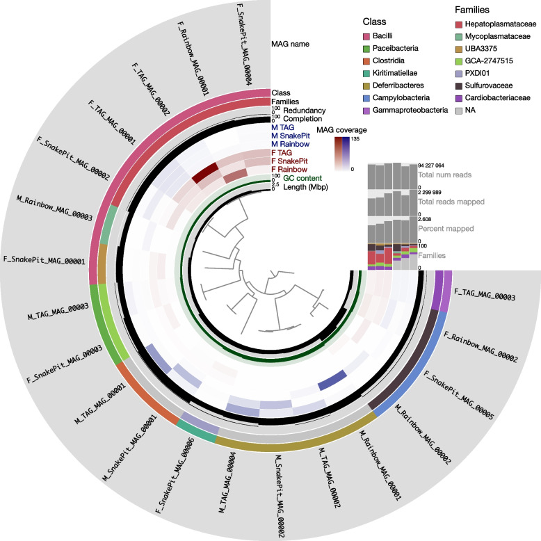
Background: In deep-sea hydrothermal vent areas, deprived of light, most animals rely on chemosynthetic symbionts for their nutrition. These symbionts may be located on their cuticle, inside modified organs, or in specialized cells. Nonetheless, many of these animals have an open and functional digestive tract. The vent shrimp Rimicaris exoculata is fueled mainly by its gill chamber symbionts, but also has a complete digestive system with symbionts. These are found in the shrimp foregut and midgut, but their roles remain unknown. We used genome-resolved metagenomics on separate foregut and midgut samples, taken from specimens living at three contrasted sites along the Mid-Atlantic Ridge (TAG, Rainbow, and Snake Pit) to reveal their genetic potential.

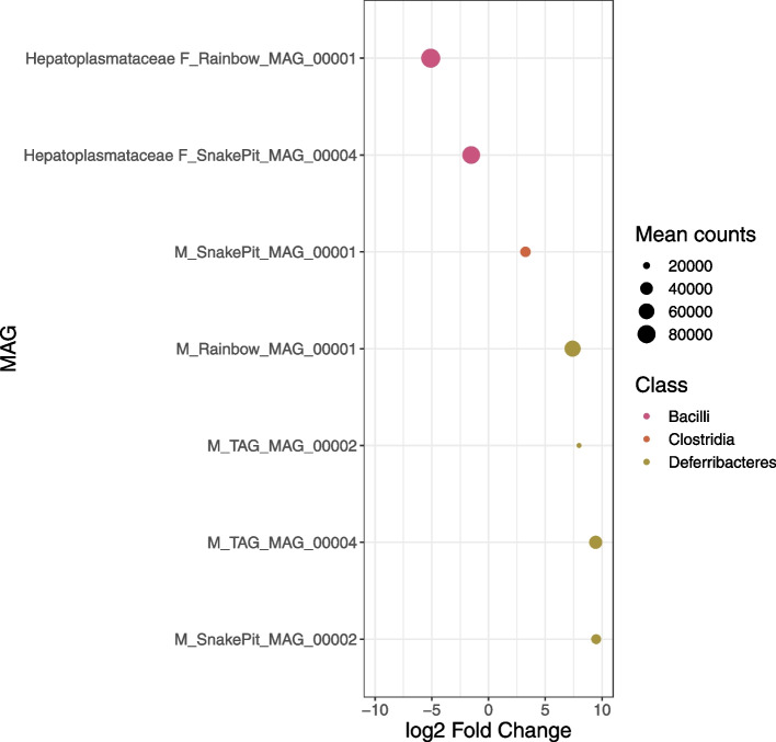
Results: We reconstructed and studied 20 Metagenome-Assembled Genomes (MAGs), including novel lineages of Hepatoplasmataceae and Deferribacteres, abundant in the shrimp foregut and midgut, respectively. Although the former showed streamlined reduced genomes capable of using mostly broken-down complex molecules, Deferribacteres showed the ability to degrade complex polymers, synthesize vitamins, and encode numerous flagellar and chemotaxis genes for host-symbiont sensing. Both symbionts harbor a diverse set of immune system genes favoring holobiont defense. In addition, Deferribacteres were observed to particularly colonize the bacteria-free ectoperitrophic space, in direct contact with the host, elongating but not dividing despite possessing the complete genetic machinery necessary for this.

Conclusion: Overall, these data suggest that these digestive symbionts have key communication and defense roles, which contribute to the overall fitness of the Rimicaris holobiont. Video Abstract.

Keywords: Deferribacteres; Digestive symbiosis; Hepatoplasmataceae; Immunity; Metagenomics; Rimicaris exoculata.


Conflict of interest statement

The authors declare that they have no competing interests.

Figures

Fig. 1
Fig. 1
Static image from the anvi’o interactive display for the six Rimicaris midgut (M) and foregut (F) samples with the 20 metagenome-assembled genomes (MAGs) reconstructed in this study. From inner to outer layers: phylogenomic tree based on concatenated protein-coding genes according to GTDB-Tk, length genome layer, GC-content information about contigs stored in the contig database auxiliary layer, six view layers with mean coverage information about MAGs across samples stored in the profile database, percentage completion and redundancy, genome family and class based on GTDB-Tk, and MAG layer. The horizontal layers show the MAG taxonomy based on GTDB-Tk for families (with the relative abundance of families noted as percentages of reads recruited to the bins for each sample), percentage of reads mapped, total number of reads mapped, and total number of reads for each sample
Fig. 2
Fig. 2
Differentially abundant MAGs between organs identified through DESeq2 of midgut vs. foregut. Each line on the y-axis indicates a MAG. MAG family is indicated on the left of the MAG name when they are assigned until this level. MAGs dots are colored according to the class level and size of the dots corresponds to mean read counts after normalization with GeTMM and DESeq2 (corresponding to baseMean). Positive and negative log2FoldChange values indicate differentially abundant MAGs in midguts and foreguts, respectively. Cutoff values for visualization in the plot were 0.01 for padj and 1.5 for log2FoldChange absolute value
Fig. 3
Fig. 3
Maximum likelihood phylogenetic tree using IQ-TREE v2.0.3 with the “WAG” general matrix model and 1000 bootstrap replicates visualized using FigTree for A Hepatoplasmataceae and B Deferribacteres MAGs and their closest relatives. Nodes represented by a dot indicate a bootstrap value of 100; lower values are specified. Ca. Hepatoplasma crinochetorum Ps and Ca. Hepatoplasma crinochetorum Av are noted A and B, respectively, in GTDB-Tk
Fig. 4
Fig. 4
Schematic representation of the predicted metabolic potentials within A the 5 Hepatoplasmataceae and B the 4 Deferribacteres MAGs using the KEGG annotations and the KEGG Mapper/Reconstruct tool. Genes present in all, at least half, or less than half are indicated in differentially contrasted colors. The metabolic potentials are focused on carbon metabolism, transporters, flagellum, and chemotaxis. Full names and gene copy numbers are detailed in Supplementary Table 4
Fig. 5
Fig. 5
FISH observation of Deferribacteres cells in the midgut of an adult male R. exoculata using newly designed probes [12]. These long filamentous bacteria (b) are inserted between the microvilli (m) of the intestine epithelium (e) of the host. Eukaryote nuclei (n) at the center of epithelial cells are labeled with DAPI (blue). The apical brush border of the epithelium with the microvilli (represented by the white line just beside the legend m) is all along the lumen (l). Bacterial cells (b) are co-hybridized with the Eubacterial probe (Eub338, colored in white on the image) and with the specific Deferribacteres symbiont probe (Def1229, colored in red on the image), so they are visible in pink on the image
Fig. 6
Fig. 6
Chromosome observation of the long filamentous bacteria using YOYO™-1 labeling. DNA (in green) was specifically labeled using the YOYO™-1 dye. The host nucleus is not visible here as being at the opposite of the microvilli of the epithelial host tissue (t). A Long filamentous bacteria (b) along the midgut brush-border cells. The white square is image B with a closer view. The scale bar represents 2 μm. B YOYO™-1 labeling reveals numerous bacterial chromosomes per cell (green balls). Cells are non-segmented and white arrows point out each chromosome. The scale bar represents 0.5 μm. C Fluorescence intensity profile within a bacterial cell revealing that chromosomes are regularly spaced by 0.6 to 0.7 μm

References

    1. McFall-Ngai MJ. Unseen forces: the influence of bacteria on animal development. Dev Biol. 2002;242:1–14. - PubMed
    1. Bordenstein SR, Theis KR. Host biology in light of the microbiome: ten principles of holobionts and hologenomes. PLoS Biol. 2015;13:e1002226. - PMC - PubMed
    1. Douglas AE. Symbiosis as a general principle in eukaryotic evolution. Cold Spring Harb Perspect Biol. 2014;6:a016113. - PMC - PubMed
    1. Zilber-Rosenberg I, Rosenberg E. Role of microorganisms in the evolution of animals and plants: the hologenome theory of evolution. FEMS Microbiol Rev. 2008;32:723–735. - PubMed
    1. Dubilier N, Bergin C, Lott C. Symbiotic diversity in marine animals: the art of harnessing chemosynthesis. Nat Rev Microbiol. 2008;6:725–740. - PubMed

Publication types

LinkOut - more resources